

Automated cross-sectional and longitudinal hippocampal volume measurement in mild cognitive impairment and Alzheimer's disease
- PMID: 20230901
- PMCID: PMC2873209
- DOI: 10.1016/j.neuroimage.2010.03.018
Automated cross-sectional and longitudinal hippocampal volume measurement in mild cognitive impairment and Alzheimer's disease
Abstract
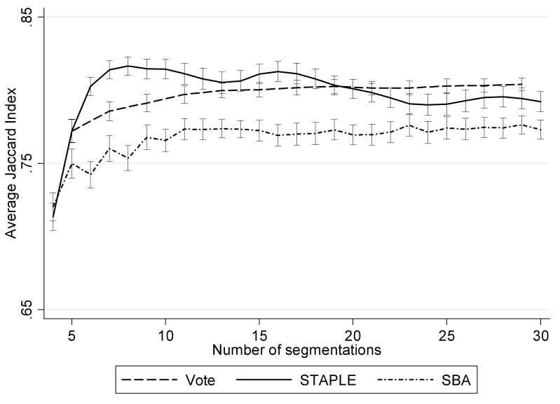
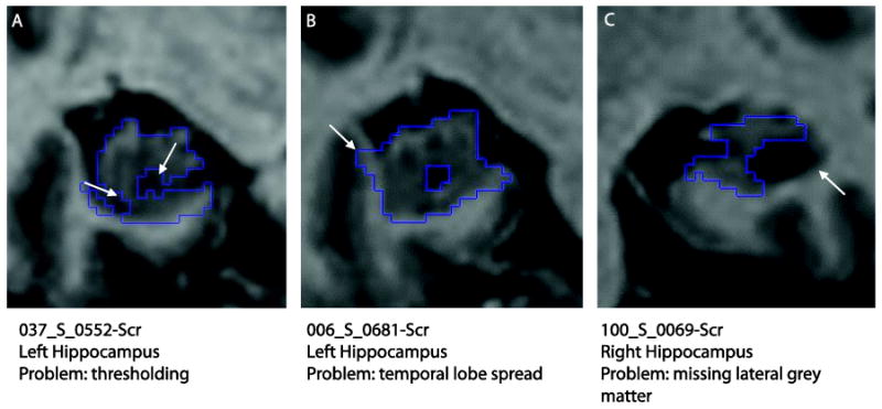
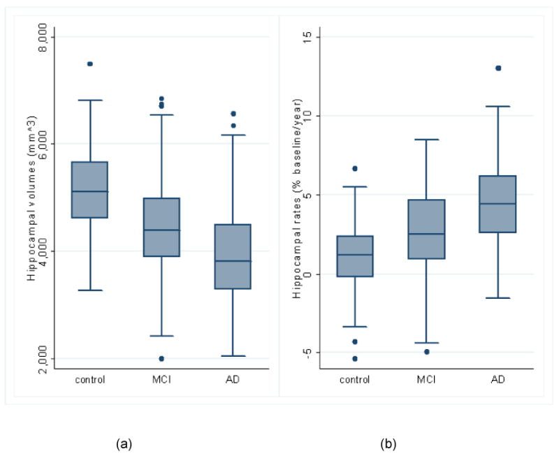
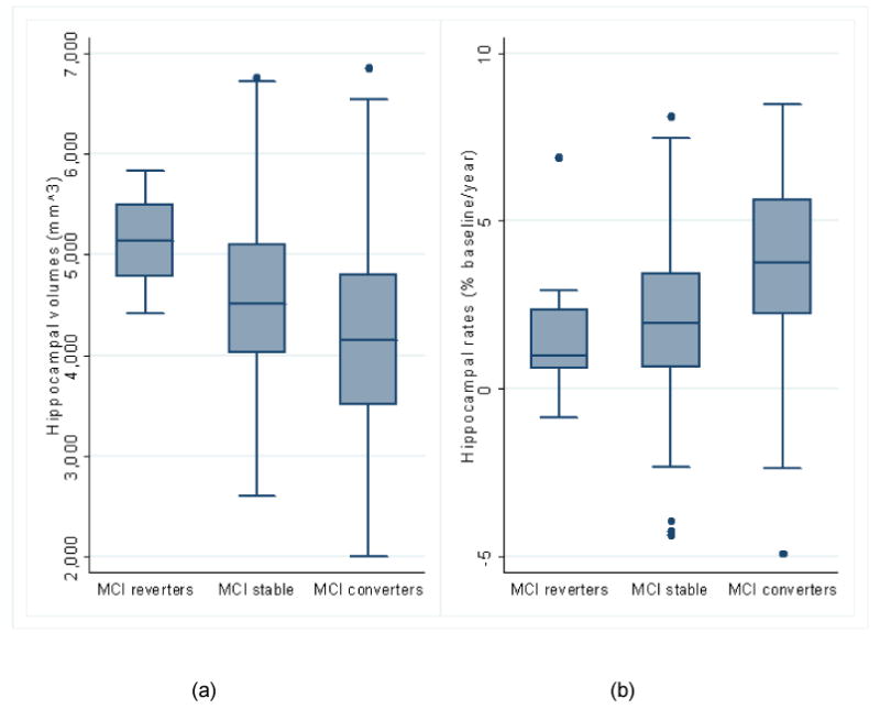
Volume and change in volume of the hippocampus are both important markers of Alzheimer's disease (AD). Delineation of the structure on MRI is time-consuming and therefore reliable automated methods are required. We describe an improvement (multiple-atlas propagation and segmentation (MAPS)) to our template library-based segmentation technique. The improved technique uses non-linear registration of the best-matched templates from our manually segmented library to generate multiple segmentations and combines them using the simultaneous truth and performance level estimation (STAPLE) algorithm. Change in volume over 12months (MAPS-HBSI) was measured by applying the boundary shift integral using MAPS regions. Methods were developed and validated against manual measures using subsets from Alzheimer's Disease Neuroimaging Initiative (ADNI). The best method was applied to 682 ADNI subjects, at baseline and 12-month follow-up, enabling assessment of volumes and atrophy rates in control, mild cognitive impairment (MCI) and AD groups, and within MCI subgroups classified by subsequent clinical outcome. We compared our measures with those generated by Surgical Navigation Technologies (SNT) available from ADNI. The accuracy of our volumes was one of the highest reported (mean(SD) Jaccard Index 0.80(0.04) (N=30)). Both MAPS baseline volume and MAPS-HBSI atrophy rate distinguished between control, MCI and AD groups. Comparing MCI subgroups (reverters, stable and converters): volumes were lower and rates higher in converters compared with stable and reverter groups (p< or =0.03). MAPS-HBSI required the lowest sample sizes (78 subjects) for a hypothetical trial. In conclusion, the MAPS and MAPS-HBSI methods give accurate and reliable volumes and atrophy rates across the clinical spectrum from healthy aging to AD.
Copyright 2010 Elsevier Inc. All rights reserved.
Figures
References
-
- Aljabar P, Heckemann RA, Hammers A, Hajnal JV, Rueckert D. Multi-atlas based segmentation of brain images: atlas selection and its effect on accuracy. Neuroimage. 2009;46:726–738. - PubMed
-
- Ashburner J, Friston KJ. Unified segmentation. Neuroimage. 2005;26:839–851. - PubMed
-
- Ashton EA, Parker KJ, Berg MJ, Chen CW. A novel volumetric feature extraction technique with applications to MR images. IEEE Trans Med Imaging. 1997;16:365–371. - PubMed
-
- Barnes J, Foster J, Boyes RG, Pepple T, Moore EK, Schott JM, Frost C, Scahill RI, Fox NC. A comparison of methods for the automated calculation of volumes and atrophy rates in the hippocampus. Neuroimage. 2008a;40:1655–1671. - PubMed
Publication types
MeSH terms
Grants and funding
LinkOut - more resources
Full Text Sources
Other Literature Sources
Medical
Molecular Biology Databases
