Control of wrist position and muscle relaxation by shifting spatial frames of reference for motoneuronal recruitment: possible involvement of corticospinal pathways
- PMID: 20231141
- PMCID: PMC2876809
- DOI: 10.1113/jphysiol.2009.186858
Control of wrist position and muscle relaxation by shifting spatial frames of reference for motoneuronal recruitment: possible involvement of corticospinal pathways
Abstract
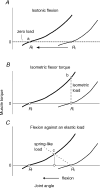
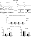
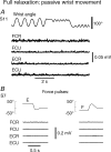
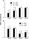
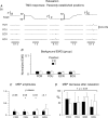
It has previously been established that muscles become active in response to deviations from a threshold (referent) position of the body or its segments, and that intentional motor actions result from central shifts in the referent position. We tested the hypothesis that corticospinal pathways are involved in threshold position control during intentional changes in the wrist position in humans. Subjects moved the wrist from an initial extended to a final flexed position (and vice versa). Passive wrist muscle forces were compensated with a torque motor such that wrist muscle activity was equalized at the two positions. It appeared that motoneuronal excitability tested by brief muscle stretches was also similar at these positions. Responses to mechanical perturbations before and after movement showed that the wrist threshold position was reset when voluntary changes in the joint angle were made. Although the excitability of motoneurons was similar at the two positions, the same transcranial magnetic stimulus (TMS) elicited a wrist extensor jerk in the extension position and a flexor jerk in the flexion position. Extensor motor-evoked potentials (MEPs) elicited by TMS at the wrist extension position were substantially bigger compared to those at the flexion position and vice versa for flexor MEPs. MEPs were substantially reduced when subjects fully relaxed wrist muscles and the wrist was held passively in each position. Results suggest that the corticospinal pathway, possibly with other descending pathways, participates in threshold position control, a process that pre-determines the spatial frame of reference in which the neuromuscular periphery is constrained to work. This control strategy would underlie not only intentional changes in the joint position, but also muscle relaxation. The notion that the motor cortex may control motor actions by shifting spatial frames of reference opens a new avenue in the analysis and understanding of brain function.
Figures




Similar articles
-
Threshold position control of anticipation in humans: a possible role of corticospinal influences.J Physiol. 2017 Aug 1;595(15):5359-5374. doi: 10.1113/JP274309. Epub 2017 Jun 28. J Physiol. 2017. PMID: 28560812 Free PMC article.
-
Subthreshold corticospinal control of anticipatory actions in humans.Behav Brain Res. 2011 Oct 10;224(1):145-54. doi: 10.1016/j.bbr.2011.05.041. Epub 2011 Jun 7. Behav Brain Res. 2011. PMID: 21672559
-
Corticospinal control strategies underlying voluntary and involuntary wrist movements.Behav Brain Res. 2013 Jan 1;236(1):350-358. doi: 10.1016/j.bbr.2012.09.008. Epub 2012 Sep 13. Behav Brain Res. 2013. PMID: 22983216
-
Dynamic changes in corticospinal control of precision grip during wrist movements.Brain Res. 2007 Aug 20;1164:32-43. doi: 10.1016/j.brainres.2007.06.014. Epub 2007 Jun 16. Brain Res. 2007. PMID: 17632089
-
Corticospinal excitability during fatiguing whole body exercise.Prog Brain Res. 2018;240:219-246. doi: 10.1016/bs.pbr.2018.07.011. Epub 2018 Sep 17. Prog Brain Res. 2018. PMID: 30390833 Free PMC article. Review.
Cited by
-
The Hand: Shall We Ever Understand How it Works?Motor Control. 2015 Apr;19(2):108-26. doi: 10.1123/mc.2014-0025. Epub 2014 Jul 16. Motor Control. 2015. PMID: 25271803 Free PMC article. Review.
-
Clinical Relevance of the Tonic Stretch Reflex Threshold and μ as Measures of Upper Limb Spasticity and Motor Impairment After Stroke.Neurorehabil Neural Repair. 2025 May;39(5):386-399. doi: 10.1177/15459683251318689. Epub 2025 Feb 13. Neurorehabil Neural Repair. 2025. PMID: 39945415 Free PMC article.
-
Cerebral activations related to ballistic, stepwise interrupted and gradually modulated movements in Parkinson patients.PLoS One. 2012;7(7):e41042. doi: 10.1371/journal.pone.0041042. Epub 2012 Jul 23. PLoS One. 2012. PMID: 22911738 Free PMC article.
-
Active sensing without efference copy: referent control of perception.J Neurophysiol. 2016 Sep 1;116(3):960-76. doi: 10.1152/jn.00016.2016. Epub 2016 Jun 15. J Neurophysiol. 2016. PMID: 27306668 Free PMC article. Review.
-
Muscle coactivation: definitions, mechanisms, and functions.J Neurophysiol. 2018 Jul 1;120(1):88-104. doi: 10.1152/jn.00084.2018. Epub 2018 Mar 28. J Neurophysiol. 2018. PMID: 29589812 Free PMC article. Review.
References
-
- Archambault PS, Mihaltchev P, Levin MF, Feldman AG. Basic elements of arm postural control analyzed by unloading. Exp Brain Res. 2005;164:225–241. - PubMed
-
- Asatrian DG, Fel’dman AG. Functional tuning of the nervous system with control of movement or maintenance of a steady posture – I. Mechanographic analysis of the work of the joint during the performance of a postural task. Biophysics. 1965;10:925–935. Engl transl of Biofizika 10, 837–846. - PubMed
-
- Battaglia D, Brunel N, Hansel D. Temporal decorrelation of collective oscillations in neural networks with local inhibition and long-range excitation. Phys Rev Lett. 2007;99:238106. - PubMed
-
- Bonnard M, Camus M, de Graaf J, Pailhous J. Direct evidence for a binding between cognitive and motor functions in humans: a TMS study. J Cogn Neurosci. 2003;15:1207–1216. - PubMed
Publication types
MeSH terms
Grants and funding
LinkOut - more resources
Full Text Sources

