. 2010 May 21;285(21):16166-83.
doi: 10.1074/jbc.M109.097725. Epub 2010 Mar 15.

Endoplasmic reticulum export, subcellular distribution, and fibril formation by Pmel17 require an intact N-terminal domain junction

Affiliations

Endoplasmic reticulum export, subcellular distribution, and fibril formation by Pmel17 require an intact N-terminal domain junction

Ralf M Leonhardt et al. J Biol Chem. .

Abstract

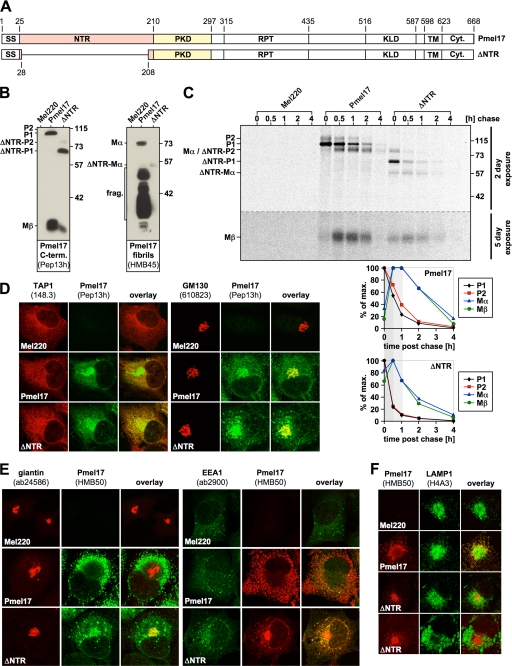
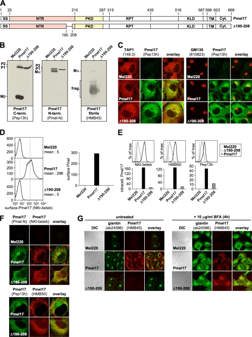
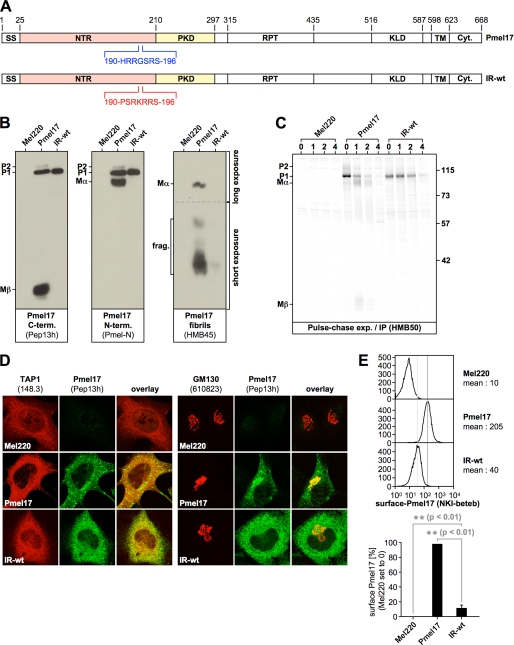
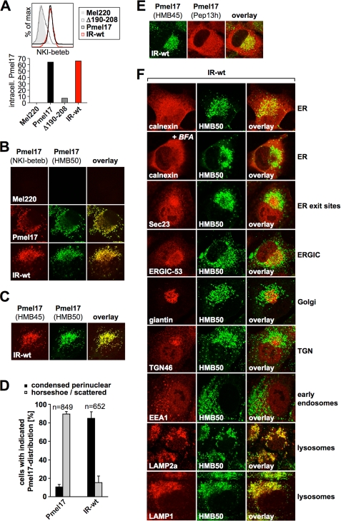
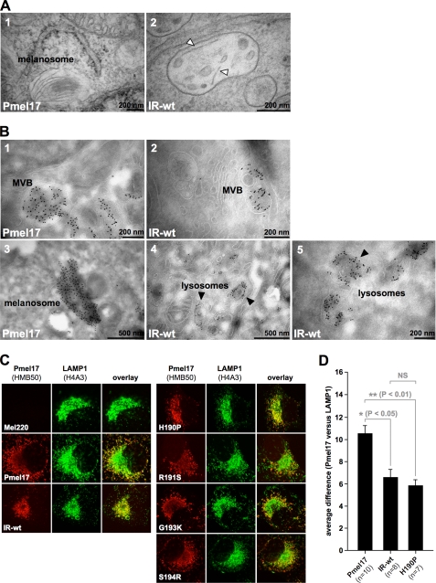
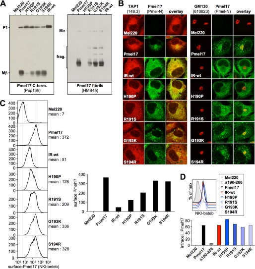
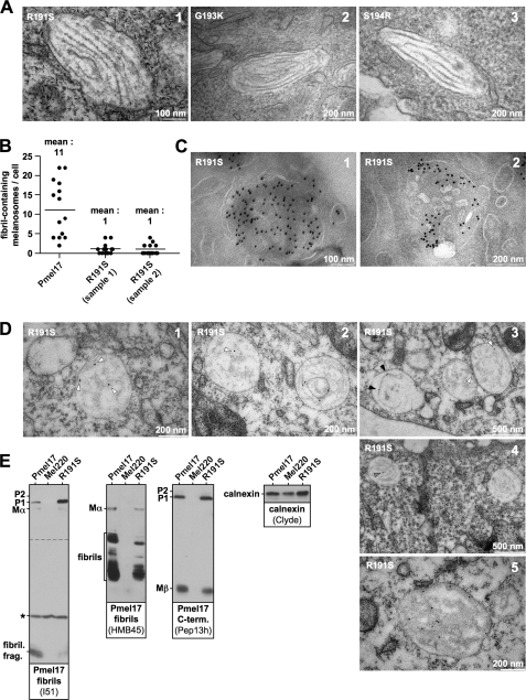
Pmel17 is a melanocyte/melanoma-specific protein that subcellularly localizes to melanosomes, where it forms a fibrillar matrix that serves for the sequestration of potentially toxic reaction intermediates of melanin synthesis and deposition of the pigment. As a key factor in melanosomal biogenesis, understanding intracellular trafficking and processing of Pmel17 is of central importance to comprehend how these organelles are formed, how they mature, and how they function in the cell. Using a series of deletion and missense mutants of Pmel17, we are able to show that the integrity of the junction between the N-terminal region and the polycystic kidney disease-like domain is highly crucial for endoplasmic reticulum export, subcellular targeting, and fibril formation by Pmel17 and thus for establishing functional melanosomes.


Figures

FIGURE 1.
FIGURE 1.
The NTR is required for normal subcellular targeting of Pmel17. A, schematic representation of the ΔNTR construct. B, ΔNTR can be processed by pPCs. A total membrane fraction derived from the indicated stable Mel220 transfectants was lysed in 1% SDS/1% β-mercaptoethanol plus protease inhibitors (Complete, Roche Applied Science) and analyzed by Western blot using Pmel17-specific antibodies. C, ΔNTR shows both accelerated pPC processing and turnover. Cells from Fig. 1B, were pulse-labeled for 30 min with 35S and subsequently chased for the indicated times. 2% Triton X-100 lysates were immunoprecipitated with Pmel17-specific antibody HMB50, eluted with 0.5% SDS under vigorous vortexing for 30 min, and analyzed by autoradiography (top panel). A 2-day exposure is shown for the upper part of the gel separated by the dashed line from a 5-day exposure for the lower part of the same gel. Quantitative PhosphorImager analysis of the pulse-chase data with maximal levels for each band set to 100% is shown (bottom panel). D, newly synthesized ΔNTR localizes to the ER and Golgi. Cells from B were analyzed by immunofluorescence using antibodies against newly synthesized Pmel17 (Pep13h) and organelle markers TAP1 (148.3) (ER) or GM130 (610823) (Golgi). E, folded HMB50-reactive ΔNTR co-localizes with Golgi and early endosomal markers. Cells from B were analyzed by immunofluorescence using antibodies against folded Pmel17 (HMB50) and organelle markers giantin (ab24586) (Golgi) or EEA1 (ab2900) (early endosomes). F, ΔNTR fails to migrate into LAMP1-positive compartments. Cells from B were analyzed by immunofluorescence using antibodies against folded Pmel17 (HMB50) and organelle marker LAMP1 (H4A3) (lysosomes/melanosomes).
FIGURE 2.
FIGURE 2.
The integrity of a small region in the vicinity of the NTR-PKD domain boundary is crucial for ER exit of Pmel17. A, schematic representation of the Δ190–208 construct. B, only the ER-associated P1 form can be detected for Δ190–208 at steady state. Membrane lysates of Mel220 transfectants stably expressing Δ190–208 were prepared as in Fig. 1B and analyzed by Western blot using Pmel17-specific antibodies. C, newly synthesized Δ190–208 is completely retained in the ER. Cells from Fig. 2B were analyzed by immunofluorescence using antibodies against newly synthesized Pmel17 (Pep13h) and organelle markers TAP1 (148.3) (ER) or GM130 (610823) (Golgi). D, folded NKI-beteb-reactive Δ190–208 is completely absent from the cell surface. Cells from Fig. 2B were surface-labeled with antibody NKI-beteb against folded Pmel17 and analyzed by flow cytometry (left panel). After background subtraction (untransfected Mel220 cells) data are represented as a bar diagram (right panel). E, low levels of folded Δ190–208 can be detected intracellularly. Cells from Fig. 2B were fixed in 2% formaldehyde, permeabilized, and stained intracellularly with antibodies reactive with folded (NKI-beteb or HMB50) or newly synthesized Pmel17 (Pep13h) and analyzed by flow cytometry (top panel). After background subtraction (untransfected Mel220 cells) data are represented as a bar diagram (bottom panel). F, folded HMB50- and NKI-beteb-reactive Δ190–208 localizes to the ER. Cells from Fig. 2B were analyzed by immunofluorescence using antibodies against folded Pmel17 (NKI-beteb (top panel) or HMB50 (bottom panel)) and newly synthesized Pmel17 (Pmel-N (top panel) or Pep13h (bottom panel)). G, Δ190–208 does not react with antibody HMB45, which specifically recognizes sialylated (post-Golgi- or Golgi-localized) Pmel17. Cells from Fig. 2B were treated or not with 10 μg/ml brefeldin A (BFA) for 4 h to shift Golgi-associated proteins back into the ER and analyzed by immunofluorescence using antibodies against sialylated (post-Golgi or Golgi-localized) Pmel17 (HMB45) and organelle marker giantin (ab24586) (Golgi).
FIGURE 3.
FIGURE 3.
Amino acid exchanges targeting the NTR-PKD domain boundary dramatically affect ER export. A, schematic representation of the IR-wt construct. B, only the ER-associated P1 form can be detected for IR-wt at steady state, when assessed with antibodies recognizing newly synthesized Pmel17. Membrane lysates of Mel220 transfectants stably expressing IR-wt were prepared as in Fig. 1B and analyzed by Western blot using Pmel17-specific antibodies. To visualize Mα better, a longer exposure of the upper part (separated by a dashed line from the lower part) of the same HMB45-blot (right panel) is shown. C, newly synthesized IR-wt shows almost no export from the ER or proprotein convertase-mediated processing. Mel220 transfectants stably expressing IR-wt were pulse-labeled for 30 min with 35S and subsequently chased for the indicated times. 2% Triton X-100 lysates were immunoprecipitated with Pmel17-specific antibody HMB50, eluted with 0.5% SDS under vigorous vortexing for 30 min, and analyzed by autoradiography. One representative out of two independent experiments is shown. D, newly synthesized IR-wt is largely retained in the ER. Cells from Fig. 3B were analyzed by immunofluorescence using antibodies against newly synthesized Pmel17 (Pep13h) and organelle markers TAP1 (148.3) (ER) or GM130 (610823) (Golgi). E, IR-wt is present at the cell surface only at minute levels. Cells from Fig. 3B were surface-labeled with antibody NKI-beteb against folded Pmel17 and analyzed by flow cytometry (top panel). After background subtraction (untransfected Mel220 cells) the data from three independent experiments are represented as a bar diagram (bottom panel). The difference in surface expression between wt-Pmel17 and IR-wt (**, p < 0.01) is statistically significant, as assessed by a one-way analysis of variance test with the Dunnett posttest.
FIGURE 4.
FIGURE 4.
Mel220 transfectants stably expressing IR-wt build up a large post-ER pool of this mutant that localizes to lysosomes. A, high levels of folded IR-wt can be detected intracellularly. Mel220 transfectants stably expressing IR-wt or Δ190–208 were fixed in 2% formaldehyde, permeabilized, and stained with antibodies reactive with folded Pmel17 (NKI-beteb) and analyzed by flow cytometry (top panel). After background subtraction (untransfected Mel220 cells) data are represented as a bar diagram (bottom panel). B, NKI-beteb- and HMB50-reactive IR-wt displays a distinct subcellular pattern as wt-Pmel17. Mel220 transfectants expressing wt-Pmel17 or IR-wt were analyzed by immunofluorescence using antibodies against folded Pmel17 (NKI-beteb and HMB50). C, HMB50-reactive IR-wt localizes to a post-Golgi compartment. Mel220 transfectants expressing IR-wt were analyzed by immunofluorescence using antibodies against sialylated (post-Golgi- or Golgi-localized) Pmel17 (HMB45) and folded Pmel17 (HMB50). D, in contrast to wt-Pmel17, HMB50-reactive IR-wt is mostly present in a condensed perinuclear pattern. Mel220 transfectants expressing wt-Pmel17 or IR-wt were analyzed by immunofluorescence using antibodies against folded Pmel17 (HMB50). Cells displaying either a condensed perinuclear pattern or a horseshoe-shaped/scattered pattern of fluorescence were blind-counted, and the results from two slides are displayed as a bar diagram. E, antibodies Pep13h and HMB45 recognize distinct populations of IR-wt. Cells from Fig. 4C were analyzed by immunofluorescence using antibodies against sialylated (post-Golgi- or Golgi-localized) Pmel17 (HMB45) and newly synthesized Pmel17 (Pep13h). F, folded HMB50-reactive post-ER IR-wt localizes to lysosomes. Cells from Fig. 4C were analyzed by immunofluorescence using antibodies against folded Pmel17 (HMB50) and organelle markers calnexin (Clyde) (ER), Sec23 (S-7696) (ER-exit sites), ERGIC-53 (E-1031) (ERGIC), giantin (ab24586) (Golgi), trans-Golgi network46 (ab16052) (TGN), EEA1 (555798) (early endosomes), LAMP2a (ab18528) (lysosomes), and LAMP1 (H4A3) (lysosomes).
FIGURE 5.
FIGURE 5.
The post-ER populations of IR-wt and H190P are non-functional and largely route to lysosomes. A, IR-wt is severely impaired or blocked in fibril formation. Electron microscopic analysis of Epon-embedded Mel220 transfectants stably expressing wt-Pmel17 or IR-wt. Panel 1 shows a typical melanosome frequently found for wt-Pmel17. Panel 2 shows an example of the very rare occurrence of immature organelles that contain individual striae-like structures (white arrowheads). Almost always such structures could be clearly identified as membrane segments as judged by a visible double layer structure. However, in a few remaining cases image quality did not allow us to fully exclude that they represent an immature fibril in the formation process. Hence, IR-wt is at least severely impaired, but more likely completely blocked in fibril formation. B, folded HMB50-reactive IR-wt gets mostly delivered to compartments with lysosomal morphology. Cells expressing wt-Pmel17 or IR-wt were fixed and examined by cryo-immunoelectron microscopy (panels 1–5) using antibody HMB50. Panels 1 and 2 display MVBs with extensive immunolabeling over intralumenal vesicles. Panel 3 shows a typical melanosome in wt-Pmel17-expressing cells. Black arrowheads in panels 4 and 5 point to lysosomal, often multilamellar compartments densely labeled with gold particles in IR-wt-expressing cells. C, wt-Pmel17 mostly distributes to LAMP1low melanosomes, whereas IR-wt and H190P extensively co-localize with LAMP1 in LAMP1high lysosomes. Mel220 cells stably expressing the indicated Pmel17 mutants were analyzed by immunofluorescence using antibodies against folded Pmel17 (HMB50) and LAMP1 (H4A3). D, quantification of the average difference (see “Experimental Procedures”) between the Pmel17 and the LAMP1 profiles in the indicated Mel220 transfectants in a statistically relevant number of cells. The pattern differences between wt-Pmel17 and either IR-wt (*, p < 0.05) or H190P (**, p < 0.01) are statistically significant, as assessed by a one-way analysis of variance test with the Dunnett post-test. Pattern differences between IR-wt and H190P are not statistically significant (NS).
FIGURE 6.
FIGURE 6.
Pmel17 missense mutants H190P and R191S display a strong and a weak ER export defect, respectively. A, H190P and R191S generate lower steady-state levels of proprotein convertase-produced fragments as well as lower levels of HMB45-reactive fibrillogenic fragments. Membrane lysates of Mel220 transfectants stably expressing Pmel17 mutants H190P, R191S, G193K, and S194R were prepared as in Fig. 1B and analyzed by Western blot using Pmel17-specific antibodies. B, newly synthesized H190P is largely retained in the ER and also R191S seems to be exported slower from this compartment. Cells from Fig. 6A were analyzed by immunofluorescence using antibodies against newly synthesized Pmel17 (Pmel-N) and organelle markers TAP1 (148.3) (ER) or GM130 (610823) (Golgi). C, H190P and R191S are present at the cell surface at abnormally low levels. Cells from Fig. 6A were surface-labeled with antibody NKI-beteb against folded Pmel17 and analyzed by flow cytometry (left panel). After background subtraction (untransfected Mel220 cells) data are represented as a bar diagram (right panel). D, high levels of folded H190P and R191S can be detected intracellularly with antibody NKI-beteb. Cells from Fig. 6A were fixed in 2% formaldehyde, permeabilized, and stained with antibodies reactive with folded Pmel17 (NKI-beteb) and analyzed by flow cytometry (top panel). After background subtraction (untransfected Mel220 cells) data are represented as a bar diagram (bottom panel).
FIGURE 7.
FIGURE 7.
Pmel17 mutant R191S is substantially affected in fibril formation. A, fibril formation by Pmel17 mutants. Electron microscopic analysis of Epon-embedded Mel220 transfectants stably expressing R191S, G193K, and S914R. Panels 2 and 3 show typical melanosomes found for G193K and S194R. Panel 1 shows one of the very rare examples of a morphologically normal melanosome in R191S-expressing cells (see also B). B, mutant R191S is quantitatively impaired in fibril formation. Fibril-containing melanosomes were counted in Epon-embedded samples of Mel220 cells stably expressing wt-Pmel17 or mutant R191S. To confirm the results with mutant R191S a second sample was independently prepared and quantitated in the same way. C, mutant R191S is frequently found in MVBs and localizes to intralumenal vesicles therein. Mel220 cells expressing Pmel17 mutant R191S were fixed and examined by cryo-immunoelectron microscopy (panels 1 and 2) using antibody HMB50. D, Pmel17 mutant R191S frequently forms somewhat amorphous structures in immature looking MVB-like organelles. Mel220 cells expressing Pmel17 mutant R191S were fixed and examined by immunolabeling of LR-gold-embedded samples with antibody HMB50 (panels 1–5). White arrowheads point to gold-labeled, somewhat amorphous structures frequently found in R191S-expressing cells. These structures may represent Pmel17 protofibrils. Black arrowheads point to individual linear filaments sometimes seen in these cells. Gray arrowheads point to gold-labeled bundles of fibrils. E, mutant R191S produces lower steady-state levels of fibrillogenic fragments. Membrane lysates of Mel220 transfectants stably expressing wt-Pmel17 or R191S were prepared as in Fig. 1B and analyzed by Western blot using Pmel17-specific antibodies. Antibody I51 detects a recently described fibril-associated fragment containing the PKD domain of Pmel17. Antibody HMB45 detects a set of fibril-associated fragments containing the RPT domain of Pmel17. Calnexin-staining was used as a loading control. To visualize the P1-band better, a longer exposure of the upper part (separated by a dashed line from the lower part) of the same I51 blot (left panel) is shown. *, a nonspecific protein recognized by I51.

References

    1. Fowler D. M., Koulov A. V., Alory-Jost C., Marks M. S., Balch W. E., Kelly J. W. (2006) PLoS Biol. 4, e6. - PMC - PubMed
    1. Hurbain I., Geerts W. J., Boudier T., Marco S., Verkleij A. J., Marks M. S., Raposo G. (2008) Proc. Natl. Acad. Sci. U.S.A. 105, 19726–19731 - PMC - PubMed
    1. Lee Z. H., Hou L., Moellmann G., Kuklinska E., Antol K., Fraser M., Halaban R., Kwon B. S. (1996) J. Invest. Dermatol. 106, 605–610 - PubMed
    1. McGlinchey R. P., Shewmaker F., McPhie P., Monterroso B., Thurber K., Wickner R. B. (2009) Proc. Natl. Acad. Sci. U.S.A. 106, 13731–13736 - PMC - PubMed
    1. Quevedo W. C., Fleischmann R. D., Dyckman J. (1981) in Phenotypic Expression in Pigment Cells (Seiji M. ed) pp. 177–184, Tokyo University Press, Tokyo

Publication types