. 2010 May 14;285(20):15500-15510.
doi: 10.1074/jbc.M110.113787. Epub 2010 Mar 15.

Platelet-derived growth factor-DD targeting arrests pathological angiogenesis by modulating glycogen synthase kinase-3beta phosphorylation

Affiliations

Platelet-derived growth factor-DD targeting arrests pathological angiogenesis by modulating glycogen synthase kinase-3beta phosphorylation

Anil Kumar et al. J Biol Chem. .

Abstract

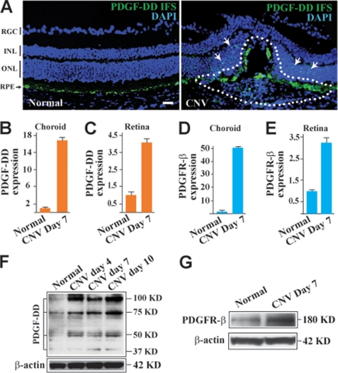
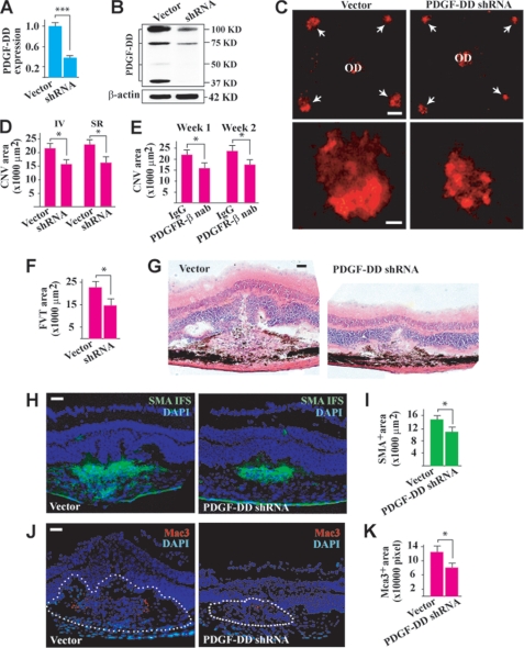
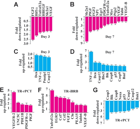
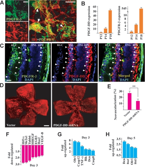
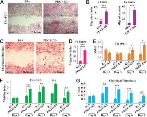
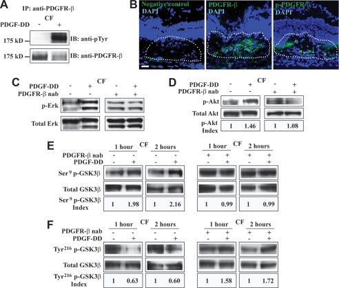
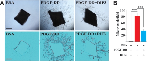
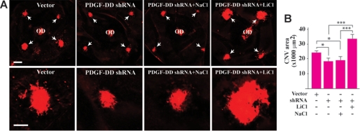
Platelet-derived growth factor-DD (PDGF-DD) is a recently discovered member of the PDGF family. The role of PDGF-DD in pathological angiogenesis and the underlying cellular and molecular mechanisms remain largely unexplored. In this study, using different animal models, we showed that PDGF-DD expression was up-regulated during pathological angiogenesis, and inhibition of PDGF-DD suppressed both choroidal and retinal neovascularization. We also demonstrated a novel mechanism mediating the function of PDGF-DD. PDGF-DD induced glycogen synthase kinase-3beta (GSK3beta) Ser(9) phosphorylation and Tyr(216) dephosphorylation in vitro and in vivo, leading to increased cell survival. Consistently, GSK3beta activity was required for the antiangiogenic effect of PDGF-DD targeting. Moreover, PDGF-DD regulated the expression of GSK3beta and many other genes important for angiogenesis and apoptosis. Thus, we identified PDGF-DD as an important target gene for antiangiogenic therapy due to its pleiotropic effects on vascular and non-vascular cells. PDGF-DD inhibition may offer new therapeutic options to treat neovascular diseases.


Figures

FIGURE 1.
FIGURE 1.
PDGF-DD and PDGFR-β expression is up-regulated in CNV. A, IFS displayed abundant PDGF-DD expression (green) within the CNV area (CNV, right, lined), while PDGF-DD expression was detected mainly in the retinal pigment epithelial cell layer (RPE, left, arrow) in normal retina. RGC, retinal ganglion cell layer; IPL, inner plexiform layer; INL, inner nuclear layer; and ONL, outer nuclear layer. Blue color, nuclei stained by 4′,6-diamidino-2-phenylindole (DAPI). Scale bar: 50 μm. B and C, real-time PCR showed up-regulated expression of PDGF-D in the choroids (B) and retinae (C) with CNV compared with normal choroids and retinae. Arbitrary units after normalizing against β-actin were used for PDGF-D expression with their normal controls set to 1. D and E, real-time PCR showed up-regulated expression of PDGFR-β in the choroids (D) and retinae (E) with CNV as compared with normal choroids and retinae. Arbitrary units after normalizing against β-actin were used for PDGFR-β expression with their normal controls set to 1. F, Western blot assay showed increased PDGF-DD protein levels in the retinae with CNV using β-actin as a loading control. Full-length (100 kDa) and differentially processed (75, 50, and 37 kDa) PDGF-DD were detected. G, Western blot assay showed an increased PDGFR-β protein level in the retinae with CNV using β-actin as a loading control.
FIGURE 2.
FIGURE 2.
PDGF-DD inhibition suppressed CNV. A, intravitreal injection of PDGF-D shRNA reduced PDGF-D expression to ∼37% of normal level in the retina 2 days after injection as measured by real-time PCR. Arbitrary unit after normalizing against β-actin was used for gene expression level. B, Western blot assay confirmed that intravitreal injection of PDGF-D shRNA reduced PDGF-DD protein level in the retina. Full-length (100 kDa) and differentially processed (75, 50, and 37 kDa) PDGF-DD were detected. C and D, PDGF-D shRNA treatment reduced CNV formation (arrows in C) 1 week after intravitreal (IV) or subretinal (SR) injection as measured by IB4 staining (red). OD: optic nerve disc. Scale bars in C: top panel, 200 μm; lower panel, 50 μm. E, intravitreal injection of PDGFR-β neutralizing antibody (nab) decreased CNV areas at different time points after treatment. F and G, histological analysis showed less fibrovascular tissue (FVT) formation in the PDGF-D shRNA-treated CNVs as revealed by hematoxylin & eosin staining. There was also less edema formation in the PDGF-D shRNA-treated CNVs as shown by the reduced hump formation and empty space around the neovascular area. H and I, immunofluorescence staining (IFS) showed that PDGF-D shRNA treatment reduced the area positive for smooth muscle cell α-actin (SMA, green, vascular smooth muscle cell marker). Scale bar in H: 50 μm. J and K, IFS showed that PDGF-D shRNA treatment reduced Mac3+ staining (red, macrophage marker) within the CNV areas. Scale bar in J: 50 μm. Blue color in H and J: nuclei stained by 4′,6-diamidino-2-phenylindole (DAPI). *, p < 0.05; ***, p < 0.001.
FIGURE 3.
FIGURE 3.
PDGF-DD regulated expression of proangiogenic and proapoptotic genes. A and B, PDGF-D shRNA treatment down-regulated the expression of many proangiogenic genes in the choroids with CNV at an early stage (A, day 3) and the peak time (B, day 7) of CNV as measured by real-time PCR. C and D, PDGF-D shRNA treatment increased the expression of many proapoptotic genes in the choroids with CNV at an early stage (C, day 3) and the peak time (D, day 7) of CNV as measured by real-time PCR. E and F, PDGF-DD protein treatment up-regulated the expression of many proangiogenic genes in rat retina-derived vascular pericytes (E, TR-rPCT) and in rat retina-derived vascular endothelial cells (F, TR-iBRB). G, PDGF-DD protein treatment inhibited the expression of many proapoptotic genes in the TR-rPCT cells.
FIGURE 4.
FIGURE 4.
PDGF-DD inhibition suppressed retinal neovascularization. A, immunofluorescence staining revealed PDGFR-β (green) expression in the neovessels (red, IB4 staining) of the neovascular retina, with a higher expression level in the neovascular tufts (arrows). Scale bars: 100 μm. B, PDGF-D and PDGFR-β expression was up-regulated at days 2 and 6 of retinal neovascularization (postnatal day (P), P14 and P18) compared with that of day 0 (P12) as measured by real-time PCR. Arbitrary unit normalized against β-actin was used for gene expression level. C, immunofluorescence staining of cross-sections of P18 neovascular retina showed that PDGFR-β expression (left, green) was found in different retinal layers, including the RGC layer, the INL (mainly on the cord-like blood vessels indicated by the arrows), and the RPE cells (indicated by arrows). PDGF-DD expression (middle, red) was mainly found in the INL and RPE layers. Co-localization of PDGF-DD with PDGFR-β was found in the RGC, INL, and RPE layers (right, yellow, arrows). Scale bar: 50 μm. D and E, intravitreal injection of a PDGF-D shRNA inhibited retinal neovascularization. Neovessels were visualized by IB4 staining (red). Scale bar: 500 μm. F, PDGF-D shRNA treatment inhibited the expression of many proangiogenic genes in the neovascular retinae as measured by real-time PCR. G and H, PDGF-D shRNA treatment up-regulated the expression of many proapoptotic genes in the neovascular retina at different time points as measured by real-time PCR. *, p < 0.05; **, p < 0.01.
FIGURE 5.
FIGURE 5.
PDGF-DD promoted vascular cell and fibroblast proliferation, survival, and migration. A and B, PDGF-DD protein promoted migration of retinal vascular pericytes (TR-rPCT) at different time points in a monolayer cell migration assay. Scale bar in A: 100 μm. C and D, PDGF-DD protein promoted choroidal fibroblast migration in a monolayer cell migration assay. Scale bar in C: 50 μm. E–G, PDGF-DD protein promoted proliferation/survival of the rat retinal derived vascular pericytes (TR-rPCT, E), rat retinal-derived vascular endothelial cells (TR-iBRB, F), and mouse choroidal fibroblasts (G) at different time points. Viability index in F: arbitrary unit with A540 values at day 0 set to 1. *, p < 0.05; **, p < 0.01; and ***, p < 0.001.
FIGURE 6.
FIGURE 6.
PDGF-DD activated PDGFR-β, Erk, Akt, and modulated GSK3β phosphorylation in vitro. A, immunoprecipitation (IP) followed by immunoblot (IB) showed that PDGF-DD protein stimulation led to PDGFR-β activation in mouse choroidal fibroblasts (CF). B, immunofluorescence staining detected abundant PDGFR-β expression (middle, lined area, green), as well as phosphorylated PDGFR-β (p-PDGFR-β, right, green, lined area) within the CNV area. Blue color: 4′,6-diamidino-2-phenylindole (DAPI)-stained nuclei. Scale bar: 50 μm. C and D, PDGF-DD protein induced Erk (C) and Akt (D) activation in mouse choroidal fibroblasts (CF, left penal). A neutralizing antibody against PDGFR-β (PDGFR-β, nab) abolished the effect of PDGF-DD on Erk and Akt activation (right penal). p-Akt index: arbitrary units of densitometry after normalization against total Akt with the controls set to 1. E, PDGF-DD protein stimulation led to GSK3β Ser9 phosphorylation in primary mouse CFs at different time points (left two panels). A PDGFR-β neutralizing antibody (PDGFR-β, nab) abolished the effect of PDGF-DD on GSK3β Ser9 phosphorylation (right two panels). Ser9 p-GSK3β index: arbitrary units of densitometry after normalization against total GSK3β with the controls set to 1. F, PDGF-DD protein induced Tyr216 dephosphorylation in primary mouse CFs at different time points (left two panels). A PDGFR-β neutralizing antibody (PDGFR-β, nab) abolished the effect of PDGF-DD on GSK3β Tyr216 dephosphorylation (right two panels). Tyr216 p-GSK3β index: arbitrary units of densitometry after normalization against total GSK3β with the controls set to 1.
FIGURE 7.
FIGURE 7.
PDGF-DD regulated GSK3β phosphorylation and expression in vivo and protected choroidal fibroblasts by regulating GSK3β phosphorylation. A, intravitreal injection of PDGF-DD protein increased GSK3β Ser9 phosphorylation in mouse retinae. Ser9 p-GSK3β index: arbitrary units of densitometry after normalization against total GSK3β with the control set to 1. B and C, PDGF-D shRNA intravitreal injection decreased GSK3β Ser9 phosphorylation (B) and increased GSK3β Tyr216 phosphorylation in mouse retinae (C). Ser9 (Tyr216) p-GSK3β index: arbitrary units of densitometry after normalization against total GSK3β with the control set to 1. D, PDGF-DD protein down-regulated GSK3β expression in different cells in vitro, and in mouse retinae in vivo. E, PDGF-DD knockdown by shRNA up-regulated GSK3β expression in the retinae with neovascularization as compared with the vector-treated samples. F, the mutant form of human GSK3β, in which the Ser9 was mutated to alanine (GSK3β-A9), was expressed in primary CFs. The wild-type GSK3β (GSK3β-WT)-transfected and non-transfected cells were used as controls. PDGF-DD protein protected CFs from H2O2-induced cell death in the GSK3β-WT-transfected as well as in the non-transfected cells. In the GSK3β-A9-transfected cells, the protective effect of PDGF-DD was abolished. **, p < 0.01.
FIGURE 8.
FIGURE 8.
GSK3β activation attenuated PDGF-DD-induced angiogenesis in aortic ring assay. A and B, PDGF-DD protein induced vascular cell proliferation, migration, and microvessel formation in an aortic ring assay (A, middle). Co-treatment of the aortic rings with the GSK3β activator DIF3 with PDGF-DD protein attenuated the PDGF-DD-induced microvessel formation. Lower panel in A: binary images of the aortic rings with branching microvessels. Scale bar: 500 μm; ***, p < 0.001.
FIGURE 9.
FIGURE 9.
GSK3β inhibition abolished the antiangiogenic effect of PDGF-DD shRNA. A and B, in the laser-induced CNV model, PDGF-D shRNA treatment inhibited CNV formation. Co-injection of the GSK3β inhibitor, LiCl, abolished the reduction of CNV formation induced by PDGF-D shRNA. Co-injection of NaCl had no effect. Scale bar in the upper panel in A: 200 μm; in the lower panel in A: 100 μm. *, p < 0.05; ***, p < 0.001.

Similar articles

Cited by

References

    1. Folkman J. (2007) Nat. Rev. Drug Discov. 6, 273–286 - PubMed
    1. Carmeliet P., De Smet F., Loges S., Mazzone M. (2009) Nat. Rev. Clin. Oncol. 6, 315–326 - PubMed
    1. Dorrell M. I., Aguilar E., Scheppke L., Barnett F. H., Friedlander M. (2007) Proc. Natl. Acad. Sci. U.S.A. 104, 967–972 - PMC - PubMed
    1. Carmeliet P. (2005) Nature 438, 932–936 - PubMed
    1. Ferrara N., Kerbel R. S. (2005) Nature 438, 967–974 - PubMed

MeSH terms

LinkOut - more resources