. 2010 May 14;285(20):14980-14989.
doi: 10.1074/jbc.M109.085696. Epub 2010 Mar 15.

Phosphoinositide 3-kinase pathway activation in phosphate and tensin homolog (PTEN)-deficient prostate cancer cells is independent of receptor tyrosine kinases and mediated by the p110beta and p110delta catalytic subunits

Affiliations

Phosphoinositide 3-kinase pathway activation in phosphate and tensin homolog (PTEN)-deficient prostate cancer cells is independent of receptor tyrosine kinases and mediated by the p110beta and p110delta catalytic subunits

Xinnong Jiang et al. J Biol Chem. .

Abstract

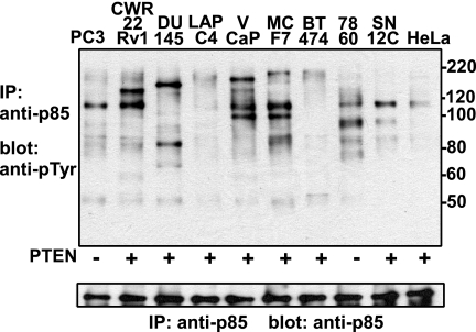
Class IA phosphoinositide 3-kinase (PI3K) p110 catalytic subunits are activated upon Src homology 2 domain-mediated binding of their p85 regulatory subunits to tyrosine-phosphorylated pYXXM motifs in receptor tyrosine kinases (RTKs) or adaptor proteins. The PI3K pathway is activated by phosphate and tensin homolog (PTEN) loss in most prostate cancers (PCa), but the contribution of upstream RTKs that may be targeted therapeutically has not been assessed. Immunoblotting of p85-associated proteins in serum-starved PTEN-deficient LNCaP and C4-2 PCa cells showed a small set of discrete tyrosine-phosphorylated proteins, but these proteins were not recognized by an anti-pYXXM motif antibody and were not found in PTEN-deficient PC3 PCa cells. LC/MS/MS using label-free proteomics and immunoblotting showed that p85 was associated primarily with p110beta and p110delta. An interaction with ErbB3 was also detected but was independent of ErbB3 tyrosine phosphorylation and was not required for basal PI3K activity. Basal tyrosine phosphorylation of p110beta and p110delta could be blocked by c-Src inhibitors, but this did not suppress PI3K activity, which was similarly independent of Ras. Basal PI3K activity was mediated by p110beta in PC3 cells and by both p110beta and p110delta in LNCaP cells, whereas p110alpha was required for PI3K activation in response to RTK stimulation by heregulin-beta1. These findings show that basal PI3K activity in PTEN-deficient PCa cells is RTK-independent and can be mediated by p110beta and p110delta. Increased p110beta expression in PCa may be required for RTK-independent PI3K pathway activation in adult prostate epithelium with genetic or epigenetic PTEN down-regulation.


Figures

FIGURE 1.
FIGURE 1.
p85 association with tyrosine-phosphorylated proteins in serum-starved PTEN-deficient PCa cells. A, PTEN-deficient LNCaP, C4-2, or PC3 cells were cultured for 1–3 days in serum-free medium and then treated for 6 h with PI3K inhibitor LY294002 (LY; 20 μm) or vehicle control (DMSO) (D) or left untreated (−). Lysates (10 μg) were then immunoblotted for pAkt (Thr308 and Ser473). B, serum-starved LNCaP or C4-2 cells (2–3 days) were lysed in TBS plus 1% Triton X-100, immunoprecipitated (IP) with anti-p85 or control rabbit Ig, and then immunoblotted with anti-Tyr(P) antibody (4G10). C, cell lysates (10 μg) from LNCaP or C4-2 cells grown in serum-free medium (SFM) or medium with 10% FBS were immunoblotted with anti-Tyr(P). D and E, LNCaP cells were either maintained in medium with 10% FBS or serum-starved for 2 days. Serum-starved cells were then stimulated with EGF (20 ng/ml, 5 min) or HRG-β1 (40 ng/ml, 15 min). Cell lysates were immunoprecipitated with anti-p85 (D) or anti-Tyr(P) (E), and the immunoprecipitates were blotted with anti-pYXXM. Molecular markers are indicated at the margins.
FIGURE 2.
FIGURE 2.
p85 interacts with ErbB3 independently of ErbB3 phosphorylation. A, lysates from serum-starved LNCaP or C4-2 cells (2 days) were immunoprecipitated (IP) with anti-p85 (p85) or control rabbit IgG (Ig), followed by blotting for ErbB3. Input is 1% of the material used for coimmunoprecipitation. B, LNCaP cells maintained in medium with 10% FBS were lysed in TBS plus 1% Triton X-100 and immunoprecipitated with anti-ErbB3 or control Ig, followed by anti-p85. The p85 immunoprecipitates were immunoblotted with anti-Tyr(P) (middle left) or anti-ErbB3 (bottom left). Top left, level of ErbB3 remaining in the anti-ErbB3 or Ig precleared lysates before the p85 immunoprecipitation. Right panels, anti-Tyr(P) (top) or anti-ErbB3 blots of initial anti-ErbB3 or Ig control immunoprecipitates. The arrow indicates a faint band at ∼190 kDa consistent with ErbB3. C and D, LNCaP cells transfected with ErbB3 or control non-targeted siRNA (C), or mock-transfected (M), were maintained in 10% FBS medium for 24 h, followed by serum starvation for 48 h. A portion of each cell lysate (10 μg) was then immunoblotted as indicated (C). The remaining lysates were immunoprecipitated with anti-p85, followed by immunoblotting with anti-ErbB3 (C) or anti-Tyr(P) (D). Molecular markers are indicated at the margins. The arrow indicates the position of proteins around 190 kDa.
FIGURE 3.
FIGURE 3.
PI3K activity and p85-ErbB3 interaction in LNCaP cells are independent of EGFR/ErbB2 activity. Serum-starved LNCaP cells (2 days) on 10-cm plates were treated with EGFR/ErbB-2 dual inhibitor lapatinib (10 μm) or vehicle (DMSO; D) for 6 h. A, cell lysates were immunoprecipitated (IP) with anti-Tyr(P), followed by immunoblotting for EGFR, ErbB2, or ErbB3. Whole cell lysates prior to immunoprecipitation were also immunoblotted as indicated for input EGFR, ErbB2, ErbB3, and Akt and for pAkt. B and C, cell lysates were immunoprecipitated with anti-p85 or rabbit IgG (Ig), followed by immunoblotting for ErbB3 (B) or Tyr(P) (C).
FIGURE 4.
FIGURE 4.
p85 is not associated with tyrosine-phosphorylated proteins in PC3 cells. Human PCa cell lines PC3, CWR22Rv1, DU145, LAPC4, and VCaP; human breast cancer lines MCF7 and BT474; human renal carcinoma cell lines 786O (PTEN deficient) and SN12C; and a human cervical cancer cell line (HeLa) grown in 10-cm plates were serum-starved for 1 day. Cell lysates were immunoprecipitated (IP) with anti-p85 and immunoblotted with anti-Tyr(P) (top) or anti-p85 (bottom). Molecular markers are indicated at the right.
FIGURE 5.
FIGURE 5.
p110 tyrosine phosphorylation is mediated by c-Src and is not required for PI3K activity. A, serum-starved LNCaP (2 days) and PC3 cells (1 day) were lysed and immunoprecipitated (IP) with anti-Tyr(P), followed by immunoblotting for anti-p110β or -δ. Input is 1% of the material used for the precipitation. B, serum-starved PC3 cells (1 day) were treated with lapatinib or Src inhibitors PP2 or dasatinib for 2 h, all at 10 μm. Cell lysates (1 mg) were then immunoprecipitated with anti-p85 and immunoblotted with anti-Tyr(P). C, serum-starved PC3 cells (1 day) were treated with PP2 or dasatinib at 10 μm for 2 h. Cell lysates were then immunoprecipitated with anti-Tyr(P), followed by immunoblotting with anti-p110β or -δ. Whole cell lysates (10 μg) were immunoblotted with pAkt, pPRAS40, or total Akt. D, serum-starved LNCaP cells (2 days) were treated with lapatinib, PP2, or dasatinib for 2 h, all at 10 μm. Cell lysates (1 mg) were immunoprecipitated with anti-p85 and immunoblotted with anti-Tyr(P). E, serum-starved LNCaP cells (2 days) were treated with PP2 or dasatinib at 10 μm for 2 h. Cell lysates were immunoprecipitated with anti-Tyr(P), followed by blotting with anti-p110β or -δ. Whole cell lysates (10 μg) were immunoblotted with pAkt, pPRAS40, or total Akt. Molecular markers are indicated at the right in B and D.
FIGURE 6.
FIGURE 6.
PI3K pathway activity in PTEN-deficient PCa cells is Ras-independent. LNCaP (left) or PC3 cells (right) plated into 24-well plates were treated with farnesylation inhibitor FTI-277 (20 or 40 μm; prepared in serum-free medium) for 2 days, and non-drug-treated wells were serum-starved for the same duration. At the end of FTI-277 treatment, cells were either treated with LY294002 (20 μm, 2 h) or stimulated with EGF (20 ng/ml, 5 min). Whole cell lysates (10 μg) were analyzed by immunoblotting for pAkt, pPRAS40, total Akt, ERK1/2, phospho-ERK1/2, Ras, or tubulin. Upper and lower bands corresponding to Ras and farnesylated Ras (fRas) are indicated.
FIGURE 7.
FIGURE 7.
Expression of PI3K p110 isoforms in PCa cell lines. A, LC/MS/MS analyses of the relative abundance by average TIC of the PI3K subunits p85α and -β and p110α, -β, and -δ in LNCaP and PC3 cells. B, expression levels of p110 isoforms in a variety of cancer cell lines based on immunoblotting. 10 μg of cell lysates from breast cancer lines (MCF-7, MB-MDA-231, BT-474, and SKBR-3) or PCa cell lines (LNCaP, PC3, VCaP, CWR22Rv1, and C4-2) were analyzed by immunoblotting with p110 isoform-specific antibodies. Intensity of each band relative to tubulin is shown. C, p110β and p110δ mRNA levels in normal prostate versus PCa or normal brain versus GBM. Expression data were generated by Oncomine based on results from Vanaja et al. (30) (PCa p110β), Dhanasekaran et al. (31) (PCa p110δ), and Sun et al. (33) (GBM).
FIGURE 8.
FIGURE 8.
Distinct p110 isoforms mediate basal versus growth factor-stimulated PI3K activity. PC3 (A) or LNCaP (C) cells in 24-well plates were transfected with siRNAs for each p110 isoform. After serum starvation for 1–2 days, cells were lysed, and 10 μg of cell lysates were subjected to immunoblotting for each p110 isoform, pAkt, total Akt, or β-tubulin. Results are quantified in bar graphs and are representative of 2–3 independent experiments for each cell type (in addition to results in E and F). PC3 (B) or LNCaP (D) transfected with siRNAs for each p110 isoform were serum-starved for 1–2 days, followed by HRG-β1 stimulation (100 ng/ml, 15 min). Cell lysates were then subjected to immunoblotting as indicated. E and F, PC3 or LNCaP cells, respectively, were transfected with siRNAs for each p110 isoform or with both p110β and -δ siRNA and were then analyzed by immunoblotting after serum starvation for 2 days.

Similar articles

Cited by

References

    1. Engelman J. A., Luo J., Cantley L. C. (2006) Nat. Rev. Genet. 7, 606–619 - PubMed
    1. Songyang Z., Shoelson S. E., Chaudhuri M., Gish G., Pawson T., Haser W. G., King F., Roberts T., Ratnofsky S., Lechleider R. J. (1993) Cell 72, 767–778 - PubMed
    1. Yu J., Zhang Y., McIlroy J., Rordorf-Nikolic T., Orr G. A., Backer J. M. (1998) Mol. Cell. Biol. 18, 1379–1387 - PMC - PubMed
    1. Carracedo A., Pandolfi P. P. (2008) Oncogene 27, 5527–5541 - PubMed
    1. Keniry M., Parsons R. (2008) Oncogene 27, 5477–5485 - PubMed

Publication types

Substances