. 2010 Mar 15;24(6):602-12.
doi: 10.1101/gad.1883410.

CDK-dependent complex formation between replication proteins Dpb11, Sld2, Pol (epsilon}, and GINS in budding yeast

Affiliations

CDK-dependent complex formation between replication proteins Dpb11, Sld2, Pol (epsilon}, and GINS in budding yeast

Sachiko Muramatsu et al. Genes Dev. .

Abstract

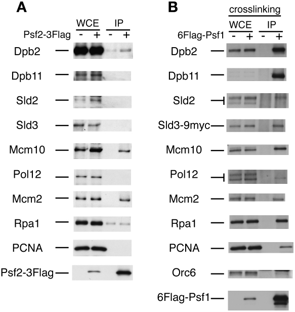
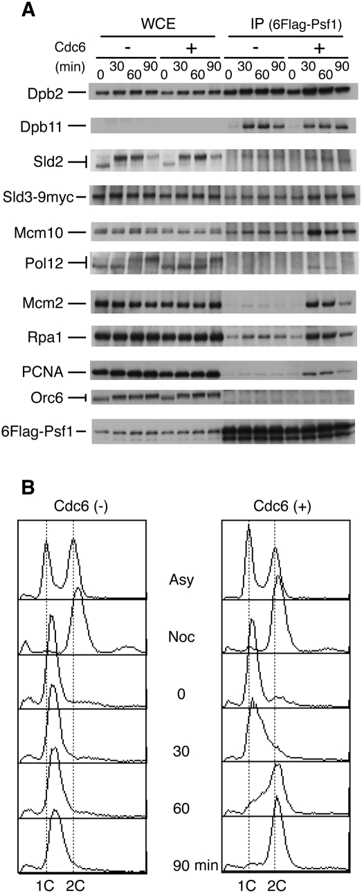
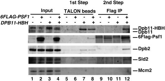
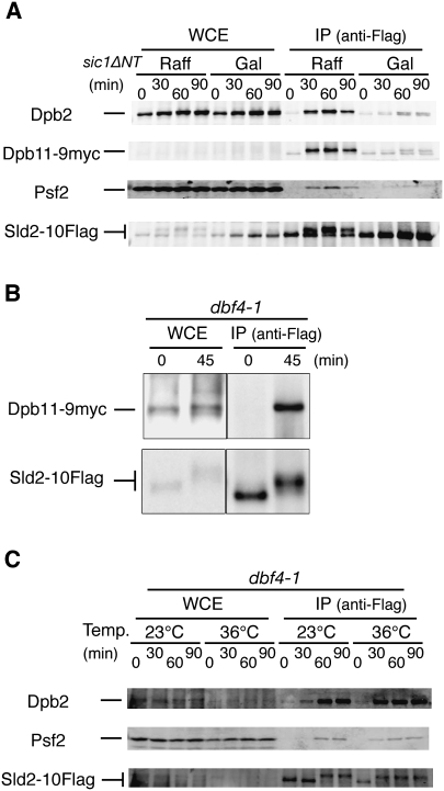
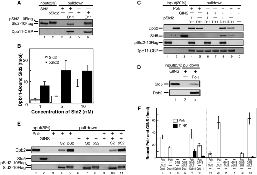
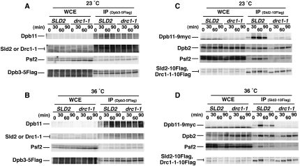
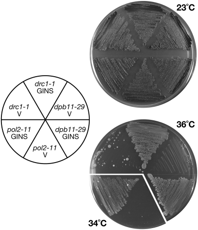
Eukaryotic chromosomal DNA replication requires cyclin-dependent kinase (CDK) activity. CDK phosphorylates two yeast replication proteins, Sld3 and Sld2, both of which bind to Dpb11 when phosphorylated. These phosphorylation-dependent interactions are essential and are the minimal requirements for CDK-dependent activation of DNA replication. However, how these interactions activate DNA replication has not been elucidated. Here, we show that CDK promotes the formation of a newly identified fragile complex, the preloading complex (pre-LC) containing DNA polymerase epsilon (Pol epsilon), GINS, Sld2, and Dpb11. Formation of the pre-LC requires phosphorylation of Sld2 by CDK, but is independent of DNA replication, protein association with replication origins, and Dbf4-dependent Cdc7 kinase, which is also essential for the activation of DNA replication. We also demonstrate that Pol epsilon, GINS, Dpb11, and CDK-phosphorylated Sld2 form a complex in vitro. The genetic interactions between Pol epsilon, GINS, Sld2, and Dpb11 suggest further that they form an essential complex in cells. We propose that CDK regulates the initiation of DNA replication in budding yeast through formation of the pre-LC.


Figures

Figure 1.
Figure 1.
Coprecipitation of replication proteins with GINS in the absence or presence of cross-linking agent. (A) YNIG20 (PSF2-3Flag-1HA) cells were arrested with hydroxyurea (HU) (0.2 M, 2 h), and cell extracts were prepared. Proteins were immunoprecipitated with the anti-Flag antibody. After the proteins were separated by SDS-PAGE, Western blotting was performed with the antibodies indicated. The samples used as whole-cell extracts (WCEs) corresponded to 2% of proteins used for immunoprecipitation. (B) Asynchronous YYK61 (6FlagPSF1 SLD3-9myc) cells were cross-linked with formaldehyde. The cell extracts were prepared using a bead shocker (Yasui Kikai) and immunoprecipitated with the anti-Flag antibody. Whole-cell extracts corresponded to 1% of the proteins used for immunoprecipitation except for 5% for Orc6 and Sld2. The Dpb11 protein was hardly detected in the formaldehyde-fixed whole-cell extracts.
Figure 2.
Figure 2.
Coprecipitation of replication proteins with GINS during the cell cycle. (A) YYK61 (6Flag-PSF1 SLD3-9myc) (+) and YYK62 (6Flag-PSF1 SLD3-9myc GAL1p-CDC6) (−) cells were arrested at G2/M phase with nocodazole (5 μg/mL) for 2.5 h in YPAGal (galactose as a carbon source), transferred to YPDA (glucose as a carbon source) containing nocodazole, released in YPDA containing α-factor (30 ng/mL), and incubated for 2.5 h at 24°C. Aliquots of the cells released from α-factor were withdrawn at the times indicated, cross-linked with formaldehyde, and immunoprecipitated with anti-Flag antibodies. Note that the Sld2 protein level fluctuated during the cell cycle and reached the lowest level at G1 (0 min). The Sld2-Flag protein level does not fluctuate significantly during the cell cycle (Masumoto et al. 2002), probably because the Flag-tagged C-terminal stabilizes the fusion protein. The amount of proteins applied as whole-cell extracts corresponds to 2% of that used for immunoprecipitation. (B) The cells were also subjected to flow cytometry analysis.
Figure 3.
Figure 3.
Two-step coprecipitation of Sld2, Dpb11, Pol ɛ, and GINS. YYK60 (Galp-CDC6), YYK62(Galp-CDC6 6Flag-PSF1), YNIG231(Galp-CDC6 DPB11-HBH) and YNIG217(Galp-CDC6 6Flag-PSF1 DPB11-HBH) cells were synchronized at 24°C in the condition that depletes Cdc6, as described in the legend for Figure 2. At 40 min after release from the G1 block, the cells were fixed with formaldehyde and disrupted. The resultant cell extracts were mixed with Dynabeads TALON (Dynal). The proteins were eluted from these beads with imidazole and precipitated with Dynabeads Protein A (Dynal) conjugated to anti-Flag (M2). The bound proteins were denatured by heat and subjected to SDS-PAGE, followed by Western blotting.
Figure 4.
Figure 4.
CDK-dependent formation of the pre-LC. (A) YNIG193 (SLD2-10Flag GAL1p-sic1ΔNT DPB11-9myc) cells were arrested at G1 phase with α-factor (30 ng/mL) in YPRaff for 1.5 h at 25°C. The culture was then split into two, and galactose (2%) was added to one half. After 45 min, the cells were released from α-factor in YPAGal (0 min). The other half was released from α-factor in YPRaff. Aliquots of cells released from α-factor were withdrawn at the times indicated, cross-linked with formaldehyde, and immunoprecipitated with the anti-Flag antibody as described in the legend for Figure 1. Western blotting was performed with the antibodies indicated. Dpb11-9myc and Sld2-10Flag were detected using anti-myc antibody 9E10 and anti-Flag antibody M2, respectively. (Raff) Raffinose; (Gal) galactose. (B) YS136 cells (dbf4-1 SLD2-10Flag DPB11-9myc) arrested with α-factor for 2.5 h at 25°C were shifted to 36°C and incubated further for 30 min at 36°C. The cells were then released (0 min) and cultivated for 45 min at 36°C. The cells were lysed with Lyticase (Sigma), and the proteins were precipitated with anti-Flag antibody M2 as described (Takayama et al. 2003). (C) YS134 (dbf4-1 SLD2-10Flag) cells were arrested at G1 phase with α-factor (30 ng/mL) in YPDA for 1.5 h at 23°C. The culture was split into two, and one half was shifted up to 36°C. After 30 min, cells were released from α-factor (0 min). Aliquots of cells were withdrawn at the times indicated, cross-linked with formaldehyde, and immunoprecipitated with anti-Flag antibody, as described in the legend for Figure 1. Western blotting was then performed with the antibodies indicated. The FACS patterns of the synchronized cells are shown in Supplemental Figure 5.
Figure 5.
Figure 5.
In vitro complex formation from purified proteins. (A) Dpb11–CBP immobilized to beads (0.250 pmol of Dpb11) was incubated with 0.125 pmol (5 nM) of Sld2-10Flag and CDK-phosphorylated Sld2-10Flag for 10 min at 4°C. The Sld2 and Dpb11 were detected by antibodies against Flag and CBP tagged to Sld2 and Dpb11, respectively. (B) Dpb11-immobilized beads (0.5 pmol of Dpb11) were mixed with various concentration of Sld2, and the amounts of phosphorylated Sld2 and bound Sld2 were measured. The results are the average of three independent experiments and standard deviation. (C) Dpb11-immobilized beads with phosphorylated Sld2 (0.5 pmol of Dpb11 and 0.1 pmol of phosphorylated Sld2) were incubated with 0.5 pmol (10 nM) of Pol ɛ and GINS for 1 h at 4°C. The Pol ɛ and GINS bound to the beads were detected by antibodies against the Dpb2 subunit of Pol ɛ and the Sld5 subunit of GINS. (D) Pol ɛ (0.5 pmol) immobilized to beads was incubated with 0.5 pmol (10 nM) of GINS for 1 h at 4°C. (E) Sld2 or CDK-phosphorylated Sld2 immobilized to beads (0.5 pmol) was incubated with 0.5 pmol (10 nM) of Pol ɛ and GINS for 1 h at 4°C. (F) The amounts of Pol ɛ and GINS bound to beads in C and E were estimated from Western blotting, and are shown as the average of three independent experiments and standard deviation.
Figure 6.
Figure 6.
Sld2 function is required for pre-LC formation. (A,B) YNIG14 (Galp-CDC6 DPB3-5Flag) and YNIG224 (Galp-CDC6 drc1-1 DPB3-5Flag) cells were synchronized in G1 phase at 23°C in the condition that depletes Cdc6, as described in the legend for Figure 2. The culture was split into two, and one half was shifted up to 36°C. After 30 min, cells were released from G1 block, withdrawn at 30 min intervals, cross-linked, and immunoprecipitated with anti-Flag antibody M2. (C,D) YNIG225 (Galp-CDC6 SLD2-10Flag DPB11-9myc) and YNIG226 (Galp-CDC6 drc1-1-10Flag DPB119myc) were treated as described in A and B.
Figure 7.
Figure 7.
Suppression of thermosensitive growth of pol2-11, dpb11-29, and drc1-1 (sld2) by increasing dosages of GINS. YHA211 (pol2-11), YNIG61 (dpb11-29), and Y799 (drc1-1) cells carrying both YEp195 and YEp181 (V) or both YEp195SLD5-PSF1 and YEp181PSF2-PSF3 (GINS) were streaked onto YPD plates and were incubated for 4 d at the temperatures indicated.

Similar articles

Cited by

References

    1. Araki H, Leem SH, Phongdara A, Sugino A. Dpb11, which interacts with DNA polymerase II (ɛ) in Saccharomyces cerevisiae, has a dual role in S-phase progression and at a cell cycle checkpoint. Proc Natl Acad Sci. 1995;92:11791–11795. - PMC - PubMed
    1. Asturias FJ, Cheung IK, Sabouri N, Chilkova O, Wepplo D, Johansson E. Structure of Saccharomyces cerevisiae DNA polymerase ɛ by cryo-electron microscopy. Nat Struct Mol Biol. 2006;13:35–43. - PubMed
    1. Bell SP, Dutta A. DNA replication in eukaryotic cells. Annu Rev Biochem. 2002;71:333–374. - PubMed
    1. Burgers PM. Polymerase dynamics at the eukaryotic DNA replication fork. J Biol Chem. 2009;284:4041–4045. - PMC - PubMed
    1. Dua R, Levy DL, Campbell JL. Analysis of the essential functions of the C-terminal protein/protein interaction domain of Saccharomyces cerevisiae pol ɛ and its unexpected ability to support growth in the absence of the DNA polymerase domain. J Biol Chem. 1999;274:22283–22288. - PubMed

Publication types

MeSH terms