Acute necrotizing encephalopathy in a child during the 2009 influenza A(H1N1) pandemia: MR imaging in diagnosis and follow-up
- PMID: 20231352
- PMCID: PMC7963992
- DOI: 10.3174/ajnr.A2058
Acute necrotizing encephalopathy in a child during the 2009 influenza A(H1N1) pandemia: MR imaging in diagnosis and follow-up
Abstract
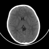
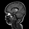
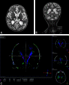
The recently emerged novel influenza A(H1N1) virus continues to spread globally. The clinical disease generally appears mild, but unfavorable outcomes have been reported. We describe a case of a 3-year-old Italian girl infected with influenza A(H1N1) virus presenting with neurologic deterioration. CT findings were negative, but MR imaging findings were consistent with ANE. To our knowledge, this is the first case reported in Europe and the second in worldwide pediatric radiology literature.
Figures


Comment in
-
Are neuroimaging findings in novel influenza A(H1N1) infection really novel?AJNR Am J Neuroradiol. 2010 Mar;31(3):393. doi: 10.3174/ajnr.A2060. AJNR Am J Neuroradiol. 2010. PMID: 20231351 Free PMC article. No abstract available.
Similar articles
-
Neurologic manifestations of pediatric novel h1n1 influenza infection.Pediatr Infect Dis J. 2011 Feb;30(2):165-7. doi: 10.1097/INF.0b013e3181f2de6f. Pediatr Infect Dis J. 2011. PMID: 20811314
-
Acute necrotizing encephalopathy in a child with H1N1 influenza infection.Pediatr Radiol. 2010 Feb;40(2):200-5. doi: 10.1007/s00247-009-1487-z. Epub 2009 Dec 18. Pediatr Radiol. 2010. PMID: 20020117
-
Are neuroimaging findings in novel influenza A(H1N1) infection really novel?AJNR Am J Neuroradiol. 2010 Mar;31(3):393. doi: 10.3174/ajnr.A2060. AJNR Am J Neuroradiol. 2010. PMID: 20231351 Free PMC article. No abstract available.
-
Acute necrotizing encephalopathy in adult patients with influenza: a case report and review of the literature.BMC Infect Dis. 2024 Sep 9;24(1):931. doi: 10.1186/s12879-024-09844-6. BMC Infect Dis. 2024. PMID: 39251995 Free PMC article. Review.
-
[Acute necrotizing encephalopathy of childhood: recent advances and future prospects].No To Hattatsu. 1998 May;30(3):189-96. No To Hattatsu. 1998. PMID: 9613149 Review. Japanese.
Cited by
-
Acute necrotizing encephalopathy: an underrecognized clinicoradiologic disorder.Mediators Inflamm. 2015;2015:792578. doi: 10.1155/2015/792578. Epub 2015 Mar 22. Mediators Inflamm. 2015. PMID: 25873770 Free PMC article. Review.
-
H1N1-Associated Encephalitis in a Child with Acute Myeloblastic Leukemia and Bacteremia due to Klebsiella Pneumoniae.Turk J Haematol. 2012 Mar;29(1):67-71. doi: 10.5505/tjh.2012.80947. Epub 2012 Mar 5. Turk J Haematol. 2012. PMID: 24744626 Free PMC article.
-
Successful Treatment of Influenza-Associated Acute Necrotizing Encephalitis in an Adult Using High-Dose Oseltamivir and Methylprednisolone: Case Report and Literature Review.Open Forum Infect Dis. 2017 Aug 7;4(3):ofx145. doi: 10.1093/ofid/ofx145. eCollection 2017 Summer. Open Forum Infect Dis. 2017. PMID: 28852683 Free PMC article. Review.
-
Two years after pandemic influenza A/2009/H1N1: what have we learned?Clin Microbiol Rev. 2012 Apr;25(2):223-63. doi: 10.1128/CMR.05012-11. Clin Microbiol Rev. 2012. PMID: 22491771 Free PMC article. Review.
-
Acute encephalopathy in childhood associated with novel influenza a h1n1 virus infection: clinical and neuroimaging findings.Ulster Med J. 2011 Jan;80(1):49-50. Ulster Med J. 2011. PMID: 22347741 Free PMC article. No abstract available.
References
-
- Mizuguchi M. Acute necrotizing encephalopathy of childhood: a novel form of acute encephalopathy prevalent in Japan and Taiwan. Brain Dev 1997;19:81–92 - PubMed
-
- Spring MD, Wright PF. Influenza-associated acute necrotizing encephalopathy in a 2-year-old American child. Int Congr Series 2004;1263:346–49
-
- Sugaya N. Influenza-associated encephalopathy in Japan: pathogenesis and treatment. Pediatr Int 2000;42:215–18 - PubMed
Publication types
MeSH terms
LinkOut - more resources
Full Text Sources
Medical
