. 2010 May;30(10):2353-64.
doi: 10.1128/MCB.00116-10. Epub 2010 Mar 15.

Separable functions of the fission yeast Spt5 carboxyl-terminal domain (CTD) in capping enzyme binding and transcription elongation overlap with those of the RNA polymerase II CTD

Affiliations

Separable functions of the fission yeast Spt5 carboxyl-terminal domain (CTD) in capping enzyme binding and transcription elongation overlap with those of the RNA polymerase II CTD

Susanne Schneider et al. Mol Cell Biol. 2010 May.

Abstract

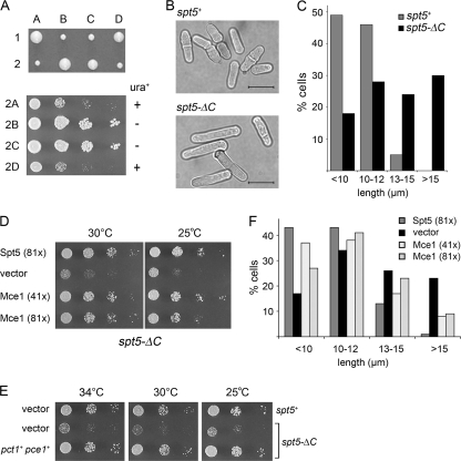
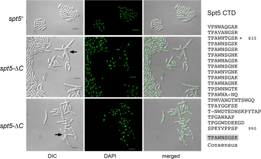
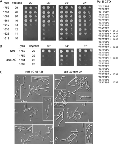
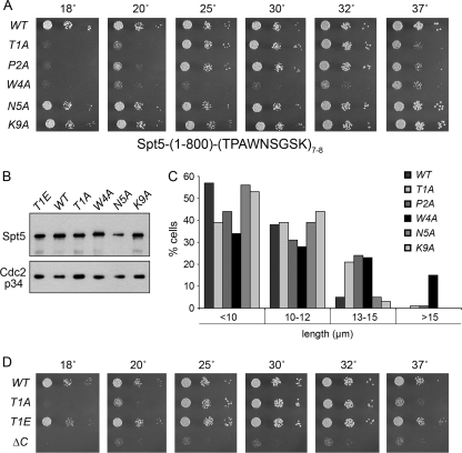
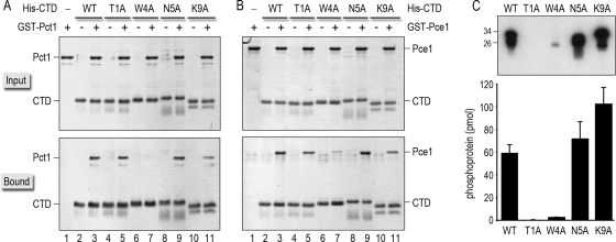
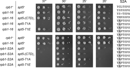
An interaction network connecting mRNA capping enzymes, the RNA polymerase II (Pol II) carboxyl-terminal domain (CTD), elongation factor Spt5, and the Cdk7 and Cdk9 protein kinases is thought to comprise a transcription elongation checkpoint. A crux of this network is Spt5, which regulates early transcription elongation and has an imputed role in pre-mRNA processing via its physical association with capping enzymes. Schizosaccharomyces pombe Spt5 has a distinctive CTD composed of tandem nonapeptide repeats of the consensus sequence (1)TPAWNSGSK(9). The Spt5 CTD binds the capping enzymes and is a substrate for threonine phosphorylation by the Cdk9 kinase. Here we report that deletion of the S. pombe Spt5 CTD results in slow growth and aberrant cell morphology. The severity of the spt5-DeltaCTD phenotype is exacerbated by truncation of the Pol II CTD and ameliorated by overexpression of the capping enzymes RNA triphosphatase and RNA guanylyltransferase. These results suggest that the Spt5 and Pol II CTDs play functionally overlapping roles in capping enzyme recruitment. We probed structure-activity relations of the Spt5 CTD by alanine scanning of the consensus nonapeptide. The T1A change abolished CTD phosphorylation by Cdk9 but did not affect CTD binding to the capping enzymes. The T1A and P2A mutations elicited cold-sensitive (cs) and temperature-sensitive (ts) growth defects and conferred sensitivity to growth inhibition by 6-azauracil that was exacerbated by partial truncations of the Pol II CTD. The T1A phenotypes were rescued by a phosphomimetic T1E change but not by capping enzyme overexpression. These results imply a positive role for Spt5 CTD phosphorylation in Pol Il transcription elongation in fission yeast, distinct from its capping enzyme interactions. Viability of yeast cells bearing both Spt5 CTD T1A and Pol II CTD S2A mutations heralds that the Cdk9 kinase has an essential target other than Spt5 and Pol II CTD-Ser2.


Figures

FIG. 1.
FIG. 1.
Deletion of the Spt5 CTD affects cells growth and morphology. (A) Heterozygous spt5+/spt5-(1-800)::ura4+ diploids were sporulated, and asci were dissected. Individual spores (A, B, C, and D) from two tetrads (1 and 2) were germinated on YES agar at 30°C for 4 days (upper panel). The four haploid progeny of tetrad 2 were grown in liquid culture. The cultures were diluted to attain an A600 of 0.1, and aliquots (3 μl) of serial 10-fold dilutions were spotted on YES agar medium. The plates were photographed after incubation for 3 days at 30°C (lower panel). The spt5-ΔC haploids 2A and 2D are ura+; the spt5+ haploids 2B and 2C are ura. (B) spt5+ and spt5-ΔC cells grown in YES medium at 30°C were examined by light microscopy. Bars, 10 μm. (C) The lengths of 270 spt5+ cells and 330 spt5-ΔC cells were measured and sorted into length bins as specified. The percentage of cells in each bin is represented in a bar graph. (D) Dosage suppression of the spt5-ΔC phenotype by mammalian capping enzyme Mce1. spt5-ΔC[spt5-(1-800)::ura4+] cells were transformed with LEU2 plasmids as specified. Serial dilutions of spt5-ΔC strains harboring a pREP81x-Spt5 plasmid (positive control), an empty vector plasmid (negative control), or plasmids for expression of Mce1 under the transcriptional control of an intermediate-strength (41x) or a low-strength (81x) nmt promoter were spotted on Leu agar medium. The plates were photographed after incubation for 6 days at 25°C or 3 days at 30°C. (E) Dosage suppression of spt5-ΔC by fission yeast capping enzymes. The spt5+ and spt5-ΔC [spt5-(1-800)::kanMX] strains harbored two plasmids marked with ura4+ and LEU2, respectively. These were either empty vector plasmids or plasmids for expression of S. pombe pct1+ (RNA triphosphatase) and pce1+ (RNA guanylyltransferase) under the transcriptional control of the low-strength nmt (81x) promoter. Cultures were grown at 30°C in minimal medium lacking uracil and leucine. Aliquots (3 μl) of serial 10-fold dilutions were spotted on Ura Leu agar medium and incubated at 25°C (4 days), 30°C (3 days), or 34°C (3 days) as specified. (F) Overexpression of Mce1 partially rescues the elongated phenotype of spt5-ΔC cells. The lengths of 300 to 500 individual cells were measured. The percentages of cells in each of the four size categories are represented by vertical bars.
FIG. 2.
FIG. 2.
Deletion of the Spt5 CTD results in aberrant cell morphology. The amino acid sequence of the Spt5 CTD is displayed at right, with the nonamer repeats aligned vertically. The consensus sequence TPAWNSGSK is shown below the alignment. Previously it was shown that truncation of the CTD to amino acid 835, leaving three nonamer repeats, had no apparent impact on cells growth. In the present study, the entire CTD was deleted in the spt5-ΔC strains of S. pombe. Cell morphology and nuclear DNA localization were assessed by light microscopy. spt5+ and spt5-ΔC cells were cultured in YES medium at 30°C. Exponentially growing cells were fixed in 70% ethanol, treated with 4′,6-diamidino-2-phenylindole (DAPI), and then visualized by differential interference contrast (DIC) and fluorescence (DAPI) microscopy (Nikon Eclipse E600 microscope equipped with a Spot camera). The bars in the DIC images are 10 μm. The arrows denote branched cells.
FIG. 3.
FIG. 3.
Genetic interactions of rpb1-ΔCTD mutants with spt5-ΔC. The amino acid sequence of the S. pombe Rpb1 CTD (aa 1551 to 1752) is shown at right; the 29 heptapeptide repeats are stacked vertically. The C termini of the rpb1-ΔCTD mutants are indicated by asterisks. (A) Truncating the Rpb1 CTD impairs growth. S. pombe strains bearing the indicated rpb1 alleles were tested for growth on YES agar. Aliquots of serial 5-fold dilutions of cultures that had been adjusted to an A600 of 0.1 were spotted onto YES agar and incubated at the indicated temperatures. The plates were photographed after incubation for 7 days at 20°C, 4 days at 25°C, 3 days at 30°C, or 2 days at 37°C. rpb1 alleles are named according to the length of the Rpb1 protein variants; the numbers of heptad repeats are indicated. (B) Phenotypic enhancement of rpb1-ΔCTD mutations by spt5-ΔC. Viable haploid double mutants harboring spt5-ΔC and the indicated CTD truncations of rpb1 were analyzed by spotting on YES agar. The plates were photographed after incubation for 3 days at 30, 34, or 37°C. (C) Aberrant morphology of spt5-ΔC rpb1-ΔCTD double mutants. Aliquots of exponentially growing cultures were spotted on slides, and the cells were visualized by differential interference contrast microscopy. All images, including the highlighted inserts, were taken at the same magnification. Bars, 10 μm.
FIG. 4.
FIG. 4.
Effects of alanine substitutions in the Spt5 CTD. (A) S. pombe strains with the indicated chromosomal spt5-(1-800)-CTD alleles—in which 7 or 8 wild-type or mutated CTD nonamer repeats were fused to Spt5-(1-800)—were grown in liquid medium until the A600 reached 0.3 to 0.5. The cultures were adjusted to equalize the A600, and aliquots of serial 5-fold dilutions were spotted on YES agar medium. The plates were photographed after incubation for 8 days at 18°C, 5 days at 20°C, 3 days at 25 and 37°C, or 2 days at 30 and 32°C. WT, wild type. (B) Western blot analysis of Spt5. Whole-cell extracts of the indicated spt5-CTD strains were resolved by SDS-PAGE. The polypeptides were transferred to a membrane and probed by serial Western blotting with affinity-purified polyclonal anti-Spt5 antibody (top panel) and then with anti-Cdc2-p34 (PSTAIRE) antibody as a loading control (bottom panel). (C) Morphological phenotypes of spt5-CTD mutants. Cultures of the indicated spt5-CTD mutant strains were grown to mid-logarithmic phase at 30°C, and 300 to 500 individual cells were measured and sorted into the length bins specified. The percentage of cells in each bin is represented in the bar graph. (D) Effects of a phosphomimetic T1E change. Aliquots (3 μl) of serial 5-fold dilutions from exponentially growing cultures of the indicated strains were spotted onto YES agar medium. The plates were photographed after incubation for 8 days at 18°C, 6 days at 20°C, or 2.5 days at 30, 34, and 37°C.
FIG. 5.
FIG. 5.
Effects of Spt5 CTD-Ala mutations on binding to capping enzymes and on CTD phosphorylation by Cdk9. (A and B) Binding of GST-tagged Pct1 (A) or Pce1 (B) to wild-type and Ala-substituted His10-Smt3-Spt5-CTD proteins was assessed by Ni-agarose affinity chromatography as described in Materials and Methods. The input proteins are specified above the lanes by “+.” Aliquots comprising 10% of the input material (top panels) and 30% of the bead-bound material (bottom panels) were analyzed by SDS-PAGE. Polypeptides were visualized by staining the gels with Coomassie blue dye. (C) Kinase reaction mixtures (20 μl) containing 50 mM Tris acetate (pH 6.0), 1 mM DTT, 2.5 mM MnCl2, 50 μM [γ32P]ATP, ∼100 ng of recombinant Cdk9T212E/Pch1 kinase (36), and 1 μg of recombinant His10-Smt3-Spt5-CTD phosphoacceptor as specified were incubated for 1 h at 20°C. The reactions were quenched by adding SDS to a 1% final concentration. Aliquots (3 μl) of the reaction mixtures were then analyzed by 12% SDS-PAGE. The 32P-labeled proteins were visualized by autoradiography of the dried gel (top panel). The positions and sizes (kDa) of marker polypeptides are indicated on the left. The extents of label transfer from [γ32P]ATP to His10-Smt3-Spt5-CTD were quantified by scanning the gel with a Molecular Dynamics Typhoon PhosphorImager. The data were normalized to the initial reaction volume and are plotted as a bar graph in the bottom panel. Each datum is the average of results from three or four separate experiments ± SD.
FIG. 6.
FIG. 6.
Spt5 CTD T1A and P2A mutations sensitize fission yeast to growth inhibition by 6-azauracil. Exponentially growing cultures of S. pombe strains with the indicated rbp1 and spt5 genotypes were adjusted to an A600 of 0.1, and aliquots (3 μl) of serial 5-fold dilutions were spotted on synthetic agar medium lacking uracil and containing 6 mM NH4OH and 0, 200, or 300 μg/ml 6-azauracil (6-AU) as specified. The plates were incubated at 30°C for 4 days (no 6-AU), 5 days (200 μg/ml 6-AU), or 5.5 days (300 μg/ml 6-AU).
FIG. 7.
FIG. 7.
Effects of spt5-CTD mutations in combination with rpb1-S2A. The amino acid sequence of the S. pombe Rpb1 CTD-S2A variant is shown at right. The heptapeptide repeats are stacked vertically with the mutated Ser2 positions shaded in gray. The indicated fission yeast strains were maintained in logarithmic growth at 30°C in YES medium. The cultures were adjusted to an A600 of 0.01, and aliquots of 10-fold serial dilutions were spotted on YES agar medium. The genotypes of the strains with respect to the rpb1 and spt5 loci are indicated on the left. The plates shown in the upper panels were incubated for either 2 days at 37 and 30°C, 3 days at 25°C, or 5 days at 20°C. The plates shown in the lower panels were incubated for either 2 days at 37 and 30°C, 4 days at 25°C, or 7 days at 20°C.

Similar articles

Cited by

References

    1. Akhtar, M. S., M. Heidermann, J. R. Tietjen, D. W. Zhang, R. D. Chapman, D. Eick, and A. Z. Ansari. 2009. TFIIH kinase places bivalent marks on the carboxy-terminal domain of RNA polymerase II. Mol. Cell 34:387-393. - PMC - PubMed
    1. Andrulis, E. D., E. Guzman, P. Dumloring, J. Werner, and J. T. Lis. 2000. High-resolution localization of Drosophila Spt5 and Spt6 at heat shock genes in vivo: roles in promoter proximal pausing and transcription elongation. Genes Dev. 14:2635-2649. - PMC - PubMed
    1. Archambault, J., F. LaCroute, A. Ruet, and J. D. Friesen. 1992. Genetic interaction between transcription elongation factor TFIIS and RNA polymerase II. Mol. Cell. Biol. 12:4142-4152. - PMC - PubMed
    1. Azuma, Y., M. Yamagashi, R. Ueshima, and A. Ishihama. 1991. Cloning and sequence determination of the Schizosaccharomyces pombe rpb1 gene encoding the largest subunit of RNA polymerase II. Nucleic Acids Res. 19:461-468. - PMC - PubMed
    1. Chen, Y., Y. Yamaguchi, Y. Tsugeno, J. Yamamoto, T. Yamada, M. Nakamura, K. Hisatake, and H. Handa. 2009. DISF, the Paf1 complex, and Tat-SF1 have nonredundant, cooperative roles in RNA polymerase II elongation. Genes Dev. 23:2765-2777. - PMC - PubMed

Publication types

MeSH terms

LinkOut - more resources