. 2010 May;30(10):2537-51.
doi: 10.1128/MCB.01282-09. Epub 2010 Mar 15.

Vasoactive intestinal peptide induces cell cycle arrest and regulatory functions in human T cells at multiple levels

Affiliations

Vasoactive intestinal peptide induces cell cycle arrest and regulatory functions in human T cells at multiple levels

Per Anderson et al. Mol Cell Biol. 2010 May.

Abstract

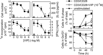
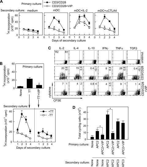
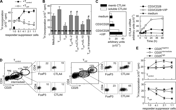
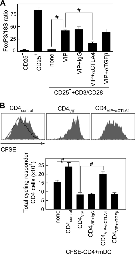
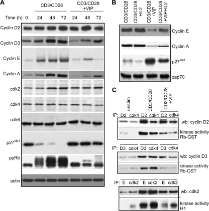
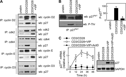
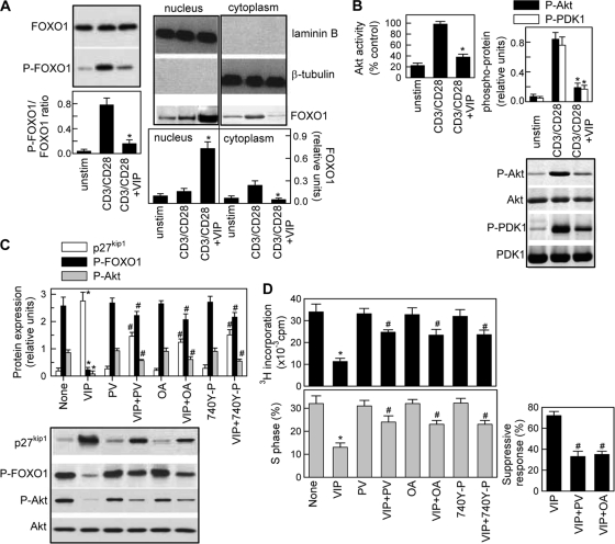
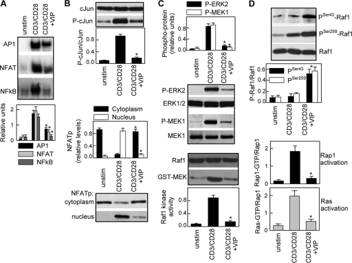
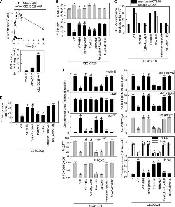
Vasoactive intestinal peptide (VIP) is a potent anti-inflammatory neuropeptide that, by inhibiting Th1-driven responses and inducing the emergence of regulatory T cells (T(reg)), has been proven successful in the induction of tolerance in various experimental models of autoimmune disorders. Here, we investigate the molecular mechanisms involved in VIP-induced tolerance. VIP treatment in the presence of T-cell receptor (TCR) signaling and CD28 costimulation induced cell cycle arrest in human T cells. VIP blocked G(1)/S transition and inhibited the synthesis of cyclins D3 and E and the activation of the cyclin-dependent kinases (CDKs) cdk2 and cdk4. This effect was accompanied by maintenance of threshold levels of the CDK inhibitor p27(kip1) and impairment of phosphatidylinositol 3-kinase (PI3K)-Akt signaling. Inhibition of interleukin 2 (IL-2) transcription and downregulation of signaling through NFAT, AP-1, and Ras-Raf paralleled the VIP-induced cell cycle arrest. Noteworthy from a functional point of view is the fact that VIP-treated T cells show a regulatory phenotype characterized by high expression of CD25, cytotoxic-T-lymphocyte-associated protein 4 (CTLA4), and Forkhead box protein 3 (FoxP3) and potent suppressive activities against effector T cells. CTLA4 appears to be critically involved in the generation and suppressive activities of VIP-induced T(reg). Finally, cyclic AMP (cAMP) and protein kinase A (PKA) activation seems to mediate the VIP-induced cell cycle arrest and T(reg) generation.


Figures

FIG. 1.
FIG. 1.
VIP induces cell cycle arrest in human T cells. Primary human T cells were cultured with medium alone (unstimulated; gray bars) or activated with anti-CD3/CD28 MAb-coated beads (CD3/CD28) in the absence or presence of different concentrations of VIP. Cytokine levels in culture supernatants were determined after 48 h by ELISA. Cell numbers were determined by counting cells excluding trypan blue at 72 h. Proliferation was determined by [3H]thymidine incorporation at 96 h. The percentages of cells in G0/G1 and S phases were determined by flow cytometry at different time points. *, P < 0.01 versus CD3/CD28 (n = 4, in duplicate). The error bars indicate SD.
FIG. 2.
FIG. 2.
VIP induces hyporesponsiveness in T cells. (A) Human T cells were CD3/CD28 activated in the absence or presence of VIP (10−7M) for 96 h. Cells were harvested and rested for 48 h, and the recovered viable cells were restimulated in a secondary culture with allogeneic mDCs in the absence or presence of IL-2 or anti-CTLA4 MAb. Proliferation was determined at different times after initiation of secondary culture. *, P < 0.01 versus CD3/CD28 (n = 4, in duplicate). The error bars indicate SD. (B) Human PBMCs (107) were activated with TT in the absence or presence of VIP (10−7 M) for 4 days, and proliferation in this primary culture was determined by [3H]thymidine incorporation (top). T cells (2 × 10⁴) were isolated from the primary culture, rested for 2 days, and restimulated in a secondary culture with syngeneic mDCs (104) in the absence or presence of TT, and proliferation was determined after 4 days (bottom). *, P < 0.01 versus TT-primed cells in the absence of VIP (n = 3, in duplicate). (C) CFSE-labeled human T cells were CD3/CD28 activated in the absence or presence of VIP (10−7 M) for 96 h. Cells were harvested and rested for 48 h, and the cytokine-producing potential of the recovered cells was determined by intracellular cytokine staining on CFSEhigh (nondividing; R1) and CFSEmild/low (cycling; R2) cells using flow cytometry. The numbers represent the percentages of cytokine-positive cells (n = 4). unstimul., unstimulated. (D) T cells from donor 1 (5 × 10⁴) were labeled with CFSE and stimulated with mDCs from donor 2 (APC2; 2 × 10⁴) in the absence or presence of VIP. After 6 days, viable cells (2 × 10⁴) were recovered and restimulated in a secondary culture with medium (None), with APC2 (104), or with mDCs from donor 3 (APC3; 104). The number of CFSElow/mild cycling cells was determined after 4 days of secondary culture. *, P < 0.01 (n = 3 or 4, in duplicate).
FIG. 3.
FIG. 3.
VIP induces regulatory functions in human T cells. (A) Human T cells isolated from donor A were CD3/CD28 activated in the absence (Tcontrol) or presence (TVIP) of VIP for 4 days. After a 48-h rest period, the suppressive potential of the recovered cells (suppressor Tcontrol or TVIP) was determined by adding increasing numbers of Tcontrol or TVIP to cocultures of responder T cells (from donor A) and allogeneic mDCs (from donor B). The proliferation of responder T cells was determined after 4 days. The black bar represents background proliferation of unstimulated responder T cells. The error bars indicate SD. (B) Tcontrol or TVIP (2.5 × 10⁴) were added to cocultures of responder T cells (donor A; 5 × 10⁴) and allogeneic mDCs (donor B) in the absence or presence of IL-2 (100 U/ml), anti-CTLA4 (10 μg/ml), or anti-IL-10 (10 μg/ml) MAb. When indicated, TVIP-mDC cocultures were separated from responder T-cell-mDC cocultures by a semipermeable membrane in transwells. The proliferation of responder T cells was determined after 4 days. The dashed line represents background proliferation of unstimulated responder T cells. (C) The human T cells isolated were CD3/CD28 activated in the absence or presence of VIP for different times. The mRNA expression of the soluble and membrane (memb) forms of CTLA4 was determined by quantitative PCR and expressed as arbitrary units normalized by β-actin transcripts. The time course corresponds to total mRNA (soluble plus membrane CTLA4). (D) CD4+ CD25 T cells isolated from donor A were CD3/CD28 activated without (Tcontrol) or with (TVIP) VIP for 96 h. Tcontrol and TVIP were analyzed in the CD4+ cell fraction for CD25 expression by flow cytometry. The subsets formed (CD25negative, CD25intermediate, and CD25high) were sorted by flow cytometry and analyzed for FoxP3 and CTLA4 expression. (E) Sorted CD25negative, CD25intermediate, and CD25high populations from Tcontrol and TVIP were added at different cell ratios to cocultures of PBMCs (5 × 10⁴) and allogeneic mDCs, and proliferation was determined. n = 3 or 4, in duplicate, for all panels, except panel C, where n = 2. *, P < 0.01 versus Tcontrol; #, P < 0.01 versus TVIP-treated cocultures.
FIG. 4.
FIG. 4.
VIP requires CTLA4 to induce FoxP3 and to generate Treg. (A) Purified CD4+ CD25 T cells were CD3/CD28 activated in the absence (none) or presence of VIP, with or without anti-CTLA4 (10 μg/ml), anti-TGF-β (10 μg/ml), or control IgG (10 μg/ml). After 96 h, FoxP3 expression was quantitated by real-time PCR and normalized to 18S rRNA. Unstimulated CD4+ CD25 and CD4+ CD25+ T cells were used as negative and positive controls, respectively. The error bars indicate SD. (B) CD4+ CD25 T cells from donor A were CD3/CD28 activated in the absence (CD4control) or presence (CD4VIP) of VIP, with or without anti-CTLA4, anti-TGF-β, or control IgG. After 96 h, the recovered cells (4 × 10⁴) were cocultured with CFSE-labeled CD4+ cells (2 × 10⁵; donor A) and mDCs (4 × 10⁴; donor B). After 96 h, the total number of cycling cells (percent CFSE-positive cells that had divided × total number of cells) was determined. The shaded histograms represent examples of each experimental group, and the solid-line histogram corresponds to CD4 cells stimulated with allogeneic mDCs. #, P < 0.01. n = 3, in duplicate.
FIG. 5.
FIG. 5.
VIP-induced suppressive T cells are arrested at the early G1 phase of the cell cycle. (A) Effects of VIP on the expression of G1 cyclins and CDK and CDKi proteins. Human T cells were CD3/CD28 activated without or with VIP (10−7 M) for different time intervals. The cell lysates were subjected to Western blot analysis for cyclins, CDKs, p27kip1, phosphorylated pRb (ppRb), or actin. (B) IL-2 partially reverses VIP effects on cyclins and p27kip1. Human T cells were CD3/CD28 activated without or with VIP (10−7 M) and IL-2 (50 U/ml) for 72 h. Western blot analysis was performed on cell lysates for cyclins, p27kip1, and Zap70. (C) VIP inhibits cyclin/CDK activity. Human T cells were CD3/CD28 activated without or with VIP (10−7 M) for 36 h. The cell lysates were immunoprecipitated (IP) with Abs against cyclin D2, cyclin D3, cyclin E, cdk2, or cdk4. Cyclin D2-cdk4, cyclin D3-cdk4, and cyclin E-cdk2 interactions were determined by Western blot (wb) analysis of precipitates with anti-cyclin D2, anti-cyclin D3, and anti-cdk2, respectively. cdk4-cyclin D2/D3 and cyclin E-cdk2 kinase activities were measured using pRb-GST and histone H1, respectively, as substrates. n = 3. (See Fig. S5 in the supplemental material for quantitative analysis.) unstim, unstimulated.
FIG. 6.
FIG. 6.
VIP maintains threshold levels of p27kip1. Human T cells were CD3/CD28 activated without or with VIP (10−7M). (A) VIP increases the interaction of p27kip1 with cyclin/CDK complexes. Cell lysates obtained after 24 h of culture were immunoprecipitated (IP) with Abs against cyclin D2, cyclin E, cdk2, or cdk4 and analyzed by Western blotting (wb) for cyclins, CDK, and p27kip1 (see Fig. S6A in the supplemental material for quantitative data). (B) VIP inhibits p27kip1 phosphorylation in activated T cells. After 48 h of culture, the cell lysates were immunoprecipitated with anti-p27kip1 Abs and analyzed by Western blotting for p27kip1 (total p27kip1) or phospho-Thr (phosphorylated p27kip1 [P-p27kip1]) and expressed as densitometric units relative to unstimulated samples. The error bars indicate SD. (C) VIP increases p27kip1 mRNA levels. p27kip1 mRNA expression was determined by Northern blotting and expressed as densitometric units relative to glyceraldehyde-3-phosphate dehydrogenase (GAPDH). When indicated, actinomycin D (ActD) was added during culture (the blot corresponds to 12 h). *, P < 0.001 versus CD3/CD28 (n = 3).
FIG. 7.
FIG. 7.
VIP inhibits FOXO1 phosphorylation and the Akt/PI3K pathway. Human T cells were cultured with medium (unstim) or CD3/CD28 activated in the absence or presence of VIP (10−7 M). (A) VIP inhibits phosphorylation and cytoplasmic translocation of FOXO1. Total cell extracts isolated at 4 h of culture (left) or nuclear and cytoplasmic protein extracts isolated at 8 h of culture (right) were analyzed by Western blotting for FOXO1 or phospho-FOXO1 and expressed as densitometric units relative to total FOXO1, laminin B, or β-tubulin. The error bars indicate SD. (B) VIP inhibits Akt/PKB phosphorylation and kinase activity in stimulated T cells. Cell lysates isolated after 2 h of culture were analyzed by Western blotting for phospho-Akt and PDK1 and expressed as densitometric units relative to total Akt or PDK1. Akt kinase activity was determined as described in Materials and Methods and expressed as percentages of the Akt activity of CD3/CD28-activated T cells. (C) The VIP effect on Akt activity is dependent on PI3K and PP2A activities. CD3/CD28-stimulated human T cells were treated with VIP and/or the PI3K activators peroxovanadate (PV) (50 μM) and 740 Y-P (20 μM) or the PP2A inhibitor okadaic acid (OA) (1 μM). Western blot analysis was performed on cell lysates isolated 2 h (for phospho-Akt), 4 h (for phospho-FOXO1), or 48 h (for p27kip1) of culture. The results are expressed as densitometric units relative to total Akt or β-tubulin. (D) Involvement of inhibition of PI3K-Akt signaling in the VIP effect on the T-cell cycle and regulatory functions. CD3/CD28-activated human T cells were treated with VIP and/or PV, OA, or 740 Y-P. The percentage of cells in S phase and proliferation were determined after 72 h. After 96 h in culture, T cells were recovered, and their suppressive potential was determined in cocultures with syngeneic responder T cells (at a suppressor/responder ratio of 1:2) and allogeneic mDCs, as described in the legend to Fig. 3A, and expressed as a percentage of inhibition of proliferation. n = 3 or 4. *, P < 0.01 versus CD3/CD28; #, P < 0.01 versus VIP-treated samples.
FIG. 8.
FIG. 8.
Mechanisms of inhibition of IL-2 expression by VIP. (A) Human T cells were cultured with medium (unstim) or CD3/CD28 activated without or with VIP (10−7 M). After 4 h, nuclear proteins were isolated and assayed for DNA binding to NF-κB, AP-1, or NFAT consensus sites by EMSA and expressed as densitometric units relative to the binding of the constitutive nuclear factor NF-Y. The error bars indicate SD. (B) After 3 h, whole-cell extracts were analyzed by Western blotting for c-Jun and phospho-c-Jun expression. NFATp levels were determined in both nuclear and cytoplasmic extracts and expressed as densitometric units relative to laminin B or β-tubulin. (C) After 1 h of culture, cell lysates were prepared, and activation of ERK1/2 and MEK1 was determined by Western blotting with phosphospecific Abs and expressed as densitometric units normalized for total ERK1/2 or MEK1. The kinase activity of Raf1 was determined by assaying the phosphotransferase activity toward GST-MEK1. (D) Activation of Rap1 and Ras was determined as described in Materials and Methods. The inactive forms of Raf1 were measured in cell lysates by Western blotting for phospho-Ser43-Raf1 and phospho-Ser259-Raf1. n = 3 or 4. *, P < 0.01 versus CD3/CD28.
FIG. 9.
FIG. 9.
VIP mediates the suppressive effect by elevating cAMP levels. (A) VIP increases cAMP levels and stimulates PKA activity in human activated T cells. Human T cells were CD3/CD28 stimulated with or without VIP (10−7 M). Intracellular cAMP levels were determined at different times (top). PKA activity was assayed by radioimmunoassay in cell lysates obtained 1 h after stimulation (bottom). The error bars indicate SD. (B to E) Human T cells were CD3/CD28 stimulated in the absence (−) or presence of forskolin (10−6 M), 8-Br-cAMP (0.1 mM), or VIP (10−7 M) with or without H89 (50 ng/ml) or Rp-cAMP (0.1 mM). (B) Cell cycle analysis was performed after 72 h. (C) Expression of the soluble and membrane forms of CTLA4 was determined at 96 h. (D) The suppressive activity on effector T cells was determined as described in the legend to Fig. 3A at an effector/suppressor cell ratio of 2:1. (E) The expression and phosphorylation statuses of cyclins, CDK, p27kip1, FOXO1, Akt, Raf1, and ERK1/2 and the activation of cyclin-CDK complexes and Ras were quantified as detailed in the legends to Fig. 5 to 8. The specificity of H89 and Rp-cAMP in our study was confirmed by their blocking effects on the actions of forskolin and Br-cAMP. Representative blots are shown in Fig. S9 in the supplemental material. n = 3 or 4. *, P < 0.001 versus CD3/CD28; #, P < 0.001 versus VIP-treated samples.

Similar articles

Cited by

References

    1. Alleaume, C., A. Eychène, E. Caigneaux, J. M. Muller, and M. Philippe. 2003. Vasoactive intestinal peptide stimulates proliferation in HT29 human colonic adenocarcinoma cells: concomitant activation of Ras/Rap1-B-Raf-ERK signalling pathway. Neuropeptides 37:98-104. - PubMed
    1. Anderson, P., and M. Delgado. 2008. Endogenous anti-inflammatory neuropeptides and proresolving lipid mediators: a new therapeutic approach for immune disorders. J. Cell Mol. Med. 12:1830-1847. - PMC - PubMed
    1. Appleman, L. J., A. A. van Puijenbroek, K. M. Shu, L. M. Nadler, and V. A. Boussiotis. 2002. CD28 costimulation mediates down-regulation of p27kip1 and cell cycle progression by activation of the PI3K/PKB signalling pathway in primary human T cells. J. Immunol. 168:2729-2736. - PubMed
    1. Appleman, L. J., A. Berezovskaya, I. Grass, and V. A. Boussiotis. 2000. CD28 costimulation mediates T cell expansion via IL-2-independent and IL-2-dependent regulation of cell cycle progression. J. Immunol. 164:144-151. - PubMed
    1. Baratelli, F., Y. Lin, L. Zhu, S. C. Yang, N. Heuze-Vourc'h, G. Zeng, K. Reckamp, M. Dohadwala, S. Sharma, and S. M. Dubinett. 2005. Prostaglandin E2 induces FOXP3 gene expression and T regulatory cell function in human CD4+ T cells. J. Immunol. 175:1483-1490. - PubMed

Publication types

MeSH terms