. 2010 May;30(10):2365-75.
doi: 10.1128/MCB.00672-09. Epub 2010 Mar 15.

The gene for aromatase, a rate-limiting enzyme for local estrogen biosynthesis, is a downstream target gene of Runx2 in skeletal tissues

Affiliations

The gene for aromatase, a rate-limiting enzyme for local estrogen biosynthesis, is a downstream target gene of Runx2 in skeletal tissues

Jae-Hwan Jeong et al. Mol Cell Biol. 2010 May.

Abstract

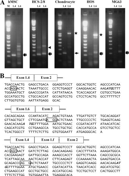
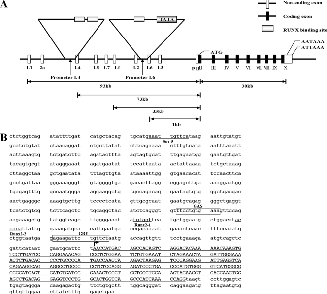
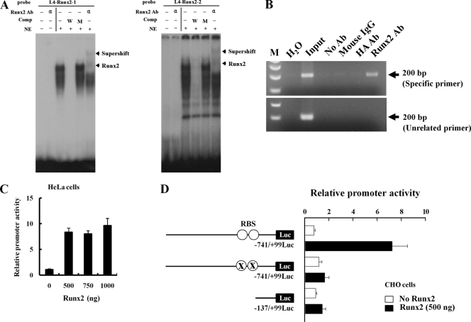
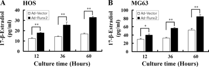
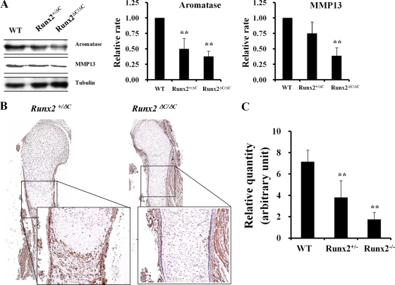
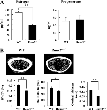
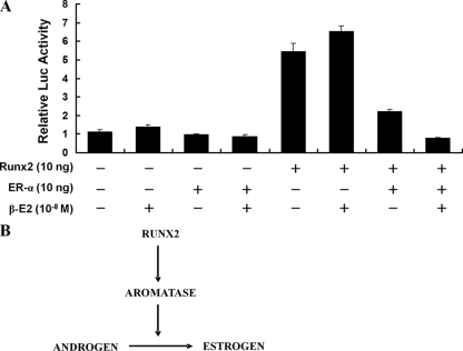
The essential osteoblast-related transcription factor Runx2 and the female steroid hormone estrogen are known to play pivotal roles in bone homeostasis; however, the functional interaction between Runx2- and estrogen-mediated signaling in skeletal tissues is minimally understood. Here we provide evidence that aromatase (CYP19), a rate-limiting enzyme responsible for estrogen biosynthesis in mammals, is transcriptionally regulated by Runx2. Consistent with the presence of multiple Runx2 binding sites, the binding of Runx2 to the aromatase promoter was demonstrated in vitro and confirmed in vivo by chromatin immunoprecipitation assays. The bone-specific aromatase promoter is activated by Runx2, and endogenous aromatase gene expression is upregulated by Runx2 overexpression, establishing the aromatase gene as a target of Runx2. The biological significance of the Runx2 transcriptional control of the aromatase gene is reflected by the enhanced estrogen biosynthesis in response to Runx2 in cultured cells. Reduced in vivo expression of skeletal aromatase gene and low bone mineral density are evident in Runx2 mutant mice. Collectively, these findings uncover a novel link between Runx2-mediated osteoblastogenic processes and the osteoblast-mediated biosynthesis of estrogen as an osteoprotective steroid hormone.


Figures

FIG. 1.
FIG. 1.
Aromatase gene expression and promoter activity in bone cells. (A) Expression of exons I.4 and I.6 in primary cultured human mesenchymal stem cells (hMSCs), the human chondrocyte HCS-2/8 cell line, primary cultured human chondrocytes (chondrocyte), and human osteosarcoma HOS cells and MG63 cells. RT-PCR was performed by using specific primers derived from the sequence of the coding region from exon I.4 or exon I.6 to exon II of the human aromatase gene. The expected PCR products (220 bp and 240 bp) were identified by 2% agarose gel electrophoresis. The top and bottom arrows indicate exon I.6 and exon I.4, respectively. The 1-kb bands seen in chondrocytes and HOS cells are contaminating genomic DNA (stars). (B) Sequences of transcripts for exons I.4 and 1.f linked to exon II. A novel transcript of the aromatase gene 5′-UTR corresponding to exons I.4 and I.f linked to exon II was observed for the human chondrocyte HCS-2/8 cell line (arrowhead in panel A). Boxes indicate exon-intron junctions, and the ATG (italic type) in each transcript indicates the translation start site. M, 1-kb molecular size ladder.
FIG. 2.
FIG. 2.
Runx2 binding sites in the aromatase promoter. (A) Promoter and coding exons of the aromatase gene. Promoter I.4 and promoter I.6 are indicated by arrows. (B) Sequences of promoter I.4 and Runx2 binding sites. Runx2 binding sites are indicated, from distal (site 1, Runx2-1) to proximal (site 2, Runx2-2). Exon sequences of the promoter I.4-derived transcript are indicated by capital letters.
FIG. 3.
FIG. 3.
Runx2 binds directly to aromatase promoter I.4. (A) EMSA was conducted with probes containing the two wild-type Runx2 motifs. 32P-labeled wild-type oligonucleotides were incubated with nuclear extracts from HOS cells in the presence of a 100-fold molar excess of unlabeled specific oligonucleotides or unlabeled mutant oligonucleotides or in the presence of anti-Runx2 antibody (α). The bottom arrows indicate the positions of the major nuclear protein-DNA complexes, and the top arrows indicate Runx2-supershifted complexes. (B) ChIP analysis was performed with formaldehyde-cross-linked chromatin isolated from HOS cells and antibody against Runx2. PCR amplification was carried out as described in Materials and Methods for a total of 28 cycles, and amplified fragments were analyzed by 2% agarose gel electrophoresis. (C) HeLa cells were cotransfected with the promoter I.4 reporter construct and Runx2 expression vector together with a β-galactosidase (β-Gal) internal control vector. The results were normalized to the protein concentration and β-Gal activity to account for transfection efficiency. (D) Runx2 did not stimulate Luc activity from a promoter with a mutant Runx2 binding site (RBS) or 0.14-kb aromatase promoter I.4 in CHO cells. Data represent the means ± SD from three independent experiments. Ab, antibody; Comp, competitor; NE, nuclear extract; W, wild-type oligonucleotide; M, mutant oligonucleotide; ○, RBS (TGTGGT and ACCACA); X, RBS mutation (TGTacT and AgtACA).
FIG. 4.
FIG. 4.
Forced expression of Runx2 increases estrogen production in bone cells. The concentration of estrogen in culture medium was measured after infection with adenovirus expressing Runx2 in two bone cell lines, HOS and MG63. The forced expression of Runx2 increased estrogen production in both HOS (A) and MG63 (B) cell lines. Ad, adenovirus. The values represent the means ± SD from three experiments. *, P < 0.05; **, P < 0.005.
FIG. 5.
FIG. 5.
Downregulation of aromatase gene expression in the long bone of Runx2ΔC/ΔC mice. (A) Expression of aromatase in vertebrae and ribs from E17.5 wild-type (WT), Runx2+/ΔC, and Runx2ΔC/ΔC mice. Total proteins were isolated from vertebrae and ribs of E17.5 embryos for Western blotting. A Runx2 downstream target gene, the MMP13 gene, was included as a positive control, and tubulin was used as an internal control. The values represent means ± SD from three mice per group. **, P < 0.005. (B) Immunohistochemical staining of tibial sections with antiaromatase antibody in Runx2+/ΔC and Runx2ΔC/ΔC mice at embryonic day 17. 5. (C) Real-time RT-PCR showed a reduced aromatase mRNA level in limbs from E17.5 Runx2 heterozygous or null mice (**, P < 0.005). The numbers of embryos in each group were 5 wild-type, 11 heterozygote, and 3 homozygote embryos. Data are expressed as means ± SD.
FIG. 6.
FIG. 6.
Estrogen production in BMSC and micro-CT analysis of Runx2 heterozygotes. (A) Primary cultured BMSC were obtained from 8-week-old wild-type (WT) and Runx2+/− mice. Estrogen and progesterone concentrations were measured by ELFA in 1-day-accumulated medium as described in Materials and Methods. The values represent means ± SD from three experiments. **, P < 0.005. (B) Five-month-old Runx2 C-terminally truncated heterozygotes showed decreased trabecular bone volume, cortical bone thickness, and total BMD. BV/TV, bone volume per tissue volume. Data are means ± SD from eight mice per group. *, P < 0.05; **, P < 0.005.
FIG. 7.
FIG. 7.
ER-α plus estrogen inhibit Runx2 transactivation function. (A) Six-Runx2-binding-site Luc reporter and Runx2 expression vectors were cotransfected with an ER-α expression vector into CHO cells, and relative Luc activity was measured at 24 h after transfection. The expression of ER-α decreased Runx2 transactivation function, and treatment with 17-β-estradiol (β-E2) enhanced this effect. Data represent the means ± SD from three independent experiments. (B) Relationship between Runx2 and the estrogen pathway through aromatase gene expression.

References

    1. Banerjee, C., L. R. McCabe, J. Y. Choi, S. W. Hiebert, J. L. Stein, G. S. Stein, and J. B. Lian. 1997. Runt homology domain proteins in osteoblast differentiation: AML3/CBFA1 is a major component of a bone-specific complex. J. Cell. Biochem. 66:1-8. - PubMed
    1. Bulun, S. E., S. Sebastian, K. Takayama, T. Suzuki, H. Sasano, and M. Shozu. 2003. The human CYP19 (aromatase P450) gene: update on physiologic roles and genomic organization of promoters. J. Steroid Biochem. Mol. Biol. 86:219-224. - PubMed
    1. Choi, J. Y., J. Pratap, A. Javed, S. K. Zaidi, L. Xing, E. Balint, S. Dalamangas, B. Boyce, A. J. van Wijnen, J. B. Lian, J. L. Stein, S. N. Jones, and G. S. Stein. 2001. Subnuclear targeting of Runx/Cbfa/AML factors is essential for tissue-specific differentiation during embryonic development. Proc. Natl. Acad. Sci. U. S. A. 98:8650-8655. - PMC - PubMed
    1. Conley, A., and M. Hinshelwood. 2001. Mammalian aromatases. Reproduction 121:685-695. - PubMed
    1. Ducy, P., R. Zhang, V. Geoffroy, A. L. Ridall, and G. Karsenty. 1997. Osf2/Cbfa1: a transcriptional activator of osteoblast differentiation. Cell 89:747-754. - PubMed

Publication types

MeSH terms

LinkOut - more resources