. 2010 Apr 12;207(4):867-80.
doi: 10.1084/jem.20091704. Epub 2010 Mar 15.

Survival effect of PDGF-CC rescues neurons from apoptosis in both brain and retina by regulating GSK3beta phosphorylation

Affiliations

Survival effect of PDGF-CC rescues neurons from apoptosis in both brain and retina by regulating GSK3beta phosphorylation

Zhongshu Tang et al. J Exp Med. .

Abstract

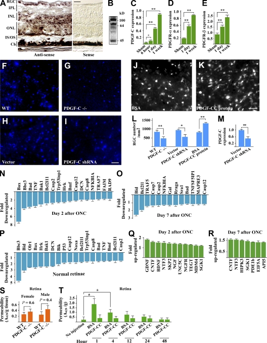
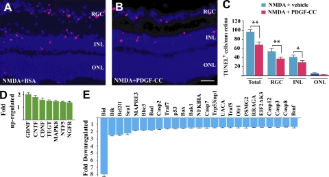
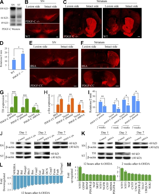
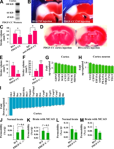
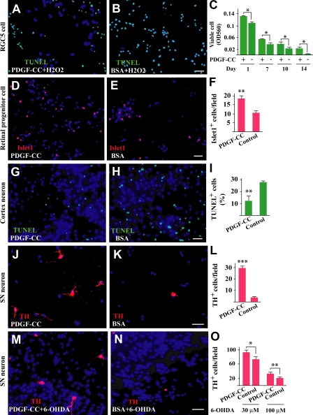
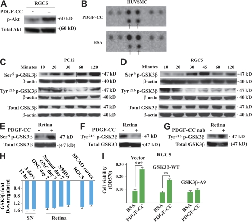
Platelet-derived growth factor CC (PDGF-CC) is the third member of the PDGF family discovered after more than two decades of studies on the original members of the family, PDGF-AA and PDGF-BB. The biological function of PDGF-CC remains largely to be explored. We report a novel finding that PDGF-CC is a potent neuroprotective factor that acts by modulating glycogen synthase kinase 3beta (GSK3beta) activity. In several different animal models of neuronal injury, such as axotomy-induced neuronal death, neurotoxin-induced neuronal injury, 6-hydroxydopamine-induced Parkinson's dopaminergic neuronal death, and ischemia-induced stroke, PDGF-CC protein or gene delivery protected different types of neurons from apoptosis in both the retina and brain. On the other hand, loss-of-function assays using PDGF-C null mice, neutralizing antibody, or short hairpin RNA showed that PDGF-CC deficiency/inhibition exacerbated neuronal death in different neuronal tissues in vivo. Mechanistically, we revealed that the neuroprotective effect of PDGF-CC was achieved by regulating GSK3beta phosphorylation and expression. Our data demonstrate that PDGF-CC is critically required for neuronal survival and may potentially be used to treat neurodegenerative diseases. Inhibition of the PDGF-CC-PDGF receptor pathway for different clinical purposes should be conducted with caution to preserve normal neuronal functions.


Figures

Figure 1.
Figure 1.
PDGF-CC protects RGCs from axotomy-induced neuronal death. (A) In situ hybridization assay detected abundant PDGF-C expression in the retina mainly in the RGC layer and INL/ONL. Bar, 50 µm. (B) Western blot assay detected PDGF-CC expression in the retina as several forms because of different proteolytic processing. (C–E) Real-time PCR showed up-regulated expression of PDGF-C, PDGFR-α, and PDGFR-β, in the retinae after ONC using β-actin as an internal control (n = 6 eyes). (F, G, and L) PDGF-C deficiency led to fewer viable RGCs (blue) after ONC (n = 9 or 11 eyes). (H, I, L, and M) PDGF-C shRNA intravitreal injection decreased PDGF-C transcript level to <50% of normal level (M, n = 8 eyes) and led to fewer viable RGCs (blue) compared with that of the control group (H, I, and L, n = 7 eyes). (J–L) PDGF-CC protein intravitreal injection increased the number of viable RGCs (white) after ONC (n = 8 eyes). (N–P) Real-time PCR validated the genome-wide gene expression profiling data. PDGF-CC treatment down-regulated the expression of numerous apoptotic/cell death–related genes in retinae after ONC at different time points (N and O, n = 8 eyes) and in normal retina (P). (Q and R) PDGF-CC treatment up-regulated the expression of many neurotrophic/survival genes in ONC-injured retinae at different time points (n = 8 eyes). (S) No difference was found in EB extravasation in the retina between PDGF-C–deficient and wild-type mice (n = 14–16 eyes). (T) Intravitreal injection of PDGF-CC protein did not affect retinal permeability at different time points, whereas the same amount of BSA increased retinal permeability transiently (n = 8–10 eyes). Bars: (F–K) 20 µm. *, P < 0.05; **, P < 0.01. The data are represented as means ± SEM of the number of determinations. All experiments were repeated independently once (A–E and M–R) or twice (F–L) with similar results. Representative images (A, B, and F–I) or pooled data (C–E and M–R) are shown. Ch, choroid; IS/OS, inner/outer segment.
Figure 2.
Figure 2.
PDGF-CC inhibits NMDA-induced neuronal apoptosis in retina. (A–C) PDGF-CC treatment reduced the number of apoptotic cells (red) in the RGC layer and INL/ONL after NMDA injury measured by TUNEL staining (n = 8 eyes). Bar, 20 µm. (D and E) Real-time PCR revealed that PDGF-CC treatment up-regulated the expression of several neurotrophic/survival genes (D) and inhibited the expression of numerous apoptotic/cell death–related genes in the NMDA-injured retina (E; n = 8 eyes). *, P < 0.05; **, P < 0.01. The data are represented as means ± SEM of the number of determinations. All experiments were repeated independently once with similar results. Representative images (A and B) or pooled data (D and E) are shown.
Figure 3.
Figure 3.
PDGF-CC protects dopaminergic neurons from apoptosis. (A) Western blotting detected PDGF-CC expression in the SN and ST in the brain as several forms because of differential proteolytic processing. (B and C) PDGF-C deficiency led to fewer TH+ cells in SN (B) and fewer TH+ fibers in ST (C) in the lesion side of the brain 7 d after 6-OHDA injury. The images shown in C represent a mosaic of several individual images. (D) PDGF-C–deficient mice displayed an increased contralateral rotation rate 1 wk after 6-OHDA injury (n = 9 or 15 mice). (E and F) PDGF-CC protein SN injection increased TH+ signals (dopaminergic neurons) in both SN (E) and ST (F) in 6-OHDA–injured brain measured by immunofluorescent staining (n = 9 or 15 mice). (G and H) PDGF-CC protein treatment increased expression levels of TH (G) and c-fos (H) in SN, whereas PDGF-C shRNA treatment decreased their expression levels (G and H) measured by real-time PCR. PDGF-C gene delivery (PDGF-C plasmid) into SN increased TH and c-fos expression levels (n = 6 mice). (I) PDGF-CC protein or gene delivery into SN decreased contralateral turning rate in the 6-OHDA–injured mice at different time points (n = 9 or 15 mice). (J) PDGF-C plasmid gene delivery (PDGF-C pls) increased TH expression level in both SN and ST at different time points measured by Western blotting (n = 6 mice). (K) PDGF-C shRNA treatment decreased TH expression level in both SN and ST in 6-OHDA–injured SN at different time points measured by Western blotting (n = 6 mice). (L) PDGF-CC protein treatment inhibited expression of numerous apoptotic/cell death–related genes in 6-OHDA–injured SN at different time points measured by real-time PCR. (M) PDGF-CC protein treatment up-regulated expression of many neurotrophic/survival factors in 6-OHDA–injured SN at different time points measured by real-time PCR. Bars: (B and E) 200 µm; (C and F) 400 µm. *, P < 0.05; **, P < 0.01; ***, P < 0.001. The data are represented as means ± SEM of the number of determinations. All experiments were repeated independently once (A, G, H, and J–M) or twice (B–F and I) with similar results. Representative images (A–C, E, F, J, and K) or pooled data (G–I, L, and M) are shown.
Figure 4.
Figure 4.
PDGF-CC protects brain cortical neurons from ischemia-induced neuronal death. (A) Western blotting detected PDGF-CC expression in brain cortex as several forms because of differential proteolytic processing. (B and C) PDGF-CC protein CSF injection decreased stroke volume, whereas CSF injection of PDGF-CC neutralizing antibody (anti–PDGF-CC) enlarged stroke volume (n = 11 or 13 mice). (D and E) PDGF-CC protein pretreatment by cortex injection decreased stroke volume significantly (n = 10 mice). (F) PDGF-C–deficient mice displayed greater stroke volume than that of wild-type mice (n = 8 mice). (G and H) Real-time PCR showed up-regulated expression of many neurotrophic/survival factors in brain cortex (G, n = 6 mice) and in isolated primary cortical neurons (H, n = 4 mice) after PDGF-CC treatment. (I) PDGF-CC treatment inhibited the expression of numerous apoptotic/cell death–related genes in brain cortex (n = 8 mice). (J and K) No difference in EB extravasation was found between PDGF-C–deficient and wild-type mice in normal brains (J, n = 10 mice) or brains with MCAO (K). (L and M) PDGF-CC CSF injection did not increase blood vessel permeability in normal brain (L, n = 8 mice) or in brains with MCAO (M, n = 8 mice). *, P < 0.05; **, P < 0.01. The data are represented as means ± SEM of the number of determinations. All experiments were repeated independently one to three times with similar results. Representative images (A, B, and D) and experiments are shown.
Figure 5.
Figure 5.
Direct protective effect of PDGF-CC on cultured neurons. (A–C) PDGF-CC treatment significantly decreased H2O2-induced apoptosis of the RGC5 cells measured by TUNEL staining (green; A and B) and increased their survival when the cells were cultured in serum-free medium (C). (D–F) PDGF-CC treatment increased survival of retinal neural progenitor cells (islet1+; red) when cultured in serum-free medium. (G–I) PDGF-CC treatment decreased hypoxia- and glucose deprivation–induced apoptosis in primary cortical neurons measured by TUNEL staining (green). (J–L) PDGF-CC treatment increased survival of TH+ neurons cultured in serum-free medium measured by TH staining (red). (M–O) PDGF-CC treatment increased survival of TH+ neurons measured by TH staining (red) when the neurons were stressed with neurotoxin 6-OHDA. Bars, 10 µm. *, P < 0.05; **, P < 0.01; ***, P < 0.001. The data are represented as means ± SEM of the number of determinations. All experiments were repeated independently twice with similar results. Representative images and experiments are shown.
Figure 6.
Figure 6.
Both PDGFR-β and PDGFR-α mediate the neuroprotective effect of PDGF-CC. (A) PDGFR-α and PDGFR-β were expressed in the retina, with the PDGFR-β expression level being more abundant as measured by real-time PCR. (B) Real-time PCR detected PDGFR-α and PDGFR-β expression at a similar level in primary cortical neurons. PDGF-CC protein up-regulated their expression. (C and D) Immunoprecipitation followed by immunoblotting (antiphosphotyrosine antibody [anti-pTyr]) detected PDGFR-β (C) and PDGFR-α (D) activation by PDGF-CC in RGC5 cells, primary cortical neurons, and SN using total PDGFR-α/β as controls (anti–tPDGFR-α/β). (E) Immunofluorescent staining using antibodies against phosphorylated PDGFR-β and PDGFR-α detected activated PDGFR-β and PDGFR-α (green) mainly in the RGC layer in the retina after PDGF-CC treatment. Bar, 50 µm. (F) PDGF-CC treatment inhibited expression of several proapoptotic genes in cortical neurons. The effect of PDGF-CC was largely abolished by both PDGFR-β and PDGFR-α neutralizing antibodies (n = 6 mice). (G) PDGF-CC treatment up-regulated the expression of many neurotrophic/survival genes in cortical neurons. This effect of PDGF-CC was abolished by both PDGFR-β and PDGFR-α neutralizing antibodies (n = 6 mice). (H and I) In the ONC model, both PDGFR-α and PDGFR-β neutralizing antibodies abolished the survival effect of PDGF-CC on RGCs (white; n = 8 eyes). Bar, 20 µm. *, P < 0.05; **, P < 0.01; ***, P < 0.001. The data are represented as means ± SEM of the number of determinations. All experiments were repeated independently once (A, B, and E–G) or twice (C, D, H, and I) with similar results. Representative images (C–E and I) and experiments are shown. IB, immunoblotting; IP, immunoprecipitation; IPL, inner plexiform layer; IS/OS, inner/outer segment; nab, neutralizing antibody.
Figure 7.
Figure 7.
Neuroprotective effect of PDGF-CC is achieved by regulating GSK3β phosphorylation. (A) PDGF-CC protein treatment activated Akt significantly in cultured RGC5 cells. (B) In a phospho-MAPK array screening assay, PDGF-CC treatment increased GSK3β Ser9 phosphorylation specifically (top, arrows) in HUVSMCs, whereas the phosphorylation of the other molecules remained unchanged. (C and D) In cultured PC12 (C) and RGC5 (D) neuronal cells, PDGF-CC protein treatment increased GSK3β Ser9 phosphorylation and decreased GSK3β Tyr216 phosphorylation, respectively, in a time-dependent manner. Black lines indicate that intervening lanes have been spliced out. (E and F) PDGF-CC protein treatment increased GSK3β Ser9 phosphorylation (E) and decreased GSK3β Tyr216 phosphorylation (F), respectively, in the retina in vivo. (G) PDGF-CC neutralizing antibody treatment increased GSK3β Tyr216 phosphorylation in the retina in vivo. (H) PDGF-CC protein treatment inhibited GSK3β expression in different types of neuronal tissues/cells, including SN, retina (normal or with ONC/NMDA injury), RGC5 cells, and brain cortex with MCAO as measured by real-time PCR. (I) PDGF-CC protein treatment protected RGC5 cells from H2O2-induced cell death in the control cells and cells expressing wild-type GSK3β (GSK3β-WT). The protective effect of PDGF-CC diminished in the RGC5 cells expressing mutant GSK3β, in which Ser9 was mutated to alanine (GSK3β-A9). **, P < 0.01; ***, P < 0.001. The data are represented as means ± SEM of the number of determinations. Experiments, except B, were repeated independently twice with similar results. Representative images (A and C–G) and experiments are shown. nab, neutralizing antibody.

Similar articles

Cited by

References

    1. Aase K., Abramsson A., Karlsson L., Betsholtz C., Eriksson U. 2002. Expression analysis of PDGF-C in adult and developing mouse tissues. Mech. Dev. 110:187–191 10.1016/S0925-4773(01)00560-3 - DOI - PubMed
    1. Andrae J., Gallini R., Betsholtz C. 2008. Role of platelet-derived growth factors in physiology and medicine. Genes Dev. 22:1276–1312 10.1101/gad.1653708 - DOI - PMC - PubMed
    1. Cao R., Bråkenhielm E., Li X., Pietras K., Widenfalk J., Ostman A., Eriksson U., Cao Y. 2002. Angiogenesis stimulated by PDGF-CC, a novel member in the PDGF family, involves activation of PDGFR-alphaalpha and -alphabeta receptors. FASEB J. 16:1575–1583 10.1096/fj.02-0319com - DOI - PubMed
    1. Dimmeler S. 2005. Platelet-derived growth factor CC–a clinically useful angiogenic factor at last? N. Engl. J. Med. 352:1815–1816 10.1056/NEJMcibr050670 - DOI - PubMed
    1. Ding H., Wu X., Kim I., Tam P.P., Koh G.Y., Nagy A. 2000. The mouse Pdgfc gene: dynamic expression in embryonic tissues during organogenesis. Mech. Dev. 96:209–213 10.1016/S0925-4773(00)00425-1 - DOI - PubMed

Publication types

MeSH terms

Associated data