. 2010 Mar 22;188(6):953-69.
doi: 10.1083/jcb.200908133. Epub 2010 Mar 15.

Joubert syndrome Arl13b functions at ciliary membranes and stabilizes protein transport in Caenorhabditis elegans

Affiliations

Joubert syndrome Arl13b functions at ciliary membranes and stabilizes protein transport in Caenorhabditis elegans

Sebiha Cevik et al. J Cell Biol. .

Erratum in

  • J Cell Biol. 2010 Apr 5;189(1):187

Abstract

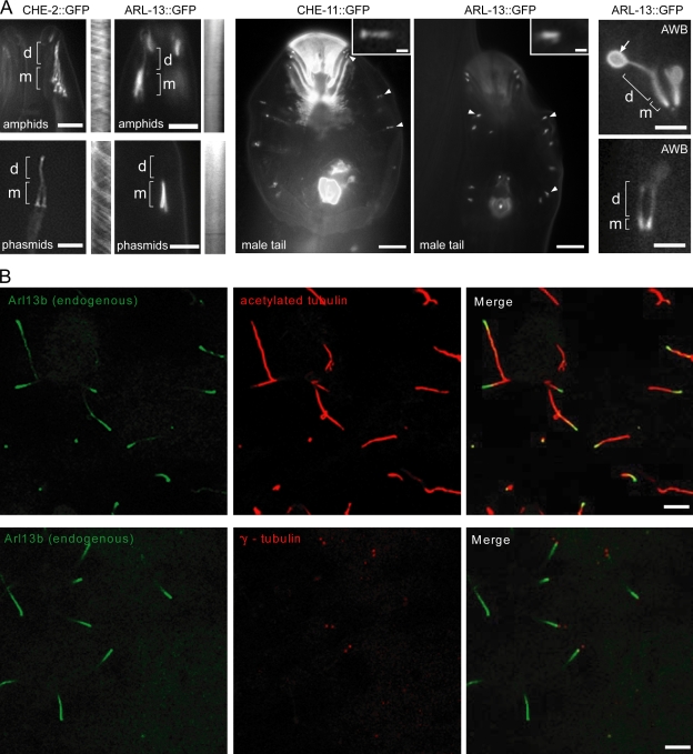
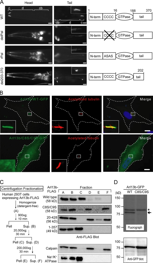
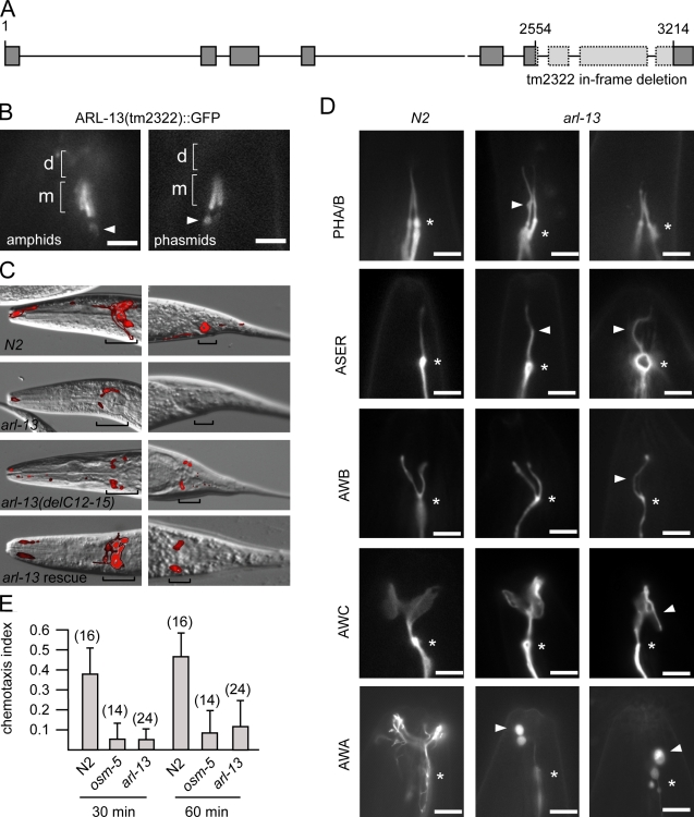
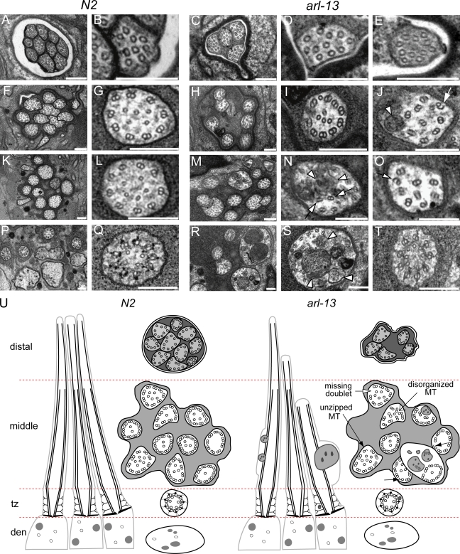
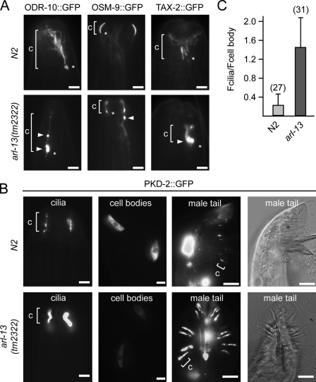
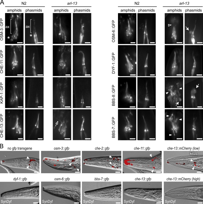
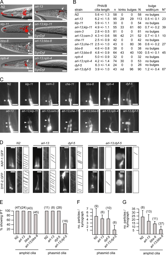
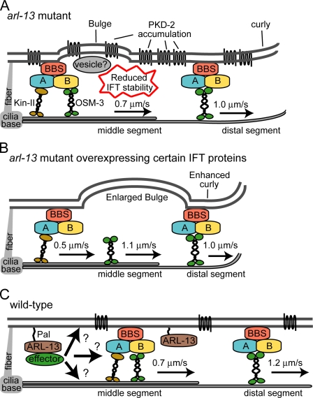
The small ciliary G protein Arl13b is required for cilium biogenesis and sonic hedgehog signaling and is mutated in patients with Joubert syndrome (JS). In this study, using Caenorhabditis elegans and mammalian cell culture systems, we investigated the poorly understood ciliary and molecular basis of Arl13b function. First, we show that Arl13b/ARL-13 localization is frequently restricted to a proximal ciliary compartment, where it associates with ciliary membranes via palmitoylation modification motifs. Next, we find that loss-of-function C. elegans arl-13 mutants possess defects in cilium morphology and ultrastructure, as well as defects in ciliary protein localization and transport; ciliary transmembrane proteins abnormally accumulate, PKD-2 ciliary abundance is elevated, and anterograde intraflagellar transport (IFT) is destabilized. Finally, we show that arl-13 interacts genetically with other ciliogenic and ciliary transport-associated genes in maintaining cilium structure/morphology and anterograde IFT stability. Together, these data implicate a role for JS-associated Arl13b at ciliary membranes, where it regulates ciliary transmembrane protein localizations and anterograde IFT assembly stability.


Figures

Figure 1.
Figure 1.
ARL-13/Arl13b is enriched in proximal regions of ciliary axonemes. (A) ARL-13::GFP localizes almost exclusively to the proximal region (MSs; m) of amphid/phasmid cilia and male tail ray cilia. Although localizing to all regions of AWB cilia, ARL-13 is enriched in proximal regions and observed at the membrane of distal fans (arrow). IFT markers (CHE-2 and -11) label amphid/phasmid channel MSs and DSs (d) and RnA/B male tail ray cilia. Kymographs, from time-lapse videos, show no IFT movement for ARL-13::GFP. Arrowheads indicate RnA/B cilia, and insets show high magnification images of RnA/B cilia. (B) Arl13b is enriched in the proximal region of MDCKII cilia. Images after immunostaining for endogenous Arl13b, acetylated α-tubulin (ciliary axoneme), and γ-tubulin (basal body) are shown. Bars: (A, left and right) 3 µm; (A, middle) 10 µm; (A, insets) 1 µm; (B) 5 µm.
Figure 2.
Figure 2.
ARL-13/Arl13b associates with ciliary membranes via palmitoyl anchors. (A) N-terminal Pal motif ARL-13 variants (delPal and rPal) fail to localize to amphid (head) and phasmid (tail) cilia (c) and diffusely mislocalize in cell bodies (cb) and dendrites (d). C-terminal deletion ARL-13(del203–370) is also mislocalized, but membrane associations are maintained (arrows). Insets show high magnification images of PHA/B cilia. (B) HsArl13b ciliary localization requires an N-terminal Pal motif. Ciliated RPE1 cells transfected with GFP-tagged Arl13b(WT) or a C8S/C9S variant and costained for ciliary axonemes using acetylated α-tubulin antibody are shown. Merged images show that Arl13b(C8S/C9S) is highly diffuse and nonmembrane associated, with weak signals in cilia. The boxed regions are shown at high magnification in the insets. Dashed lines outline the cell. (C) Loss of Pal motif shifts HsArl13b to cytosolic fractions. A subcellular fractionation scheme for Flag-tagged Arl13b (WT and variants) transiently expressed in 293T cells is shown. Supernatants (Sup.) and pellets (Pell) were probed for Arl13b::Flag using Western blotting and an anti-Flag antibody. Calpain and Na+/K+ ATPase proteins mark cytosol and membrane fractions, respectively. Arl13b(WT) and Arl13b(1–357) pellet with Na+/K+ ATPase membrane proteins after 20,000 g spins, whereas Arl13b(C8S/C9S) remains in calpain-enriched cytosolic supernatants even after 200,000 g spins. (D) Arl13b is palmitoylated in 293T cells. 293T cells transiently transfected with GFP-tagged Arl13b(WT) or Arl13b(C8S/C9S) were metabolically labeled with [3H]palmitate, and proteins were separated by SDS-PAGE. The top panel shows a fluorograph; the bottom panel shows a Western blot with anti-GFP antibody. The arrow indicates palmitoylated Arl13b-GFP, and the asterisk indicates an unknown endogenous palmitoylated protein. Bars: (A) 5 µm; (B) 10 µm.
Figure 3.
Figure 3.
C. elegans arl-13 mutants possess defective cilium structure, morphology, and functions. (A) arl-13 gene schematic, showing genomic position of in-frame tm2322 deletion. (B) tm2322 encodes a ciliary ARL-13 protein. Fluorescence images of amphid/phasmid channel cilia in worms expressing arl-13(tm2322)::gfp are shown. GFP signals are restricted to MSs (m) and excluded from DSs (d). Some abnormal accumulations are found beneath cilia (arrowheads). (C) tm2322 mutants are dye-filling (Dyf) defective. Merged differential interference contrast (DIC)–fluorescence images after a DiI uptake assay are shown. Dye uptake into amphid (head) and phasmid (tail) neuron cell bodies (denoted by brackets) is strongly reduced in tm2322 mutants and weakly reduced in WT animals expressing arl-13(delPal)::gfp but restored in tm2322 animals expressing arl-13(WT)::gfp. (D) Cilium morphologies are defective in tm2322 worms. Fluorescence images of cilia from N2 and tm2322 animals expressing ciliated cell–specific transcriptional GFP markers srb-6p::gfp (PHA/B), gcy-5p::gfp (ASER), str-1p::gfp (AWB), str-2p::gfp (AWC), and gpa-6p::gfp (AWA) are shown. All images are similarly orientated, with ciliary base denoted (asterisks). Arrowheads indicate morphology defects such as kinks (ASER), bulges (PHA/B and AWA), curls (AWB,) and ectopic projections (AWC). (E) tm2322 mutants are chemosensory defective. Indices obtained from 30- and 60-min chemoattraction assays toward isoamyl alcohol for tm2322, N2, and osm-5(p802) worms are shown. Assay numbers are shown in parentheses. Error bars indicate SEM. Bars: (B and D) 3 µm; (C) 10 µm.
Figure 4.
Figure 4.
arl-13(tm2322) mutant cilia possess ultrastructural defects. Transmission electron microscopy serial cross sections of amphid pore from N2 and tm2322 worms. (A–E) Distal pore showing 10 axonemes in N2 worms (A and B) but only 4 axonemes in tm2322 mutants (C–E). (F–J) 2 µm proximal to A–E (through MSs) showing axonemes still missing in tm2322 worms (H). Also, abnormal accumulation of electron-dense material (J, arrowhead) and unzipping of MTs (J, arrow) are observed. (K–O) 3 µm proximal to A–E (through MSs) showing most ciliary axonemes are present in tm2322 mutants (M). Further abnormal axonemal accumulations of amorphous electron-dense material (N, arrowheads) and missing, misplaced, and unzipped doublet MTs (N and O, arrows) are also shown. (P–T) 5 µm proximal to A and B, through MSs, transition zones, and transitional fibers. Some tm2322 cilia are abnormally enlarged (R and S), filled with electron-dense material that is amorphous, or contained within vesicle-type structures (R and S). (S) Arrowheads indicate amorphous and vesicular-like accumulations. Transition zones appear normal (T). (U) Schematics of amphid channel cilia from N2 and tm2322 mutants, showing the major ultrastructural defects observed. tz, transition zone. Bars, 200 µm.
Figure 5.
Figure 5.
Ciliary transmembrane protein localization is disrupted in arl-13(tm2322) mutants. (A and B) Representative fluorescence images of the distal head region (nose) of worms expressing gfp-tagged ODR-10, OSM-9, TAX-2, and PKD-2 are shown. In tm2322 mutants, abnormal accumulations (arrowheads) are found in ciliary axonemes (ODR-10), near the ciliary base (ODR-10 and TAX-2; asterisks), or within the distal dendrite (OSM-9; arrowhead). In tm2322 mutants, PKD-2::GFP ciliary abundance is elevated in CEM and RnB cells, with cell body levels reduced (shown for CEMs). c, cilium. (C) Analysis of PKD-2::GFP ciliary abundance in CEM cells. The ratio of PKD-2::GFP signal intensities in individual CEM cilia (Fcilia) and individual CEM cell bodies (Fcell body) is shown. All images were captured and analyzed using identical settings. The number of cilia analyzed is shown in parentheses. Error bars indicate SEM. Bars: (A and B [first and second columns]) 2 µm; (B, third and fourth columns) 10 µm.
Figure 6.
Figure 6.
IFT transgene overexpression enhances the structure/morphology defects of tm2322 mutant cilia. (A) Fluorescence images of amphid/phasmid cilia in N2 and arl-13(tm2322) animals expressing gfp-tagged IFT transgenes show that IFT proteins localize normally to ciliary axonemes (ax) and accumulate normally at ciliary bases (asterisks) in tm2322 worms. In tm2322 mutants overexpressing che-13::gfp, osm-6::gfp, and bbs-7/8::gfp, amphid/phasmid cilia morphologies are more severely defective than other transgenic strains. Enhanced defects include missing DS staining (open arrowhead), axonemes that are less tightly bunched and highly disorganized (closed arrowheads; open arrow), and increased frequency of large axonemal bulges (closed arrows). (B) arl-13 animals overexpressing osm-3::gfp, che-2::gfp, che-11::gfp, and che-13::mCherry(low expression level) display similar levels of DiI incorporation to nontransgenic controls (arrowheads indicate dye uptake in cell bodies). However, tm2322 mutants overexpressing che-13::gfp, osm-6::gfp, bbs-7::gfp, dyf-1::gfp, and che-13::mCherry(high expression level) are SynDyf, failing to take dye. Merged DIC–fluorescence images (head) after a DiI uptake assay are shown. Bars: (A) 2 µm; (B) 10 µm.
Figure 7.
Figure 7.
arl-13 interacts synthetically with ciliopathy/ciliary transport genes to maintain cilium structure/morphology and IFT. (A) Compared with single mutants, arl-13;klp-11, arl-13;bbs-8, and arl-13;nph-4 mutants are SynDyf, failing to take up dye. Merged DIC–fluorescence head images from single and double mutants after a DiI uptake assay (arrows denote dye uptake) are shown. (B and C) PHA/B cilia morphology defects are enhanced in double mutants of arl-13 and ciliopathy/ciliary transport genes. B shows cilium morphology data from worms expressing a PHA/B cilium marker (srb-6p::gfp). Data for kinks/bulges represent the percentage of cilia with these defects. n, number of animals assayed for cilium length; N, number of cilia assessed for ciliary kinks/bulges; N*, number of cilia assessed for bulges. C shows fluorescence images of PHA/B cilia in single and double mutants. Arrowheads indicate axonemal bulges, and asterisks indicate the cilia base. (D) Anterograde IFT particle formation (marked by KAP-1::GFP and DYF-2::GFP) is defective in arl-13;dyf-5 double mutants compared with single mutants. Fluorescence images, kymographs, and kymograph schematics of single and double mutants expressing kap-1::gfp or dyf-2::gfp are shown. (E–G) Quantitative analysis of anterograde IFT kymographs (from animals expressing dyf-2::gfp). Unlike single mutants, in many arl-13;dyf-5 double mutants, moving IFT assemblies are not found (E) or are severely reduced in number (F). Fewer moving IFT particles also found in arl-13;bbs-8 double mutants compared with single mutants (G). (F and G) Error bars indicate SEM. Bars: (A) 10 µm; (C) 3 µm; (D) 2 µm.
Figure 8.
Figure 8.
Model of ARL-13/Arl13b function. (A) Summary of arl-13(tm2322) ciliary phenotypes showing disrupted cilium ultrastructure/morphology and ciliary membrane protein localization and weakly destabilized anterograde IFT assemblies (e.g., reduced DS speeds). (B) Overexpression of IFT transgenes in tm2322 further destabilizes anterograde IFT assemblies, causing decoupling of OSM-3 and enhancement of cilium structure/morphology defects. OSM-3 retains the ability to dock with IFT/BBS assemblies in DSs. (C) Model of ARL-13/Arl13b function in WT cilia. Arl13b associates with ciliary membranes via palmitoyl anchors, where it regulates the function or functions of unknown effectors required to stabilize ciliary protein transport processes. Effectors may interact directly with transport machinery or, alternatively, regulate ciliary membrane or axonemal MT processes (e.g., membrane biogenesis/turnover or MT stabilization), which indirectly facilitate protein transport.

Similar articles

Cited by

References

    1. Baala L., Romano S., Khaddour R., Saunier S., Smith U.M., Audollent S., Ozilou C., Faivre L., Laurent N., Foliguet B., et al. 2007. The Meckel-Gruber syndrome gene, MKS3, is mutated in Joubert syndrome. Am. J. Hum. Genet. 80:186–194 10.1086/510499 - DOI - PMC - PubMed
    1. Bae Y.K., Qin H., Knobel K.M., Hu J., Rosenbaum J.L., Barr M.M. 2006. General and cell-type specific mechanisms target TRPP2/PKD-2 to cilia. Development. 133:3859–3870 10.1242/dev.02555 - DOI - PubMed
    1. Beck R., Sun Z., Adolf F., Rutz C., Bassler J., Wild K., Sinning I., Hurt E., Brügger B., Béthune J., Wieland F. 2008. Membrane curvature induced by Arf1-GTP is essential for vesicle formation. Proc. Natl. Acad. Sci. USA. 105:11731–11736 10.1073/pnas.0805182105 - DOI - PMC - PubMed
    1. Blacque O.E., Leroux M.R. 2006. Bardet-Biedl syndrome: an emerging pathomechanism of intracellular transport. Cell. Mol. Life Sci. 63:2145–2161 10.1007/s00018-006-6180-x - DOI - PMC - PubMed
    1. Blacque O.E., Reardon M.J., Li C., McCarthy J., Mahjoub M.R., Ansley S.J., Badano J.L., Mah A.K., Beales P.L., Davidson W.S., et al. 2004. Loss of C. elegans BBS-7 and BBS-8 protein function results in cilia defects and compromised intraflagellar transport. Genes Dev. 18:1630–1642 10.1101/gad.1194004 - DOI - PMC - PubMed

Publication types

Substances