. 2010 May;78(5):1990-2007.
doi: 10.1128/IAI.01340-09. Epub 2010 Mar 15.

Multiple host proteins that function in phosphatidylinositol-4-phosphate metabolism are recruited to the chlamydial inclusion

Affiliations

Multiple host proteins that function in phosphatidylinositol-4-phosphate metabolism are recruited to the chlamydial inclusion

Andrew M Moorhead et al. Infect Immun. 2010 May.

Abstract

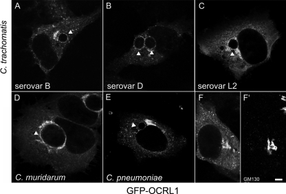
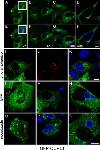
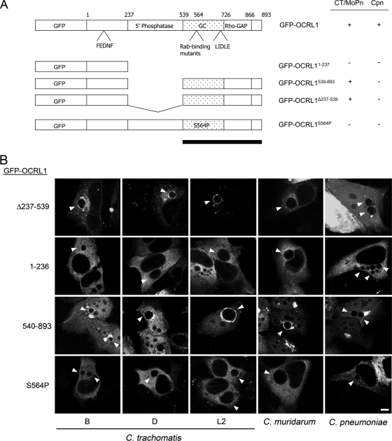
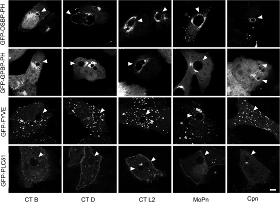
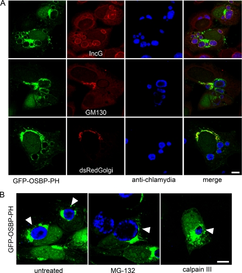
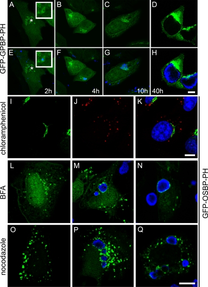
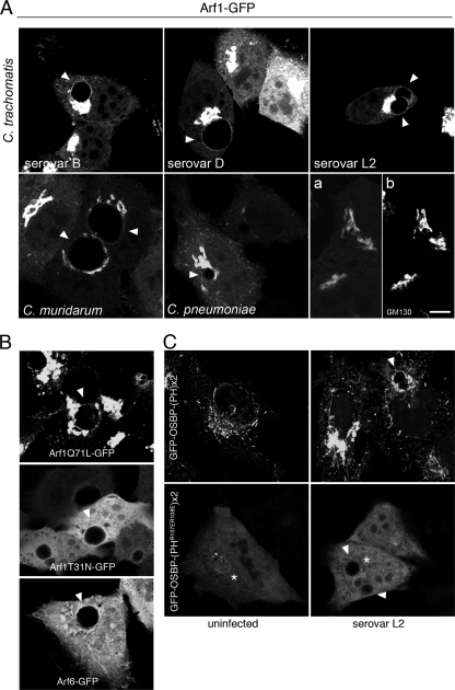
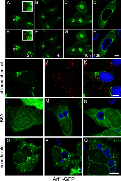
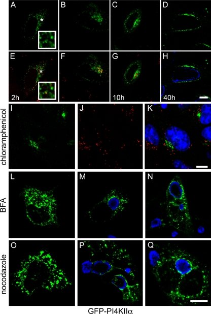
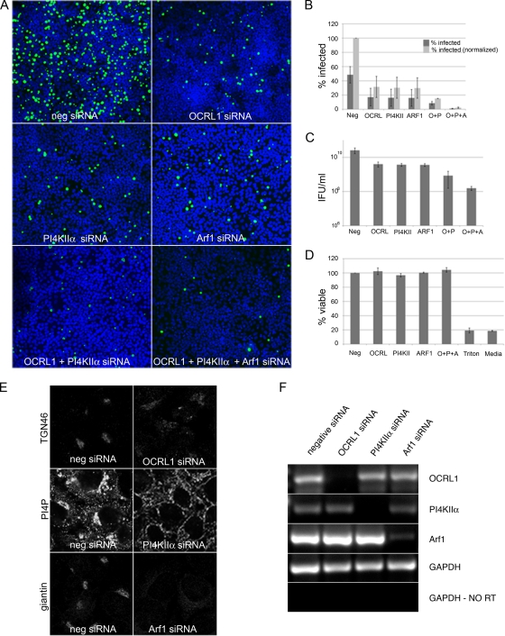
Chlamydiae replicate within a nonacidified vacuole, termed an inclusion. As obligate intracellular bacteria, chlamydiae actively modify their vacuole to exploit host signaling and trafficking pathways. Recently, we demonstrated that several Rab GTPases are actively targeted to the inclusion. To define the biological roles of inclusion localized Rab GTPases, we have begun to identify inclusion-localized Rab effectors. Here we demonstrate that oculocerebrorenal syndrome of Lowe protein 1 (OCRL1), a Golgi complex-localized phosphatidylinositol (PI)-5-phosphatase that binds to multiple Rab GTPases, localizes to chlamydial inclusions. By examining the intracellular localization of green fluorescent protein (GFP) fusion proteins that bind to unique phosphoinositide species, we also demonstrate that phosphatidylinositol-4-phosphate (PI4P), the product of OCRL1, is present at the inclusion membrane. Furthermore, two additional host proteins, Arf1, which together with PI4P mediates the recruitment of PI4P-binding proteins to the Golgi complex, and PI4KII alpha, a major producer of Golgi complex-localized PI4P, also localize to chlamydial inclusions. Depletion of OCRL1, Arf1, or PI4KII alpha by small interfering RNA (siRNA) decreases inclusion formation and the production of infectious progeny. Infectivity is further decreased in cells simultaneously depleted for all three host proteins, suggesting partially overlapping functions in infected cells. Collectively, these data demonstrate that Chlamydia species create a unique replication-competent vacuolar environment by modulating both the Rab GTPase and the PI composition of the chlamydial inclusion.


Figures

FIG. 1.
FIG. 1.
GFP-OCRL1 is recruited to chlamydial inclusions in a species-independent fashion. HeLa cells transiently expressing GFP-OCRL1 were infected with C. trachomatis serovar B (24 h) (A), C. trachomatis serovar D (18 h) (B), C. trachomatis serovar L2 (18 h) (C), C. muridarum (18 h) (D), and C. pneumoniae (48 h) (E). (F and F′) Uninfected HeLa cells transiently expressing GFP-OCRL1 were stained with GM130 antibody to confirm Golgi complex localization of GFP-OCRL1. Cells were fixed and viewed by LSCM. Arrowheads indicate inclusion. Scale bar, 5 μm.
FIG. 2.
FIG. 2.
Association of GFP-OCRL1 with the chlamydial inclusion. (A to H) HeLa cells transiently expressing GFP-OCRL1 were infected with C. trachomatis serovar L2 at an MOI of approximately 25 to 50. At the indicated time points p.i. (2 h, 4 h, 10 h, and 40 h), cells were fixed and stained with antichlamydial antiserum (blue) and viewed by LSCM. (E to H) Merged images of GFP-expressing cells (green) and chlamydia staining (blue). The insets are selected regions denoted by asterisks. Scale bar, 10 μm. (I to K) Inhibition of chlamydial gene expression with chloramphenicol prevents recruitment of GFP-OCRL1 to chlamydia-containing vacuoles. HeLa cells transiently expressing GFP-OCRL1 (I; green) were infected with C. trachomatis serovar L2 in the presence of 25 μg/ml chloramphenicol and incubated for 18 h at 37°C. Cells were fixed and stained with antichlamydial antisera (J; red) and viewed by LSCM. (K) Merged image of panels I and J. (L to N) Golgi complex-dependent trafficking is not essential for the trafficking and association of GFP-OCRL1 with the inclusion. (L) Uninfected HeLa cells transiently expressing GFP-OCRL1 (green) were treated with 1 μg/μl BFA for 30 min at 37°C. (M) HeLa cells transiently expressing GFP-OCRL1 were infected with C. trachomatis serovar D. Eighteen hours p.i., cells were treated with 1 μg/μl BFA for 30 min at 37°C. (N) HeLa cells transiently expressing GFP-OCRL1 were pretreated with 1 μg/μl BFA for 30 min prior to infection with C. trachomatis serovar D. Cells were incubated for an additional 18 h in the continued presence of BFA. Fixed cells were stained with antichlamydial antisera (blue) and viewed by LSCM. (O to Q) Microtubules are not essential for the trafficking or association of GFP-OCRL1 with inclusions. (O) Uninfected HeLa cells expressing GFP-OCRL1 were treated with 20 μM nocodazole for 3 h at 37°C. (P) HeLa cells transiently expressing GFP-OCRL1 were infected with C. trachomatis serovar D. Eighteen hours p.i., cells were treated with 20 μM nocodazole for 3 h at 37°C. (Q) HeLa cells transiently expressing GFP-OCRL1 were pretreated with 20 μm nocodazole for 2 h prior to infection with C. trachomatis serovar D. Cells were incubated for an additional 18 h in the presence of nocodazole. (O to Q) Fixed cells were stained with antichlamydial antisera (blue) and viewed by LSCM. Scale bar, 10 μm.
FIG. 3.
FIG. 3.
The carboxy-terminal Rab binding domain mediates the recruitment of OCRL1 to C. trachomatis and C. muridarum inclusions. (A) GFP-OCRL1 fusion proteins utilized in this study. Amino acids 1 to 236 contain an AP-2 binding site (FEDNF); amino acids 237 to 539 encode the catalytic 5-phosphatase domain; and amino acids 540 to 893 are required for Golgi complex localization (GC; stippled box) and contain separate Rab GTPase, clathrin heavy-chain binding domains (LIDLE), and the Rho-GAP-like domain. The black bar indicates the inclusion-targeting domain for C. trachomatis and C. muridarum. + indicates localization of GFP fusion protein to inclusion; − indicates no localization of the GFP fusion protein to the inclusion. CT, C. trachomatis; MoPn, C. muridarum; Cpn, C. pneumoniae. (B) HeLa cells transiently expressing GFP-OCRL1Δ237-539, GFP-OCRL11-236, GFP-OCRL1540-893, or GFP-OCRL1S564P were infected with C. trachomatis serovar B (24 h), C. trachomatis serovar D (18 h), C. trachomatis serovar L2 (18 h), C. muridarum (18 h), and C. pneumoniae (48 h). Cells were fixed and viewed by LSCM. Arrowheads indicate inclusions. Scale bar, 5 μm.
FIG. 4.
FIG. 4.
PI4P is present on the chlamydial inclusion membrane. HeLa cells transiently expressing GFP-OSBP-PH, GFP-GPBP-PH, GFP-PLCδ1-PH, or GFP-FYVE were infected with C. trachomatis serovar B (24 h), C. trachomatis serovar D (18 h), C. trachomatis serovar L2 (18 h), C. muridarum (18 h), and C. pneumoniae (48 h). Infected cells were fixed and viewed by LSCM. Arrowheads indicate inclusions. CT, C. trachomatis; MoPn, C. muridarum; Cpn, C. pneumoniae. Scale bar, 5 μm.
FIG. 5.
FIG. 5.
GFP-OSBP-PH localizes to the inclusion membrane in the absence of Golgi complex markers. (A) HeLa cells transiently expressing GFP-OSBP-PH (green) were infected with C. trachomatis serovar D. Eighteen hours p.i., cells were fixed and dually labeled with either anti-IncG (red) and antichlamydial antisera (blue) or anti-GM130 (red) and antichlamydial antisera (blue). HeLa cells transiently coexpressing GFP-OSBP-PH (green) and dsRed-Golgi (red) (Clontech) were fixed and stained with antichlamydial antisera (blue). (B) HeLa cells transiently expressing GFP-OSBP-PH (green) were infected with C. trachomatis serovar L2. Eight hours p.i., cells were mock treated or treated with 2.5 μM MG132 or 100 μM calpain III and incubated for an additional 18 h at 37°C. Cells were fixed and stained with antichlamydial antisera (blue). Arrowheads indicated GFP-OSBP-PH localization on selected inclusions. Cells were viewed by LSCM. Scale bar, 10 μm.
FIG. 6.
FIG. 6.
Association of the PI4P-binding PH domain with the chlamydial inclusion. (A to H) HeLa cells transiently expressing GFP-GPBP-PH were infected with C. trachomatis serovar L2 at an MOI of approximately 25 to 50. At the indicated time points p.i. (2 h, 4 h, 10 h, and 40 h), cells were fixed and stained with antichlamydial antiserum (blue) and viewed by LSCM. (E to H) merged images of GFP-expressing cells (green) and chlamydial staining (blue). The insets are selected regions denoted by asterisks. Scale bar, 10 μm. (I to K) Inhibition of chlamydial gene expression with chloramphenicol prevents recruitment of GFP-OSPB-PH to chlamydia-containing vacuoles. HeLa cells transiently expressing GFP-OSPB-PH (I; green) were infected with C. trachomatis serovar L2 in the presence of 25 μg/ml chloramphenicol and incubated for 18 h at 37°C. Cells were fixed and stained with antichlamydial antisera (J; red) and viewed by LSCM. (K) Merged image of panels I and J. (L to N) Golgi complex-dependent trafficking is not necessary for the trafficking and association of GFP-OSBP-PH with the inclusion. (L) Uninfected HeLa cells transiently expressing GFP-OSBP-PH (green) were treated with 1 μg/μl BFA for 30 min at 37°C. (M) HeLa cells transiently expressing GFP-OSBP-PH were infected with C. trachomatis serovar D. Eighteen hours p.i., cells were treated with 1 μg/μl BFA for 30 min at 37°C. (N) HeLa cells transiently expressing GFP-OSBP-PH were pretreated with 1 μg/μl BFA for 30 min prior to infection with C. trachomatis serovar D. Cells were incubated for an additional 18 h in the continued presence of BFA. (O to Q) Microtubules are not essential for the trafficking or association of GFP-OSBP-PH with inclusions. (O) Uninfected HeLa cells expressing GFP-OSBP-PH were treated with 20 μM nocodazole for 3 h at 37°C. (P) HeLa cells transiently expressing GFP-OSBP-PH were infected with C. trachomatis serovar D. Eighteen hours p.i., cells were treated with 20 μM nocodazole for 3 h at 37°C. (Q) HeLa cells transiently expressing GFP-OSBP-PH were pretreated with 20 μM nocodazole for 2 h prior to infection with C. trachomatis serovar D. Cells were incubated for an additional 18 h in the presence of nocodazole. (L to Q) Fixed cells were stained with antichlamydial antisera (blue) and viewed by LSCM. Scale bar, 10 μm.
FIG. 7.
FIG. 7.
Arf1-GFP localizes to chlamydial inclusions but is not sufficient to mediate recruitment of GFP-OSBP-PH. (A) HeLa cells transiently expressing Arf1-GFP were infected with C. trachomatis serovar B (24 h), C. trachomatis serovar D (18 h), C. trachomatis serovar L2 (18 h), C. muridarum (18 h), and C. pneumoniae (48 h). Cells were fixed and viewed by LSCM. (a and b) Uninfected HeLa cells expressing Arf1-GFP (a) were fixed and stained with anti-GM130 (b) to confirm Golgi complex localization of the fusion protein. Scale bar, 10 μm. (B) HeLa cells transiently expressing constitutively active Arf1Q71L-GFP, dominant-negative Arf1T31N-GFP, or Arf6-GFP were infected with C. trachomatis serovar L2 for 18 h. Cells were fixed, stained with antichlamydial antisera (data not shown), and viewed by LSCM. (C) HeLa cells transiently expressing GFP-OSBP-(PH)×2 and GFP-OSBP-(PHR107ER108E)×2, a PI4P-binding-defective mutant, were mock infected or infected with C. trachomatis serovar L2 for 18 h. Cells were fixed and viewed by LSCM. Residual Golgi complex localization of the PI4P-binding deficient mutant is depicted by asterisks. Arrowheads indicate inclusions.
FIG. 8.
FIG. 8.
Association of Arf1-GFP with the chlamydial inclusion. (A to H) HeLa cells transiently expressing Arf1-GFP were infected with C. trachomatis serovar L2 at an MOI of approximately 25 to 50. At the indicated time points p.i. (2 h, 4 h, 10 h, and 40 h), cells were fixed and stained with antichlamydial antiserum (blue or red) and viewed by LSCM. (E to H) Merged images of GFP-expressing cells (green) and chlamydia staining (blue or red). The insets are selected regions denoted by asterisks. Scale bar, 10 μm. (I to K) Inhibition of chlamydial gene expression with chloramphenicol prevents recruitment of Arf1-GFP to chlamydia-containing vacuoles. HeLa cells transiently expressing Arf1-GFP (I; green) were infected with C. trachomatis serovar L2 in the presence of 25 μg/ml chloramphenicol and incubated for 18 h at 37°C. Cells were fixed and stained with antichlamydial antisera (J; red) and viewed by LSCM. (K) Merged image of I and J. (L to N) The trafficking and association of Arf1-GFP with the inclusion are sensitive to BFA. (L) Uninfected HeLa cells transiently expressing Arf1-GFP (green) were treated with 1 μg/μl BFA for 30 min at 37°C. (M) HeLa cells transiently expressing Arf1-GFP were infected with C. trachomatis serovar D. Eighteen hours p.i., cells were treated with 1 μg/μl BFA for 30 min at 37°C. (N) HeLa cells transiently expressing Arf1-GFP were pretreated with 1 μg/μl BFA for 30 min prior to infection with C. trachomatis serovar D. Cells were incubated for an additional 18 h in the continued presence of BFA. (O to Q) Microtubules are not essential for trafficking or association of Arf1-GFP with the inclusion. (O) Uninfected HeLa cells expressing Arf1-GFP were treated with 20 μM nocodazole for 3 h at 37°C. (P) HeLa cells transiently expressing Arf1-GFP were infected with C. trachomatis serovar D. Eighteen hours p.i., cells were treated with 20 μM nocodazole for 3 h at 37°C. (Q) HeLa cells transiently expressing Arf1-GFP were pretreated with 20 μM nocodazole for 2 h prior to infection with C. trachomatis serovar D. Cells were incubated for an additional 18 h in the presence of nocodazole. (L to Q) Fixed cells were stained with antichlamydial antisera (blue) and viewed by LSCM. Scale bar, 10 μm.
FIG. 9.
FIG. 9.
PI4KIIα localizes to chlamydial inclusions. HeLa cells transiently expressing GFP-PI4KIIα, GFP-PI4KIIβ, or GFP-PI4KIIIβ were infected with C. trachomatis serovar B (24 h), C. trachomatis serovar D (18 h), C. trachomatis serovar L2 (18 h), C. muridarum (18 h), and C. pneumoniae (48 h). Cells were fixed and viewed by LSCM. CT, C. trachomatis; MoPn, C. muridarum; Cpn, C. pneumoniae. Arrowheads indicate inclusions. Scale bar, 10 μm.
FIG. 10.
FIG. 10.
Association of GFP-PI4KIIα with the chlamydial inclusion. (A to H) HeLa cells transiently expressing GFP-PI4KIIα were infected with C. trachomatis serovar L2 at an MOI of approximately 25 to 50. At the indicated time points p.i. (2 h, 4 h, 10 h, and 40 h), cells were fixed and stained with antichlamydial antiserum (blue or red) and viewed by LSCM. (E to H) Merged images of GFP-expressing cells (green) and chlamydia staining (blue or red). The insets are selected regions denoted by asterisks. Scale bar, 10 μm. (I to K) Inhibition of chlamydial gene expression with chloramphenicol prevents recruitment of GFP-PI4KIIα to chlamydia-containing vacuoles. HeLa cells transiently expressing GFP-PI4KIIα (I; green) were infected with C. trachomatis serovar L2 in the presence of 25 μg/ml chloramphenicol and incubated for 18 h at 37°C. Cells were fixed and stained with antichlamydial antisera (J; red) and viewed by LSCM. (K) Merged image of panels I and J. (L to N) Golgi complex-dependent trafficking is not necessary for the trafficking and association of GFP-PI4KIIα with the inclusion. (L) Uninfected HeLa cells transiently expressing GFP-PI4KIIα (green) were treated with 1 μg/μl BFA for 30 min at 37°C. (M) HeLa cells transiently expressing GFP-PI4KIIα were infected with C. trachomatis serovar D. Eighteen hours p.i., cells were treated with 1 μg/μl BFA for 30 min at 37°C. (N) HeLa cells transiently expressing GFP-PI4KIIα were pretreated with 1 μg/μl BFA for 30 min prior to infection with C. trachomatis serovar D. Cells were incubated for an additional 18 h in the continued presence of BFA. (O to Q) Microtubules are not essential for the association or trafficking of GFP-PI4KIIα to the inclusion. (O) Uninfected HeLa cells expressing GFP-PI4KIIα were treated with 20 μM nocodazole for 3 h at 37°C. (P) HeLa cells transiently expressing GFP-PI4KIIα were infected with C. trachomatis serovar D. Eighteen hours p.i., cells were treated with 20 μM nocodazole for 3 h at 37°C. (Q) HeLa cells transiently expressing GFP-PI4KIIα were pretreated with 20 μM nocodazole for 2 h prior to infection with C. trachomatis serovar D. Cells were incubated for an additional 18 h in the presence of nocodazole. (L to Q) Fixed cells were stained with antichlamydial antisera (blue) and viewed by LSCM. Scale bar, 10 μm.
FIG. 11.
FIG. 11.
OCRL1, Arf1, and PI4KIIα are required for optimal chlamydial infection. siRNA-induced silencing was utilized to knock down OCRL1 (O), Arf1 (A), and PI4KIIα (P) expression in infected cells. HeLa cells were transfected with gene-specific or negative control siRNA oligonucleotide for 72 h. (F) RT-PCR was performed on total RNA isolated from OCRL1, PI4KIIα, Arf1, or negative (neg) siRNA-treated cells using gene-specific oligonucleotides as indicated. To control for equivalent amounts of total RNA, RT-PCR was performed with GAPDH-specific oligonucleotides. RT, reverse transcriptase. (E) Uninfected HeLa cells treated with indicated siRNAs for 72 h were fixed and stained as follows. OCRL1 siRNA-treated cells were stained with anti-TGN46, Arf1-siRNA-treated cells were stained with antigiantin, and PI4KIIα-siRNA-treated cells were stained with anti-PI4KIIα. (A, B, C, and D) Seventy-two hours posttransfection, siRNA-treated cells were infected with C. trachomatis serovar L2 for 18 h (A and B) or for 36 h (C). (A and B) To quantify primary inclusion formation, at 18 h p.i., cells were fixed and stained with antichlamydial antiserum. Inclusions were counted by indirect immunofluorescence microscopy. Percent infection was normalized such that the negative siRNA control cells were set to 100% infection. Experiments were performed in triplicate and repeated at least 3 times. Error bars indicate standard deviation. (C) To quantify infectivity, IFU were enumerated by lysing cells in distilled water (dH2O) at the indicated time points p.i. and titrating lysates on fresh HeLa cell monolayers. Experiments were performed in triplicate and repeated at least 3 times. Error bars indicate standard deviation. Statistics were performed with Microsoft Excel 2003 software using two-tailed t tests. (D) Cell viability was determined using WST-1 reagent. Cells were lysed with Triton X-100 as a control for inviable cells..

Similar articles

Cited by

References

    1. Attree, O., I. M. Olivos, I. Okabe, L. C. Bailey, D. L. Nelson, R. A. Lewis, R. R. McInnes, and R. L. Nussbaum. 1992. The Lowe's oculocerebrorenal syndrome gene encodes a protein highly homologous to inositol polyphosphate-5-phosphatase. Nature 358:239-242. - PubMed
    1. Balana, M. E., F. Niedergand, A. Subtil, A. Alcover, P. Chavrier, and A. Dautry-Varsat. 2005. Arf6 GTPase controls bacterial invasion by actin remodeling. J. Cell Sci. 118:2201-2210. - PubMed
    1. Balla, A., and T. Balla. 2006. Phosphatidylinositol 4-kinases: old enzymes with emerging functions. Trends Cell Biol. 16:351-361. - PubMed
    1. Balla, A., G. Tuymetova, A. Tsiomenko, P. Varnai, and T. Balla. 2005. A plasma membrane pool of phosphatidylinositol 4-phosphate is generated by phosphatidylinositol 4-kinase type-III alpha: studies with the PH domains of the oxysterol binding protein and FAPP1. Mol. Biol. Cell 16:1282-1295. - PMC - PubMed
    1. Beatty, W. L. 2006. Trafficking from CD63-positive late endocytic multivesicular bodies is essential for intracellular development of Chlamydia trachomatis. J. Cell.Sci. 119:350-359. - PubMed

Publication types

MeSH terms

Substances