. 2010 Jun 17;115(24):5026-36.
doi: 10.1182/blood-2009-09-243071. Epub 2010 Mar 15.

Complement receptor 2/CD21- human naive B cells contain mostly autoreactive unresponsive clones

Affiliations

Complement receptor 2/CD21- human naive B cells contain mostly autoreactive unresponsive clones

Isabelle Isnardi et al. Blood. .

Abstract

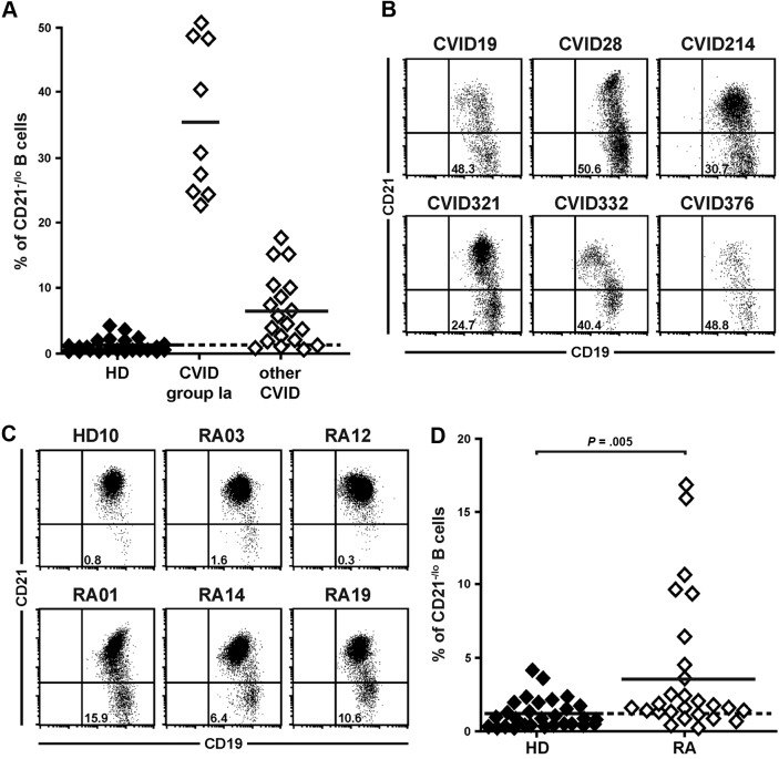
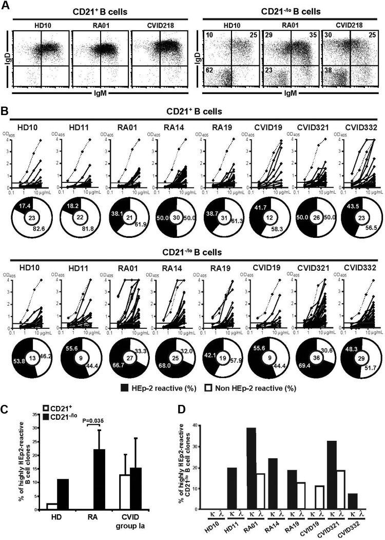
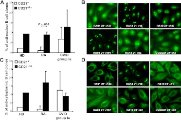
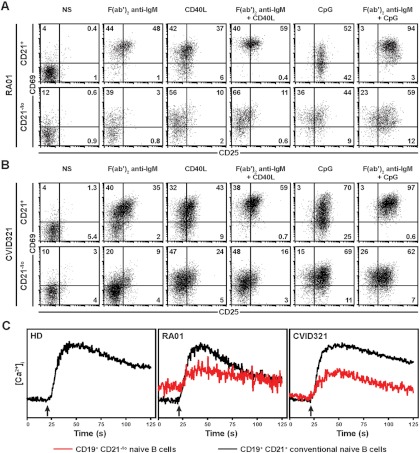
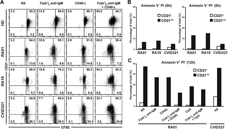
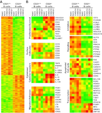
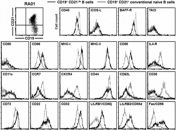
Complement receptor 2-negative (CR2/CD21(-)) B cells have been found enriched in patients with autoimmune diseases and in common variable immunodeficiency (CVID) patients who are prone to autoimmunity. However, the physiology of CD21(-/lo) B cells remains poorly characterized. We found that some rheumatoid arthritis (RA) patients also display an increased frequency of CD21(-/lo) B cells in their blood. A majority of CD21(-/lo) B cells from RA and CVID patients expressed germline autoreactive antibodies, which recognized nuclear and cytoplasmic structures. In addition, these B cells were unable to induce calcium flux, become activated, or proliferate in response to B-cell receptor and/or CD40 triggering, suggesting that these autoreactive B cells may be anergic. Moreover, gene array analyses of CD21(-/lo) B cells revealed molecules specifically expressed in these B cells and that are likely to induce their unresponsive stage. Thus, CD21(-/lo) B cells contain mostly autoreactive unresponsive clones, which express a specific set of molecules that may represent new biomarkers to identify anergic B cells in humans.


Figures

Figure 1
Figure 1
CVID and RA patients may display an increased frequency of CD21−/lo B cells in their blood. (A) CVID group Ia patients were previously defined as patients displaying a frequency of CD21−/lo naive B cells > 20% of total B cells. Each diamond represents a person and (B) dot plots show CD19 and CD21 expression on CD27-gated peripheral B cells from representative CVID group Ia patients. (C) Increased CD21−/lo B-cell frequency in RA patients. Dot plots show CD19 and CD21 expression on CD27-depleted peripheral B cells from a representative healthy donor (HD10) and RA patients. (D) The frequencies of CD21−/lo naive B cells are compared between healthy donors and RA patients. Each diamond represents a person and statistically significant differences are indicated.
Figure 2
Figure 2
CD21−/lo naive B cells express autoreactive BCRs. (A) A majority of CD21−/lo B cells from RA and CVID patients express IgM and IgD. Dot plots show IgM and IgD expression gated on CD19+CD21+(left) and CD19+CD21−/lo (right) CD27-depleted B cells. (B) Most CD21−/lo naive B cells express autoreactive antibodies. Antibodies cloned from single CD21+CD27CD10 and CD21−/loCD27CD10 B cells from 2 healthy donors (HD10 and 11), 3 RA, and 3 CVID patients were tested by ELISA for reactivity against HEp-2 lysates. Dotted lines show ED38-positive control. Horizontal lines show cutoff OD405 for positive reactivity. For each B-cell population, the frequency of HEp-2–reactive and non–HEp-2–reactive clones is summarized in pie charts, with the number of antibodies tested indicated in the centers. The frequency of HEp-2–reactive B cells was higher in CD21−/lo than in CD21+ B cells in all persons. (C) CD21−/lo B cells express highly autoreactive antibodies. The frequency of highly HEp-2–reactive antibodies, arbitrarily defined by ELISA OD405 > 2, expressed by CD21+ and CD21−/lo B cells from HD, RA, and CVID patients is represented. A large fraction of CD21−/lo B cells from most persons was highly HEp-2 reactive. P values calculated using a paired t test are indicated when significant. (D) A majority of highly autoreactive antibodies from CD21−/lo B cells express kappa light chains. The frequency of highly HEp-2–reactive CD21−/lo B cells from HD, RA, and CVID patients is represented subdivided among kappa and lambda clones. Many highly HEp-2–reactive CD21−/lo B cells express kappa light chains, suggesting that receptor editing using lambda light chains was not induced to silence these clones.
Figure 3
Figure 3
CD21−/lo B cells contain antinuclear and anticytoplasmic clones. The frequencies of antinuclear (A) and anticytoplasmic (C) clones in CD21+ and CD21−/lo B cells from HD, RA, and CVID patients are shown. P values calculated using a paired t test are indicated when significant. (B) ANAs expressed by CD21−/lo B cells showed a diversity of nucleolar (RA01 21 κ181, RA01 21 κ137, RA14 21 κ15, and RA14 21 κ83), nucleolar and speckled (RA19 21 κ127), and speckled (CVID 21 κ12) antinuclear staining patterns. (D) Anticytoplasmic CD21−/lo B cells can react with well-defined intracellular structures, which include mitochondria (RA01 21 κ140, and CVID321 21 κ82), the Golgi (RA14 21 κ18), and cytoplasmic fibers (RA19 21 κ61, RA14 21 κ62, and RA01 21 κ157 that is likely to recognize actin).
Figure 4
Figure 4
CD21−/lo naive B cells fail to properly respond to BCR triggering. CD21−/lo naive B cells from (A) RA01 and (B) CVID321 patients fail to up-regulate CD69 and CD25 expression after stimulation with F(ab′2) anti-IgM, recombinant human CD40L, and TLR9 agonist CpG. Total naive B cells containing both CD21−/lo and CD21+ B cells were plated and dot plots show CD69 and CD25 expression after 2 days according to gated CD21 expression. (C) BCR triggering in CD21−/lo naive B cells does not properly increase [Ca2+]i. [Ca2+]i was assessed by flow cytometry in CD21−/lo and CD21+ naive B cells from a healthy donor (HD) and RA01 and CVID321 patients. Both B-cell populations were loaded with Fluo-3 and expressed similar levels of surface IgM expression when experiments were performed. Arrow indicates when F(ab′)2 anti-IgM was added. Data from each group of persons are representative of 2-3 independent experiments.
Figure 5
Figure 5
CD21−/lo naive B cells do not proliferate after BCR and CD40 cotriggering and are prone to cell death. (A) Total naive B cells from healthy donors (HD), and RA and CVID patients containing both CD21−/lo and CD21+ B cells were labeled with CFSE and stimulated with F(ab′2) anti-IgM and/or recombinant human CD40L. Dot plots show CD21 expression according to CFSE dilution after 3 days. CFSE dilution in CD21+ but not in CD21−/lo B cells stimulated with F(ab′2) anti-IgM and recombinant human CD40L reveals that some CD21+ B cells underwent up to 2 divisions, whereas CD21−/lo B cells did not divide. (B) Increased frequency of annexin V+ propidium iodide–negative (PI) early apoptotic, and annexin V+ PI+ late apoptotic and dead cells in freshly isolated CD21−/lo naive B cells from RA and CVID patients compared with their CD21+ B-cell counterparts. (C) BCR, CD40, and/or TLR9 triggering do not prevent the apoptosis and cell death of CD21−/lo naive B cells. Total naive B cells containing both CD21−/lo and CD21+ B cells from RA01 or CVID321 patients were plated, and annexin V and PI staining was assessed after 12 hours. BCR, CD40, and TLR9 stimulation prevents late apoptosis and cell death in CD21+ but not in CD21−/lo B cells.
Figure 6
Figure 6
Gene array comparisons of CD21−/lo and CD21+ naive B cells from RA and CVID patients using the Affymetrix Human Genome U133 Plus 2.0 Array. (A) Transcripts differentially expressed by CD21−/lo and CD21+ naive B cells from 2 RA and 3 CVID group Ia patients are shown. (B) Selected transcripts well known to B-cell biology or of potential interest are presented. Up- and down-regulated transcripts are indicated in red and green, respectively. The magnitude of expression is depicted by the color bar.
Figure 7
Figure 7
CD21−/lo B cells from RA and CVID patients display a phenotype reflecting gene array profiling data. Overlays for the indicated markers were obtained when CD27-depleted B cells isolated from the blood of patient RA01 were gated on either CD19+CD21−/lo (bold line) or CD19+CD21+ (dashed line) B cells. The similar phenotypes of CD21−/lo B cells from CVID patients and healthy donors are shown in supplemental Figures 7 and 8.

Comment in

Similar articles

Cited by

References

    1. Nemazee D. Does immunological tolerance explain the waste in the B-lymphocyte immune system? experiment and theory. Ann N Y Acad Sci. 1995;764:397–401. - PubMed
    1. Radic MZ, Weigert M. Origins of anti-DNA antibodies and their implications for B-cell tolerance. Ann N Y Acad Sci. 1995;764:384–396. - PubMed
    1. Goodnow CC, Sprent J, Fazekas de St Groth B, Vinuesa CG. Cellular and genetic mechanisms of self tolerance and autoimmunity. Nature. 2005;435(7042):590–597. - PubMed
    1. Goodnow CC, Crosbie J, Adelstein S, et al. Altered immunoglobulin expression and functional silencing of self-reactive B lymphocytes in transgenic mice. Nature. 1988;334(6184):676–682. - PubMed
    1. Erikson J, Radic MZ, Camper SA, Hardy RR, Carmack C, Weigert M. Expression of anti-DNA immunoglobulin transgenes in non-autoimmune mice. Nature. 1991;349(6307):331–334. - PubMed

Publication types

MeSH terms