. 2010 Mar 30;107(13):5967-71.
doi: 10.1073/pnas.0912496107. Epub 2010 Mar 15.

Plasmodium vivax clinical malaria is commonly observed in Duffy-negative Malagasy people

Affiliations

Plasmodium vivax clinical malaria is commonly observed in Duffy-negative Malagasy people

Didier Ménard et al. Proc Natl Acad Sci U S A. .

Abstract

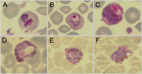
Malaria therapy, experimental, and epidemiological studies have shown that erythrocyte Duffy blood group-negative people, largely of African ancestry, are resistant to erythrocyte Plasmodium vivax infection. These findings established a paradigm that the Duffy antigen is required for P. vivax erythrocyte invasion. P. vivax is endemic in Madagascar, where admixture of Duffy-negative and Duffy-positive populations of diverse ethnic backgrounds has occurred over 2 millennia. There, we investigated susceptibility to P. vivax blood-stage infection and disease in association with Duffy blood group polymorphism. Duffy blood group genotyping identified 72% Duffy-negative individuals (FY*B(ES)/*B(ES)) in community surveys conducted at eight sentinel sites. Flow cytometry and adsorption-elution results confirmed the absence of Duffy antigen expression on Duffy-negative erythrocytes. P. vivax PCR positivity was observed in 8.8% (42/476) of asymptomatic Duffy-negative people. Clinical vivax malaria was identified in Duffy-negative subjects with nine P. vivax monoinfections and eight mixed Plasmodium species infections that included P. vivax (4.9 and 4.4% of 183 participants, respectively). Microscopy examination of blood smears confirmed blood-stage development of P. vivax, including gametocytes. Genotyping of polymorphic surface and microsatellite markers suggested that multiple P. vivax strains were infecting Duffy-negative people. In Madagascar, P. vivax has broken through its dependence on the Duffy antigen for establishing human blood-stage infection and disease. Further studies are necessary to identify the parasite and host molecules that enable this Duffy-independent P. vivax invasion of human erythrocytes.


Conflict of interest statement

The authors declare no conflict of interest.

Figures

Fig. 1.
Fig. 1.
Frequency distribution of P. vivax infections and clinical cases identified in Duffy-positive and -negative Malagasy people. Pie graphs show the prevalence of Duffy-positive (dark/light green) and Duffy-negative (red/pink quadrants) phenotypes in the eight Madagascar study sites. Prevalence of P. vivax infection observed in the survey of school-age children is shown in red and dark green; population subsets not infected with P. vivax are pink and light green. Study sites identified by a red star indicate that clinical vivax malaria was observed in Duffy-negative individuals (additional data classify clinical cases as numbers of people diagnosed with monoinfection P. vivax/mixed P. vivax + P. falciparum). A green star indicates that vivax malaria was observed in Duffy-positive individuals only (Ejeda). In Ihosy clinical malaria was observed in one individual with a mixed P. vivax/P. falciparum infection. P. vivax malaria was not observed in Andapa and Farafangana (black star). Malaria transmission strata are identified as tropical (lightest gray), subdesert (light gray), equatorial (middle gray), and highlands (dark gray).
Fig. 2.
Fig. 2.
Standard Giemsa-stained thin smear preparations of P. vivax infection and development in human Duffy-negative erythrocytes. A–C originated from a 4-year-old female, genotyped as Duffy negative (FY*BES/*BES), who presented at the Tsiroanomandidy health center (June 26, 2006) with fever (37.8 °C), headache, and sweating without previous antimalarial treatment. Standard blood smear diagnosis revealed a mixed infection with P. vivax [parasitemia = 3,040 parasitized red blood cells (pRBC)/μL] and P. falciparum (parasitemia = 980 pRBC/μL). PCR-based Plasmodium species diagnosis confirmed the blood smear result; P. malariae and P. ovale were not detected. A shows an undifferentiated P. vivax trophozoite with enlarged erythrocyte volume, clear evidence of Schüffner stippling, and amoeboid morphology. B shows a P. vivax early stage trophozoite with condensed chromatin, enlarged erythrocyte volume, Schüffner stippling, and irregular ring-shaped cytoplasm. C shows a P. vivax gametocyte: Lavender parasite, larger pink chromatin mass, and brown pigment scattered throughout the cytoplasm are characteristics of microgametocytes (male). D originated from a 12-year-old Duffy-negative (FY*BES/*BES) male, who presented at the Miandrivazo health center (June 27, 2006) with fever (37.5 °C) and shivering without previous antimalarial treatment. Standard blood smear diagnosis and light microscopy revealed infection with only P. vivax (parasitemia = 3,000 pRBC/μL). PCR-based Plasmodium species diagnosis confirmed this blood smear result; P. falciparum, P. malariae, and P. ovale were not detected. The parasite featured shows evidence of a P. vivax gametocyte: Large blue parasite, smaller pink chromatin mass, and brown pigment scattered throughout the cytoplasm are characteristics of macrogametocytes (female). E and F originated from a 3-year-old Duffy-negative (FY*BES/*BES) female, who presented at the Moramanga health center (April 11, 2006) with fever (37.8 °C) without previous antimalarial treatment. Standard blood smear diagnosis and light microscopy revealed infection with only P. vivax (parasitemia = 3,368 pRBC/μL). PCR-based Plasmodium species diagnosis confirmed this blood smear result; P. falciparum, P. malariae, and P. ovale were not detected. The parasites featured show additional evidence of P. vivax gametocytes.
Fig. 3.
Fig. 3.
Flow cytometry and serological correlation of Duffy-negative and Duffy-positive phenotypes with respective genotypes in Malagasy school children from Tsiroanomandidy, Madagascar. (A) Flow cytometric analysis of Duffy blood group genotypes. Flow cytometry histograms show MFI that reflect binding of the Duffy antigen-specific anti-Fy6 antibody (NaM185-2C3) for one Malagasy genotyped as FY*BES/*BES [Fy(a−b−)] (red) and five well-characterized control donors who are FY*BES/*BES [Fy(a−b−)] (solid black), FY*BES/*X [Fy(a−bweak)] (light green), FY*X/*X [Fy(a−bweak)] (green), FY*A/*BES [Fy(a+b−)] (light blue), and FY*A/*B [Fy(a+b+)] (blue), respectively. Results include fluorescence of a Duffy-positive blood sample incubated with an isotype control antibody (dotted black line). (B) Flow cytometry of Duffy antigen expression on erythrocytes from 40 Malagasy study participants. Flow cytometry results show MFI that reflect binding of the Duffy antigen-specific anti-Fy6 antibody for 30 FY*BES/*BES [Fy(a−b−)] Malagasy samples (mean = 48, SD = 1.2), 6 FY*A/*BES [Fy(a+b−)] Malagasy samples (mean = 1,025, SD = 22.4), 2 FY*A/*A [Fy(a+b−)] Malagasy samples (mean = 1,937, SD = 54) and 2 FY*A/*B [Fy(a+b+)] Malagasy samples (mean = 1,896, SD = 8).

References

    1. Boyd MF, Stratman-Thomas WK. Studies on benign tertian malaria. 4. On the refractoriness of negroes to inoculation with Plasmodium vivax. Am J Hyg. 1933;18:485–489.
    1. Bray RS. The susceptibility of Liberians to the Madagascar strain of Plasmodium vivax. J Parasitol. 1958;44(4, Sect 1):371–373. - PubMed
    1. O’Leary PA. Treatment of neurosyphilis by malaria: Report on the three years’ observation of the first one hundred patients treated. J Am Med Assoc. 1927;89:95–100.
    1. Zimmerman PA. In: Infectious Disease and Host-Pathogen Evolution. Dronamraju KR, editor. New York: Cambridge Univ Press; 2004. pp. 141–172.
    1. Roychoudhury AK, Nei M. Human Polymorphic Genes: World Distribution. New York: Oxford Univ Press; 1988.

Publication types

MeSH terms

Associated data

LinkOut - more resources