. 2010 Mar 30;107(13):5983-8.
doi: 10.1073/pnas.0912293107. Epub 2010 Mar 15.

Elongated fibrillar structure of a streptococcal adhesin assembled by the high-affinity association of alpha- and PPII-helices

Affiliations

Elongated fibrillar structure of a streptococcal adhesin assembled by the high-affinity association of alpha- and PPII-helices

Matthew R Larson et al. Proc Natl Acad Sci U S A. .

Abstract

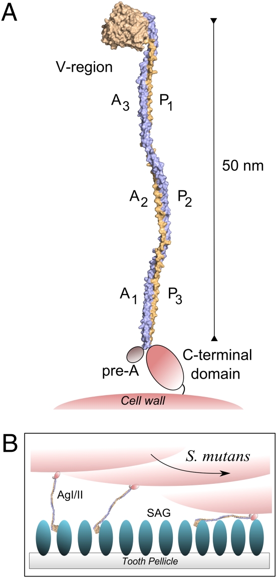
Streptococcus mutans antigen I/II (AgI/II) is a cell surface-localized protein adhesin that interacts with salivary components within the salivary pellicle. AgI/II contributes to virulence and has been studied as an immunological and structural target, but a fundamental understanding of its underlying architecture has been lacking. Here we report a high-resolution (1.8 A) crystal structure of the A(3)VP(1) fragment of S. mutans AgI/II that demonstrates a unique fibrillar form (155 A) through the interaction of two noncontiguous regions in the primary sequence. The A(3) repeat of the alanine-rich domain adopts an extended alpha-helix that intertwines with the P(1) repeat polyproline type II (PPII) helix to form a highly extended stalk-like structure heretofore unseen in prokaryotic or eukaryotic protein structures. Velocity sedimentation studies indicate that full-length AgI/II that contains three A/P repeats extends over 50 nanometers in length. Isothermal titration calorimetry revealed that the high-affinity association between the A(3) and P(1) helices is enthalpically driven. Two distinct binding sites on AgI/II to the host receptor salivary agglutinin (SAG) were identified by surface plasmon resonance (SPR). The current crystal structure reveals that AgI/II family proteins are extended fibrillar structures with the number of alanine- and proline-rich repeats determining their length.


Conflict of interest statement

The authors declare no conflict of interest.

Figures

Fig. 1.
Fig. 1.
(A) The primary sequence layout of AgI/II is shown at the Top. The alanine-rich repeats are highlighted in shades of blue, and the proline-rich repeats are in shades of purple. Shown Below are the fragments of AgI/II used in this study, including the crystallized fragment A3VP1. Residue numbers correspond to the primary sequence of AgI/II from S. mutans strain NG8 (GenBank accession GQ456171). (B) The sequence alignments of A1, A2, and A3 repeats, where the 19 heptad motifs are highlighted in red. (C) The alignments of P1, P2, and P3 repeats, where the proline residues are shown in red.
Fig. 2.
Fig. 2.
Antigen I/II layout and A3VP1 structure overview. (A) Ribbon diagram of the crystal structure of A3VP1 generated by PyMol (48). The α-helix from A3 and the PPII helix from the P1 regions interact to form the 155 Å stalk. (B) Electrostatic map of A3VP1 generated using MolMol (49). The stalk formed by the association of the A3 and P1 helices displays an extensive hydrophobic surface. (C) Superposition of the unique molecules from the P21 and P212121 crystal forms by strictly anchoring over the V region shows ~17 Å displacement at the termini of the helices.
Fig. 3.
Fig. 3.
Interactions between the A3 and P1 helices. (A) The helical wheel diagram (Top) shows the interaction between the A3’s heptad motif “AxYxAx[LV]” and P1’s “PxxP” motif. The helical net of the interactions between the α- and PPII helices is also shown, where the A3-repeat heptad motifs are highlighted in blue and the P1 repeat PxxP motifs are highlighted in orange. Asparagine residues within the A3 region, intervening the heptads, that are involved in hydrogen bonding with the P1 PPII helix are highlighted in red. The conserved tyrosines and leucines (highlighted in yellow) of the heptad sequences are nestled between prolines of the P repeat. Additionally, the phenolic oxygen atom of the tyrosine residues participate in water-mediated hydrogen bonding (red-dashed lines). (B) The stereo diagram of the heptad interactions shows that tyrosine side chains nestle between the prolines in a knobs-in-holes interaction, which is highlighted by surface plots for the A and P repeats. (C) The stereo diagram of the region intervening the heptads shows a dominant direct hydrogen bonding between the asparagine side chains of A3 and the main chain oxygen and nitrogen of the PPII helix. Two prolines (Pro855 and Pro858) break this pattern and face outward.
Fig. 4.
Fig. 4.
Calorimetry, ELISA, and BIACore studies. (A) Calorimetry of the A/P interaction. (Upper) Calorimetric measurements at 20 °C, 25 injections of 10 μL A1–3 into the cell containing VP3. (Lower) The energy (kcal/mol) released during each injection. (B) Inhibition of binding of anti-AgI/II MAbs to S. mutans whole cells. Twofold serial dilutions of AgI/II polypeptides (5–0.05 μM) were incubated with each indicated MAb. Competition ELISA results are shown as the percent inhibition of binding of the indicated MAb to S. mutans whole cells in the presence of each polypeptide. Error bars indicate standard deviations. CG14 (yellow squares), A3VP1 (red diamonds), A1VP3 (blue squares), and C-terminal construct (green triangles). (C) BIAcore studies of AgI/II fragments with immobilized SAG. Full-length CG14, A3VP1, C-terminal, or V-region fragments (analytes) were flowed over a CM5 chip coated with covalently attached SAG. Analyte proteins ranged from 0.25 μM to 4 μM concentrations. Arrows indicate injection starting and stopping points. During the 4-min injection of each polypeptide, an increase in the RU is observed as AgI/II polypeptides bind to SAG. CG14 and A3VP1 showed the maximal binding responses (1 RU is equivalent to 1 pg per square millimeter of sensor surface).
Fig. 5.
Fig. 5.
Model of the AgI/II structure and predicted binding with SAG. (A) The crystal structure of the AgI/II A3VP1 region revealed an extended stalk formed by the A3 and P1 repeats. Ultracentrifugation studies predict that the A2–P2 and A1–P3 repeats extend the stalk to a length over 50 nm. (B) Adherence studies indicated the presence of two sites, one within A3VP1 and another within the C-terminal region, which are widely separated in the AgI/II structure. The cartoon represents a possible model for AgI/II binding to SAG, where interactions occur at both the distal end through the A3VP1 region, and at a secondary adherence site mediated by the C-terminal domain.

Similar articles

Cited by

References

    1. Loesche WJ. Role of Streptococcus mutans in human dental decay. Microbiol Rev. 1986;50:353–380. - PMC - PubMed
    1. Russell MW, Childers NK, Michalek SM, Smith DJ, Taubman MA. A caries vaccine? The state of the science of immunization against dental caries. Caries Res. 2004;38:230–235. - PubMed
    1. Ma JK, Kelly CG, Munro G, Whiley RA, Lehner T. Conservation of the gene encoding streptococcal antigen I/II in oral streptococci. Infect Immun. 1991;59:2686–2694. - PMC - PubMed
    1. Zhang S, Green NM, Sitkiewicz I, Lefebvre RB, Musser JM. Identification and characterization of an antigen I/II family protein produced by group A Streptococcus. Infect Immun. 2006;74:4200–4213. - PMC - PubMed
    1. Jenkinson HF, Demuth DR. Structure, function and immunogenicity of streptococcal antigen I/II polypeptides. Mol Microbiol. 1997;23:183–190. - PubMed

Publication types

MeSH terms

LinkOut - more resources