. 2010 Mar 23;107(12):5587-92.
doi: 10.1073/pnas.0912403107. Epub 2010 Mar 15.

Polymorphonuclear leukocytes mediate Staphylococcus aureus Panton-Valentine leukocidin-induced lung inflammation and injury

Affiliations

Polymorphonuclear leukocytes mediate Staphylococcus aureus Panton-Valentine leukocidin-induced lung inflammation and injury

Binh An Diep et al. Proc Natl Acad Sci U S A. .

Abstract

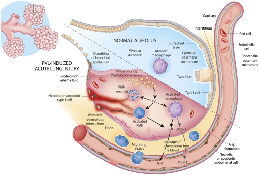
Community-associated methicillin-resistant Staphylococcus aureus (CA-MRSA) is epidemic in the United States, even rivaling HIV/AIDS in its public health impact. The pandemic clone USA300, like other CA-MRSA strains, expresses Panton-Valentine leukocidin (PVL), a pore-forming toxin that targets polymorphonuclear leukocytes (PMNs). PVL is thought to play a key role in the pathogenesis of necrotizing pneumonia, but data from rodent infection models are inconclusive. Rodent PMNs are less susceptible than human PMNs to PVL-induced cytolysis, whereas rabbit PMNs, like those of humans, are highly susceptible to PVL-induced cytolysis. This difference in target cell susceptibility could affect results of experimental models. Therefore, we developed a rabbit model of necrotizing pneumonia to compare the virulence of a USA300 wild-type strain with that of isogenic PVL-deletion mutant and -complemented strains. PVL enhanced the capacity of USA300 to cause severe lung necrosis, pulmonary edema, alveolar hemorrhage, hemoptysis, and death, hallmark clinical features of fatal human necrotizing pneumonia. Purified PVL instilled directly into the lung caused lung inflammation and injury by recruiting and lysing PMNs, which damage the lung by releasing cytotoxic granule contents. These findings provide insights into the mechanism of PVL-induced lung injury and inflammation and demonstrate the utility of the rabbit for studying PVL-mediated pathogenesis.


Conflict of interest statement

The authors declare no conflict of interest.

Figures

Fig. 1.
Fig. 1.
Pore-forming and cytolytic activities of PVL toward human and rabbit PMNs. (A) PMN pore formation [percent ethidium bromide (EtBr)-positive cells] and (B) PMN lysis [percent lactate dehydrogenase (LDH) release] assays using LukS-PV and LukF-PV purified from culture supernatants of a PVL-producing USA300 strain as described in SI Text. Results in A and B are mean of independent experiments with six human and six rabbit blood donors. (C) Pore formation caused by 1:2,000 dilutions of 8-h CCY culture supernatants of USA300 wild-type strain, Δpvl mutant, and compΔpvl strain as indicated. Results in C are mean of independent experiments with 10 human and 4 rabbit blood donors. Statistically significant differences are indicated by asterisk (P < 0.05); all other comparisons are nonsignificant.
Fig. 2.
Fig. 2.
PVL contributes to virulence of USA300 in a rabbit model of necrotizing pneumonia. (A) Kaplan–Meier survival curves for comparison of mortality in rabbits infected via endotracheal instillation with increasing number of cfu of a SF8300 wild-type (wt) strain and its isogenic Δpvl mutant strain (n = 88 total). Fifteen rabbits were randomized to receive one of the six inocula, which were blinded. Two rabbits had anesthesia-related deaths after randomization but before bacterial inoculation; these rabbits were excluded from subsequently analysis. Log-rank test was used to compute P values for comparison of wild-type and Δpvl mutant strains at the same inocula. (B) Comparison of bacterial densities and (C) LW/BW ratio. (D) Kaplan–Meier survival curves for comparison of mortality in rabbits randomized for infection with blinded inocula containing wild-type, Δpvl mutant, or compΔpvl strains (n = 45 total). (E) Corresponding data on bacterial densities in the lung, (F) LW/BW ratio, (G) IL-8 per lung, and (H) MCP-1 per lung. In B, C, and E–H, filled symbols represent data from dead rabbits, and open symbols represent data from surviving rabbits that were killed 48 h postinfection. Unpaired Student's t test was used to compute two-sided P values for comparison of wild-type and Δpvl. (I) LukS-PV (in ng) per gram of lung tissue, as measured by ELISA, after each extraction (open symbols, dashed lines) and final cumulative LukS-PV (in μg) per lung (filled symbols, solid lines); data represent mean of three necrotic lungs harvested from rabbits 12–39 h postinfection with either the wild-type strain (blue) or compΔpvl strain (green). LukS-PV was not detected in lungs from rabbits infected with Δpvl mutant.
Fig. 3.
Fig. 3.
Time-course of PVL-induced acute lung injury after endotracheal instillation with SF8300 wild-type (wt) strain, isogenic Δpvl mutant, or vehicle control. Twenty-four rabbits each were randomized to receive either the wild-type or Δpvl mutant strain, which were blinded, and then eight rabbits chosen at random from each group were killed at 3, 6, or 9 h postinfection (n = 57 total). Three vehicle control-instilled rabbits were included for each time point. (A) Bacterial cfu per lung. (B) LW/BW ratio. (C) total protein concentration in BAL fluid. (D) IL-8 (in ng) per lung. (E) IL-8 (pg/mL) in plasma. (F) MCP-1 (in ng) per lung. (G) MCP-1 (ng/mL) in plasma. Unpaired Student's t test was used to compute two-sided P values for between-group comparisons; statistical significant differences compared with wild type are indicated by an asterisk (P < 0.05); all other comparisons were nonsignificant. Photograph depicts lungs (H) and corresponding H&E-stained sections (magnification 200×) harvested from rabbits at 9 h after endotracheal instillation of wild-type strain (I), isogenic Δpvl mutant (J), or vehicle control (K).
Fig. 4.
Fig. 4.
PVL-induced acute lung injury is mediated by PMNs. Normal rabbits were treated with endotracheal instillation of 12 μg of LukS-PV alone (n = 3), 12 μg of LukF-PV alone (n = 3), or 12 μg each of LukS-PV and LukF-PV (n = 9); neutropenic rabbits were treated with 12 μg each of LukS-PV and LukF-PV (n = 6). All rabbits were killed 3 h after instillation to determine (A) LW/BW ratio, (B) total protein concentration in BAL fluid, (C) IL-8 (in ng) per lung, and (D) MCP-1 (in ng) per lung. Unpaired Student's t test was used to compute two-sided P values for between-group comparisons; differences that are statistically significant compared with wild type are indicated by an asterisk (P < 0.05); all other comparisons were nonsignificant. H&E-stained sections (E–H; magnification 200×) or immunohistochemistry for IL-8 (I–L; magnification 400×) of lungs from normal rabbits instilled with LukS-PV only (E and I), LukF-PV only (F and J), LukS-PV and LukF-PV (G and K), or neutropenic rabbits instilled with LukS-PV and LukF-PV (H and L). Results in K showed deposition of brown reaction product (IL-8) in PMNs and to a lesser extent in alveolar macrophages.
Fig. 5.
Fig. 5.
Mechanisms of PVL-induced acute lung injury and lung inflammation. Black arrows indicate observed events; gray arrows indicate postulated events.

Similar articles

Cited by

References

    1. Klevens RM, et al. Active Bacterial Core surveillance (ABCs) MRSA Investigators. Invasive methicillin-resistant Staphylococcus aureus infections in the United States. JAMA. 2007;298:1763–1771. - PubMed
    1. Gillet Y, et al. Association between Staphylococcus aureus strains carrying gene for Panton-Valentine leukocidin and highly lethal necrotising pneumonia in young immunocompetent patients. Lancet. 2002;359:753–759. - PubMed
    1. Hageman JC, et al. Severe community-acquired pneumonia due to Staphylococcus aureus, 2003-04 influenza season. Emerg Infect Dis. 2006;12:894–899. - PMC - PubMed
    1. Hidron AI, Low CE, Honig EG, Blumberg HM. Emergence of community-acquired meticillin-resistant Staphylococcus aureus strain USA300 as a cause of necrotising community-onset pneumonia. Lancet Infect Dis. 2009;9:384–392. - PubMed
    1. Francis JS, et al. Severe community-onset pneumonia in healthy adults caused by methicillin-resistant Staphylococcus aureus carrying the Panton-Valentine leukocidin genes. Clin Infect Dis. 2005;40:100–107. - PubMed

Publication types

MeSH terms