. 2010 Mar 30;107(13):5931-6.
doi: 10.1073/pnas.0911877107. Epub 2010 Mar 15.

Dendritic cells control T cell tonic signaling required for responsiveness to foreign antigen

Affiliations

Dendritic cells control T cell tonic signaling required for responsiveness to foreign antigen

Kristin Hochweller et al. Proc Natl Acad Sci U S A. .

Abstract

Dendritic cells (DCs) are key components of the adaptive immune system contributing to initiation and regulation of T cell responses. T cells continuously scan DCs in lymphoid organs for the presence of foreign antigen. However, little is known about the functional consequences of these frequent T cell-DC interactions without cognate antigen. Here we demonstrate that these contacts in the absence of foreign antigen serve an important function, namely, induction of a basal activation level in T cells required for responsiveness to subsequent encounters with foreign antigens. This basal activation is provided by self-recognition of MHC molecules on DCs. Following DC depletion in mice, T cells became impaired in TCR signaling and immune synapse formation, and consequently were hyporesponsive to antigen. This process was reversible, as T cells quickly recovered when the number of DCs returned to a normal level. The extent of T cell reactivity correlated with the degree of DC depletion in lymphoid organs, suggesting that a full DC compartment guarantees optimal T cell responsiveness. These findings indicate that DCs are specialized cells that not only present foreign antigen, but also promote a "tonic" state in T cells for antigen responsiveness.


Conflict of interest statement

The authors declare no conflict of interest.

Figures
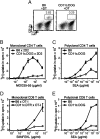
Fig. 1.
Fig. 1.
cDCs control the sensitivity of CD4 and CD8 T cells to foreign antigen. (A) Flow cytometry analysis of splenocytes from WT (B6) and CD11c.DOG mice 1 day after two daily DT injections. Numbers next to the gate indicate the percentage of cDCs. (B–E) Proliferation of splenic CD4 T cells (B and C) and CD8 T cells (D and E) isolated from mice with normal (B6) or reduced (CD11c.DOG and CD11c.DTR) numbers of cDCs. After 2 days of DT administration, purified splenic CD4 T cells were stimulated with APCs with titrated amounts of MOG35-55 peptide (B) or SEA (C). Purified CD8 T cells were stimulated with APCs with titrated amounts of SIINFEKL peptide (D) or SEA (E). Data in C–F are presented as mean ± SEM and are representative of five experiments.
Fig. 2.
Fig. 2.
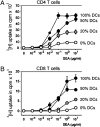
Titration of T cell antigen sensitivity depending on the size of the cDC compartment. Mixed BM chimeras were generated as depicted in Fig. S3 to obtain ~100%, 50%, 20%, and 0% of cDCs in the spleen of mice treated with two daily DT injections before isolation of splenic T cells and stimulation with APCs/SEA. (A and B) Proliferation of CD4 (A) and CD8 (B) T cells isolated from the spleen of the indicated BM chimeras in response to APCs/SEA. Numbers on the right of each graph indicate the number of cDCs in the spleen after two daily DT injections. Data are presented as mean ± SEM (n = 3).
Fig. 3.
Fig. 3.
Rescue of T cell sensitivity after a short interaction with cDCs in the absence of foreign antigen. (A) Numbers of cDCs (CD11chi MHC-II+) in the spleen of CD11c.DOG mice treated with a single DT injection on day 0. Three mice were analyzed at each time point. (B) Proliferation of polyclonal CD4 T cells purified from mice with normal (B6; two daily DT injections), depleted (CD11c.DOG; two daily DT injections), and recovered (CD11c.DOG; 4 days after DT administration) cDC levels in response to APCs and 0.5 μg/mL of SEA. (C) Proliferation of splenic CD4 T cells isolated from cDC-depleted and normal mice and preincubated with cDCs at a ratio of 5:1 immediately before (ex vivo; Left) or 30 min before (Right) stimulation with APCs and SEA. Data are representative of two or three independent experiments (mean ± SEM).
Fig. 4.
Fig. 4.
Recognition of self-MHC class I and II on cDCs is required for maintenance of T cell sensitivity to foreign antigen. (A) Biochemical analysis of TCRζ chain phosphorylation in CD4 T cells from the spleen of mice with normal (DC+) or reduced (DC) cDC levels. Anti–ZAP-70 immunoprecipitates were blotted for ZAP-70 and phosphotyrosine content (Left), and bands were semiquantified by densitometry (Right). The density of the phosphorylated TCRζ band in the DC+ sample was set at 100%. IP, immunoprecipitation. (B) Proliferation of CD4 (Left) and CD8 (Right) T cells purified from unmanipulated mice and preincubated at a ratio of 5:1 with cDCs from WT (B6 DC) or Aβ−/− β2m−/− (MHC KO DC) mice or without cDCs for 18 h before stimulation with APCs with titrated amounts of SEA. Data are representative of three independent experiments (mean ± SEM).
Fig. 5.
Fig. 5.
Impaired immune synapse formation. (A) CD4 T cells isolated from 2D2 mice with a normal (Left) or deficient (Right) cDC compartment were incubated with B cells with (Lower) or without (Upper) cognate MOG35-55 peptide. Fluorescence images of T cell–APC conjugates were acquired using the high-throughput ImageStream system and analyzed with IDEAS software. Shown is an exemplary conjugate for each sample 20 min after initiation of cellular contacts. BF, bright field. (B) Quantification of TCRβ and LFA-1 enrichment in the contact area between T cells and APCs induced by recognition of cognate peptide at the indicated times after initiation of cellular contacts. TCRβ and LFA-1 enrichment at each time point in the absence of peptide was set at a value of 1. Data are presented as mean ± SEM from two pooled experiments.
Fig. 6.
Fig. 6.
Normal numbers of T cells in DC-depleted mice. WT (B6) and CD11c.DOG mice were treated with DT for 10 consecutive days and analyzed the next day. (A) FACS dot plots of total spleen cells. Numbers next to the gate indicate the percentage of cDCs. (B and C) Frequency (B) and number (C) of splenic CD4 and CD8 T cells of the indicated mice. Data are representative of three independent experiments (mean ± SEM; n = 3). (D) Proliferative response of splenic CD4 T cells in response to SEA. Data are presented as mean ± SEM (n = 4).

References

    1. Banchereau J, Steinman RM. Dendritic cells and the control of immunity. Nature. 1998;392:245–252. - PubMed
    1. Steinman RM. Lasker Basic Medical Research Award. Dendritic cells: Versatile controllers of the immune system. Nat Med. 2007;13:1155–1159. - PubMed
    1. Shortman K, Liu YJ. Mouse and human dendritic cell subtypes. Nat Rev Immunol. 2002;2:151–161. - PubMed
    1. Villadangos JA, Schnorrer P. Intrinsic and cooperative antigen-presenting functions of dendritic-cell subsets in vivo. Nat Rev Immunol. 2007;7:543–555. - PubMed
    1. Jenkins MK, et al. In vivo activation of antigen-specific CD4 T cells. Annu Rev Immunol. 2001;19:23–45. - PubMed

Publication types

MeSH terms

Substances