. 2010 Mar 30;107(13):6058-63.
doi: 10.1073/pnas.0909586107. Epub 2010 Mar 15.

Locus ceruleus controls Alzheimer's disease pathology by modulating microglial functions through norepinephrine

Affiliations

Locus ceruleus controls Alzheimer's disease pathology by modulating microglial functions through norepinephrine

Michael T Heneka et al. Proc Natl Acad Sci U S A. .

Abstract

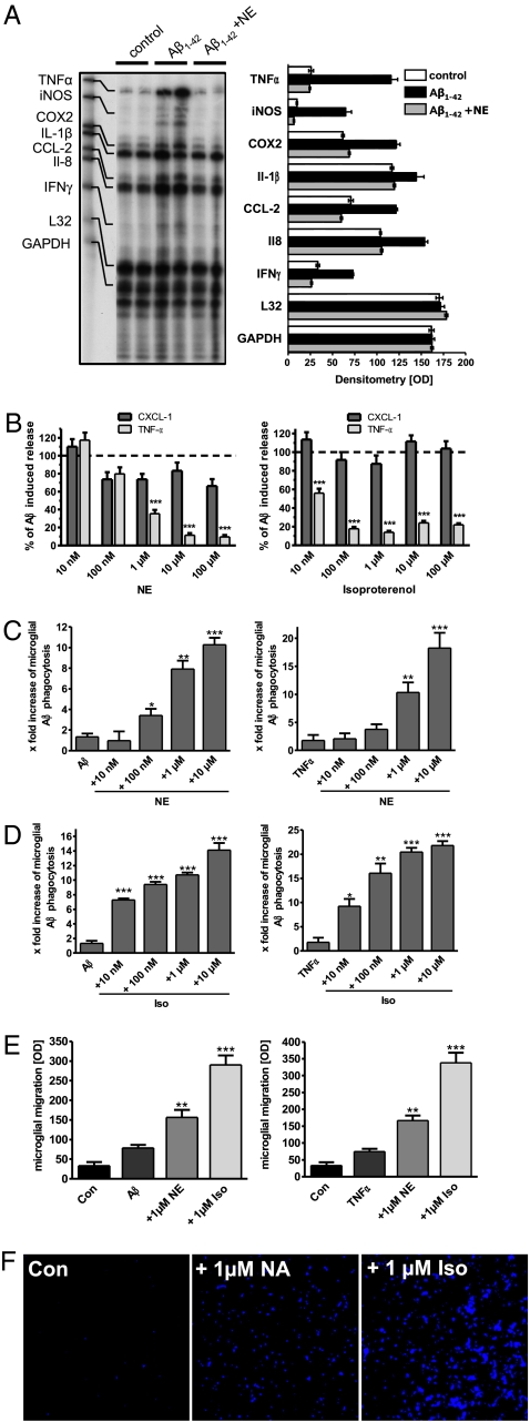
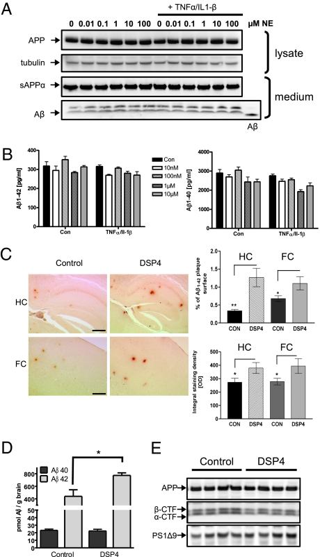
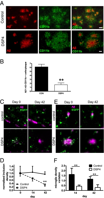
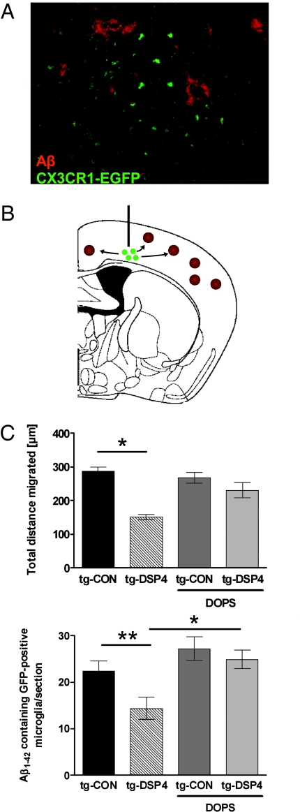
Locus ceruleus (LC)-supplied norepinephrine (NE) suppresses neuroinflammation in the brain. To elucidate the effect of LC degeneration and subsequent NE deficiency on Alzheimer's disease pathology, we evaluated NE effects on microglial key functions. NE stimulation of mouse microglia suppressed Abeta-induced cytokine and chemokine production and increased microglial migration and phagocytosis of Abeta. Induced degeneration of the locus ceruleus increased expression of inflammatory mediators in APP-transgenic mice and resulted in elevated Abeta deposition. In vivo laser microscopy confirmed a reduced recruitment of microglia to Abeta plaque sites and impaired microglial Abeta phagocytosis in NE-depleted APP-transgenic mice. Supplying the mice the norepinephrine precursor L-threo-DOPS restored microglial functions in NE-depleted mice. This indicates that decrease of NE in locus ceruleus projection areas facilitates the inflammatory reaction of microglial cells in AD and impairs microglial migration and phagocytosis, thereby contributing to reduced Abeta clearance. Consequently, therapies targeting microglial phagocytosis should be tested under NE depletion.


Conflict of interest statement

The authors declare no conflict of interest.

Figures

Fig. 1.
Fig. 1.
Norepinephrine suppresses Aβ1–42-induced activation of primary murine microglial cells, and potentiates phagocytosis of Aβ and microglial cell migration. (A) Ribonuclease protection assay analysis of microglia for the expression of TNFα, iNOS, COX2, IL-1β, CCL2, IL-8, and INFγ under control conditions and stimulation with Aβ1–42 with or without NE coincubation for 2 h. L32 and GAPDH served as housekeeping control (n = 8 ± SE, performed in duplicate; **, P < 0.01; ***, P < 0.001, one-way ANOVA, Tukey's post hoc test). (B) Microglial cells were stimulated with 1 μM Aβ1–42 in the presence of increasing concentrations of NE (Left) or isoproterenol (Right). After 18 h, release of TNFα and CXCL1 was measured by ELISA (n = 7 ± SE; **, P < 0.01; ***, P < 0.001, one-way ANOVA, Tukey's post hoc test). (C) Microglia was either stimulated with 150 nM Aβ1–42 (Left) or 2 ng/mL TNFα (Right) for 30 min and thereafter exposed to a fluorescence-labeled, fibrillar Aβ1–42 (FITC-Aβ) with increasing concentrations of NE or isoproterenol (Iso) (D). Phagocytosis was assessed by FACS analysis (n = 12 ± SE; *, P < 0.05; **, P < 0.01; ***, P < 0.001, one-way ANOVA, Tukey's post hoc test). (E) Microglial migration was examined by Boyden chamber assay. Cells were either unstimulated (Con) or immunostimulated by Aβ1–42 or TNFα with or without NE or Iso (n = 8 ± SE; **, P < 0.01; ***, P < 0.001, one-way ANOVA, Tukey's post hoc test). (F) Visualization of migrated cells using DAPI. (Scale bar: 200 μm.)
Fig. 2.
Fig. 2.
Norepinephrine does not modify APP processing but modulates Aβ deposition in Alzheimer's disease mouse models. (A) N2a APPsw cells were incubated for 18 h with increasing concentrations of norepinephrine (NE) in the absence and presence of 1 ng/mL TNFα/IL-1β. Conditioned media were immunoblotted for Aβ and sAPPα by using antibody 6E10. APP and tubulin were detected in cell lysates by immunoblot with antibody 140 and E7, respectively. (B) Amounts of Aβ1–40 and Aβ1–42 in the conditioned media were quantified by sandwich ELISA. (C) Immunohistochemical analysis of Aβ deposition in 12-month-old APPV717I mice in response to noradrenergic depletion in the hippocampus (HC) and frontal cortex (FC) (n = 10 ± SE; *, P < 0.05; **, P < 0.01, Student's t test). (Scale bars: HC, 250 μm; FC, 100 μm.) (D) Noradrenergic depleted 6-month-old APP/PS-1 transgenic mice were analyzed for insoluble Aβ1–40 and Aβ1–42 by sandwich ELISA (n = 7 ± SE; *, P < 0.05, Student's t test). (E) Lysates of control and DSP4-treated mice were immunoblotted using antibodies against APP and presenilin 1.
Fig. 3.
Fig. 3.
NE depletion decreases microglial Aβ phagocytosis and results in reduced recruitment of microglia to Aβ plaques. (A) Confocal laser scanning microscopy detected a higher number of Aβ-plaque-associated and Aβ-containing microglial cells in 12-month-old APPV717I-transgenic mice compared to DSP4-treated mice. (Scale bars: 20 μm.) (B) Fifty randomly chosen plaques on 10 serial sections having a defined distance were evaluated for Aβ/Cd11b colocalization per animal (n = 6 ± SE; **, P < 0.01, Student's t test). (C) Example of plaques labeled with methoxy-X04 surrounded by EGFP expressing microglia at day 0 (Upper Left) and 42 (Upper Right). (Lower) Mouse treated with DSP4 at day 0 (Lower Left) and day 42 (Lower Right). Images are maximum intensity projection (MIPs) along the z axis. (Scale bar: 10 μm.) (D) Quantification of the temporal variation of the number of amyloid inclusions in microglia in different regions of control and DSP4-treated mice, respectively. Values are normalized by the median at day 0 (n = 5 for control and n = 3 for Dsp4-treated mice ± SE; **, P < 0.01, one-way ANOVA, Tukey's post hoc test). (E) Inclusions in microglia in the same region at day 0 (Upper Left) and day 42 (Upper Right). (Lower) Mouse treated with DSP4 at day 0 (Lower Left) and day 42 (Lower Right). Images are single optical sections. (Scale bar: 10 μm.) (F) Quantification of the variation of microglia density around plaques (in comparison with day 0) in control and DSP4-treated mice (n = 5 for control and n = 2 for Dsp4-treated mice ± SE; **, P < 0.01, Student's t test).
Fig. 4.
Fig. 4.
Decrease of microglial migration after NE depletion in vivo is rescued by the NE-precursor L-threo-DOPS. (A) APPV717I-transgenic mice treated with DSP4 or solvent control received a single injection of primary murine microglia derived from CX3CR1-EGFP-transgenic mice. A subgroup of animals in both DSP4-treated and control groups received three i.p. injections of the NE precursor L-threo-DOPS (DOPS) over 24 h to increase NE levels within the neocortex. Shown is confocal laser scanning microscopy of Aβ plaques and EGFP-positive microglial cells. (Scale bar: 50 μM.) (B) Scheme of the intracerebral injection site depicting the migration of EGFP-positive microglial cells (green) toward amyloid plaques (brown). (C) The total distance migrated and the number of EGFP-positive microglial cells per section was evaluated by analyzing serial sections with a defined distance to each other and the injection site (n = 6 ± SE; *, P < 0.05; **, P < 0.01, one-way ANOVA, Tukey's post hoc test).

Comment in

References

    1. Forno L. Pathology of Parkinsonism: A preliminary report of 24 cases. J Neurosurg. 1966;(Supplement, Part II):266–271.
    1. Iversen LL, et al. Loss of pigmented dopamine-beta-hydroxylase positive cells from locus coeruleus in senile dementia of Alzheimer's type. Neurosci Lett. 1983;39:95–100. - PubMed
    1. Bondareff W, et al. Neuronal degeneration in locus ceruleus and cortical correlates of Alzheimer disease. Alzheimer Dis Assoc Disord. 1987;1:256–262. - PubMed
    1. Matthews KL, et al. Noradrenergic changes, aggressive behavior, and cognition in patients with dementia. Biol Psychiatry. 2002;51:407–416. - PubMed
    1. Grudzien A, et al. Locus coeruleus neurofibrillary degeneration in aging, mild cognitive impairment and early Alzheimer's disease. Neurobiol Aging. 2007;28:327–335. - PubMed

Publication types

MeSH terms