

Direct structural insight into the substrate-shuttling mechanism of yeast fatty acid synthase by electron cryomicroscopy
- PMID: 20231485
- PMCID: PMC2889056
- DOI: 10.1073/pnas.0913547107
Direct structural insight into the substrate-shuttling mechanism of yeast fatty acid synthase by electron cryomicroscopy
Abstract
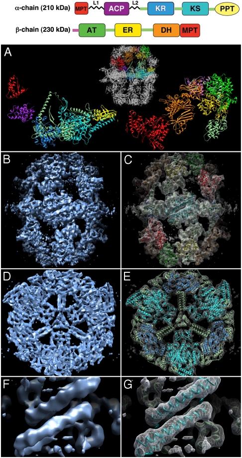
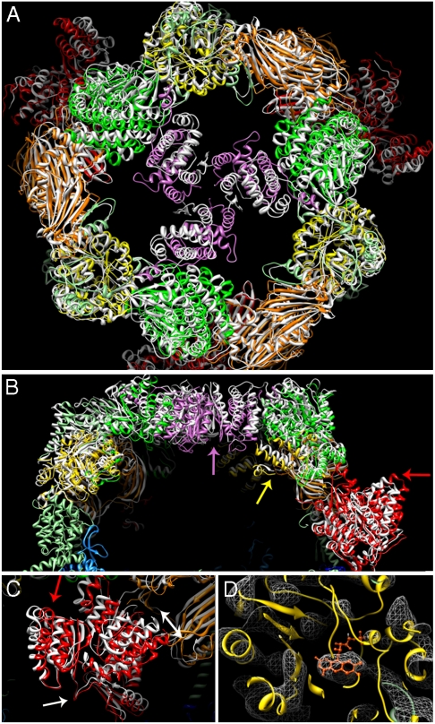
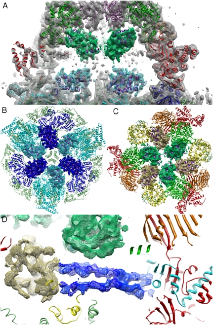
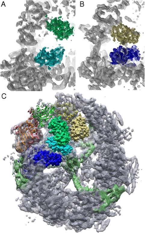
Yeast fatty acid synthase (FAS) is a 2.6-MDa barrel-shaped multienzyme complex, which carries out cyclic synthesis of fatty acids. By electron cryomicroscopy of single particles we obtained a three-dimensional map of yeast FAS at 5.9-A resolution. Compared to the crystal structures of fungal FAS, the EM map reveals major differences and new features that indicate a considerably different arrangement of the complex in solution compared to the crystal structures, as well as a high degree of variance inside the barrel. Distinct density regions in the reaction chambers next to each of the catalytic domains fitted the substrate-binding acyl carrier protein (ACP) domain. In each case, this resulted in the expected distance of approximately 18 A from the ACP substrate-binding site to the active site of the catalytic domains. The multiple, partially occupied positions of the ACP within the reaction chamber provide direct structural insight into the substrate-shuttling mechanism of fatty acid synthesis in this large cellular machine.
Conflict of interest statement
The authors declare no conflict of interest.
Figures
Similar articles
-
Direct structural analysis of a single acyl carrier protein domain in fatty acid synthase from the fungus Saccharomyces cerevisiae.Commun Biol. 2024 Jan 12;7(1):92. doi: 10.1038/s42003-024-05777-7. Commun Biol. 2024. PMID: 38216676 Free PMC article.
-
Interactions of the acyl chain with the Saccharomyces cerevisiae acyl carrier protein.Biochemistry. 2015 Apr 7;54(13):2205-13. doi: 10.1021/bi5014563. Epub 2015 Mar 25. Biochemistry. 2015. PMID: 25774789
-
Structural basis for substrate delivery by acyl carrier protein in the yeast fatty acid synthase.Science. 2007 Apr 13;316(5822):288-90. doi: 10.1126/science.1138249. Science. 2007. PMID: 17431182
-
Using modern tools to probe the structure-function relationship of fatty acid synthases.Chembiochem. 2015 Mar 2;16(4):528-547. doi: 10.1002/cbic.201402578. Epub 2015 Feb 10. Chembiochem. 2015. PMID: 25676190 Free PMC article. Review.
-
Acyl carrier protein: structure-function relationships in a conserved multifunctional protein family.Biochem Cell Biol. 2007 Dec;85(6):649-62. doi: 10.1139/o07-109. Biochem Cell Biol. 2007. PMID: 18059524 Review.
Cited by
-
CryoDRGN-ET: deep reconstructing generative networks for visualizing dynamic biomolecules inside cells.Nat Methods. 2024 Aug;21(8):1537-1545. doi: 10.1038/s41592-024-02340-4. Epub 2024 Jul 18. Nat Methods. 2024. PMID: 39025970
-
Analysis of the co-translational assembly of the fungal fatty acid synthase (FAS).Sci Rep. 2020 Jan 21;10(1):895. doi: 10.1038/s41598-020-57418-8. Sci Rep. 2020. PMID: 31964902 Free PMC article.
-
Atomic model of the F420-reducing [NiFe] hydrogenase by electron cryo-microscopy using a direct electron detector.Elife. 2014 Feb 25;3:e01963. doi: 10.7554/eLife.01963. Elife. 2014. PMID: 24569482 Free PMC article.
-
Structural definition of the lysine swing in Arabidopsis thaliana PDX1: Intermediate channeling facilitating vitamin B6 biosynthesis.Proc Natl Acad Sci U S A. 2016 Oct 4;113(40):E5821-E5829. doi: 10.1073/pnas.1608125113. Epub 2016 Sep 19. Proc Natl Acad Sci U S A. 2016. PMID: 27647886 Free PMC article.
-
Visualizing transiently associated catalytic domains in assembly-line biosynthesis using cryo-electron microscopy.J Struct Biol. 2021 Dec;213(4):107802. doi: 10.1016/j.jsb.2021.107802. Epub 2021 Oct 1. J Struct Biol. 2021. PMID: 34606906 Free PMC article.
References
-
- White SW, Zheng J, Zhang Y-M, Rock CO. The structural biology of type II fatty acid biosynthesis. Annu Rev Biochem. 2005;74:791–831. - PubMed
-
- Wakil SJ, Stoops JK, Joshi VC. Fatty acid synthesis and its regulation. Ann Rev Biochem. 1983;52:537–579. - PubMed
-
- Smith S, Witkowski A, Joshi AK. Structural and functional organization of the animal fatty acid synthase. Prog Lipid Res. 2003;42(4):298–317. - PubMed
-
- Asturias FJ, et al. Structure and molecular organization of mammalian fatty acid synthase. Nat Struct Mol Biol. 2005;12(3):225–232. - PubMed
Publication types
MeSH terms
Substances
Grants and funding
LinkOut - more resources
Full Text Sources
Molecular Biology Databases
Research Materials
Miscellaneous
