

Review
doi: 10.1161/CIRCULATIONAHA.109.896357.
A-kinase anchoring proteins: getting to the heart of the matter
Affiliations
- PMID: 20231544
- PMCID: PMC3014256
- DOI: 10.1161/CIRCULATIONAHA.109.896357
Item in Clipboard
Review
A-kinase anchoring proteins: getting to the heart of the matter
Circulation.
.
No abstract available
Figures
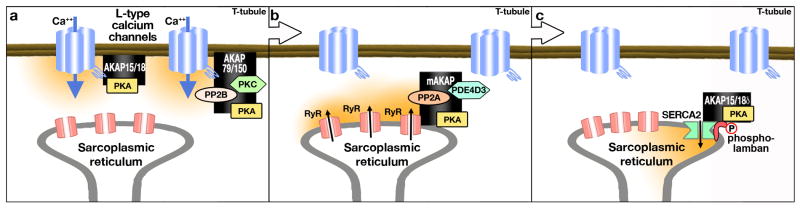
a) Phase 1 of EC coupling: AKAP79/150 and the short forms of AKAP15/18 (α and β) maintain signaling complexes tethered to L-type calcium channels to enable calcium influx. b) Phase 2 of EC coupling: Ryanodine receptors at the sarcoplasmic reticulum are bound to the mAKAP signaling complex as it responds to the increased cytosolic Ca++. mAKAP clusters PKA, PDE4D3 and PP2A to regulate the phosphorylation status of the Ryr and the resultant Ca++ release from the SR. c) Phase 3 of EC coupling: Active transport of calcium back into the SR via SERCA2 is regulated by phospholamban. The AKAP15/18δ long isoform brings PKA into complex with phospholamban and SERC2 where it can augment the phosphorylation of phospholamban and the reuptake of calcium into the SR.
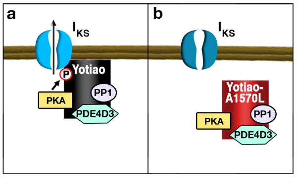
a) In myocytes Yotiao associates with the KCNQ1 subunit of the Iκs potassium channel. Anchored PKA phosphorylation of Yotiao enhances activation of the channel. b) The S1570L Yotiao mutant has reduced interaction with KCNQ1 and delayed repolarization of ventricular action potential.
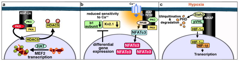
a) An upregulation of AKAP-Lbc facilitates PKD activation and phosphorylation of HDAC5, leading to transcriptional activation of the MEF pathway and the onset of hypertrophy. b) Calcium mediated activation of AKAP150-targeted PP2B leads to dephosphorylation of NFATc3 in arterial myocytes. Dephosphorylated NFATc3 accumulates in the nucleus where it leads to decreased gene expression of channel subunits. c) mAKAP organizes ubiquitin E3 ligases that managed the stability of the transcription factor HIF-1α During hypoxia, HIF1α translocated to the nucleus initiates transcription of proangiogenic, metabolic, and antiapoptotic genes that promote cell survival during hypoxia.
Similar articles
-
Phosphoinositide 3-kinase regulates excitation-contraction coupling in neonatal cardiomyocytes.Am J Physiol Heart Circ Physiol. 2004 Feb;286(2):H796-805. doi: 10.1152/ajpheart.00546.2003. Epub 2003 Oct 16. Am J Physiol Heart Circ Physiol. 2004. PMID: 14563664
-
Phosphodiesterase 4D regulates baseline sarcoplasmic reticulum Ca2+ release and cardiac contractility, independently of L-type Ca2+ current.Circ Res. 2011 Oct 14;109(9):1024-1030. doi: 10.1161/CIRCRESAHA.111.250464. Epub 2011 Sep 8. Circ Res. 2011. PMID: 21903937 Free PMC article.
-
Nuclear targeting of Akt enhances ventricular function and myocyte contractility.Circ Res. 2005 Dec 9;97(12):1332-41. doi: 10.1161/01.RES.0000196568.11624.ae. Epub 2005 Nov 17. Circ Res. 2005. PMID: 16293788
-
Regulation of sarcoplasmic reticulum Ca2+ ATPase pump expression and its relevance to cardiac muscle physiology and pathology.Cardiovasc Res. 2008 Jan 15;77(2):265-73. doi: 10.1093/cvr/cvm056. Epub 2007 Oct 30. Cardiovasc Res. 2008. PMID: 18006443 Review.
-
T-tubules and ryanodine receptor microdomains: on the road to translation.Cardiovasc Res. 2013 May 1;98(2):159-61. doi: 10.1093/cvr/cvt077. Epub 2013 Apr 2. Cardiovasc Res. 2013. PMID: 23554456 Review. No abstract available.
Cited by
-
The Popeye domain containing gene family encoding a family of cAMP-effector proteins with important functions in striated muscle and beyond.J Muscle Res Cell Motil. 2019 Jun;40(2):169-183. doi: 10.1007/s10974-019-09523-z. Epub 2019 Jun 13. J Muscle Res Cell Motil. 2019. PMID: 31197601 Free PMC article. Review.
-
Using cAMP Sensors to Study Cardiac Nanodomains.J Cardiovasc Dev Dis. 2018 Mar 13;5(1):17. doi: 10.3390/jcdd5010017. J Cardiovasc Dev Dis. 2018. PMID: 29533995 Free PMC article. Review.
-
The Role of Cyclic AMP Signaling in Cardiac Fibrosis.Cells. 2019 Dec 26;9(1):69. doi: 10.3390/cells9010069. Cells. 2019. PMID: 31888098 Free PMC article. Review.
-
Functions of PDE3 Isoforms in Cardiac Muscle.J Cardiovasc Dev Dis. 2018 Feb 6;5(1):10. doi: 10.3390/jcdd5010010. J Cardiovasc Dev Dis. 2018. PMID: 29415428 Free PMC article. Review.
-
cGMP signals modulate cAMP levels in a compartment-specific manner to regulate catecholamine-dependent signaling in cardiac myocytes.Circ Res. 2011 Apr 15;108(8):929-39. doi: 10.1161/CIRCRESAHA.110.230698. Epub 2011 Feb 17. Circ Res. 2011. PMID: 21330599 Free PMC article.
References
-
- Bers DM. Calcium cycling and signaling in cardiac myocytes. Annu Rev Physiol. 2008;70:23–49. - PubMed
-
- Oldham WM, Hamm HE. Heterotrimeric G protein activation by G-protein-coupled receptors. Nat Rev Mol Cell Biol. 2008;9:60–71. - PubMed
-
- Sutherland EW. Studies on the mechanism of hormone action. Science. 1972;171:401–408. - PubMed
-
- Beavo JA, Brunton LL. Cyclic nucleotide research -- still expanding after half a century. Nat Rev Mol Cell Biol. 2002;3:710–718. - PubMed
Publication types
MeSH terms
Substances
Grants and funding
LinkOut - more resources
Full Text Sources
Other Literature Sources
