. 2010 Apr 15;184(8):4338-48.
doi: 10.4049/jimmunol.0903824. Epub 2010 Mar 15.

Protein-DNA complex is the exclusive malaria parasite component that activates dendritic cells and triggers innate immune responses

Affiliations

Protein-DNA complex is the exclusive malaria parasite component that activates dendritic cells and triggers innate immune responses

Xianzhu Wu et al. J Immunol. .

Abstract

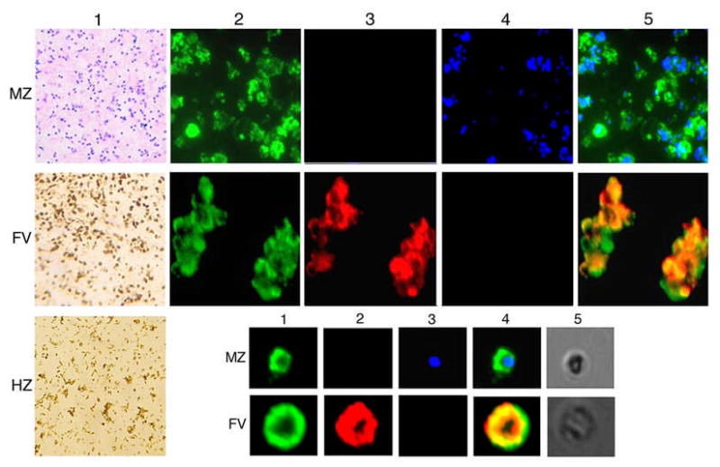
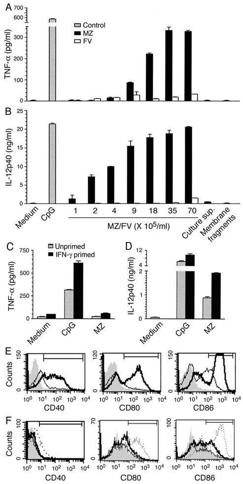
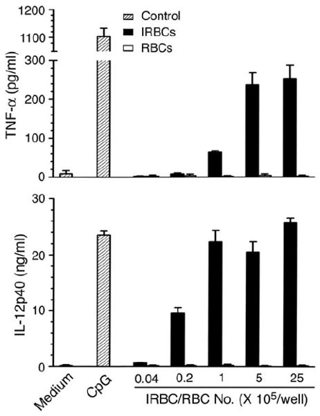
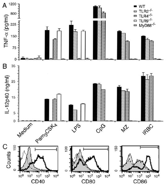
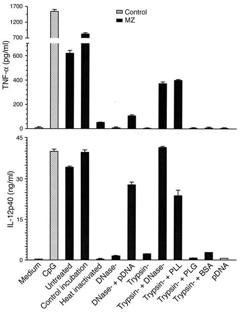
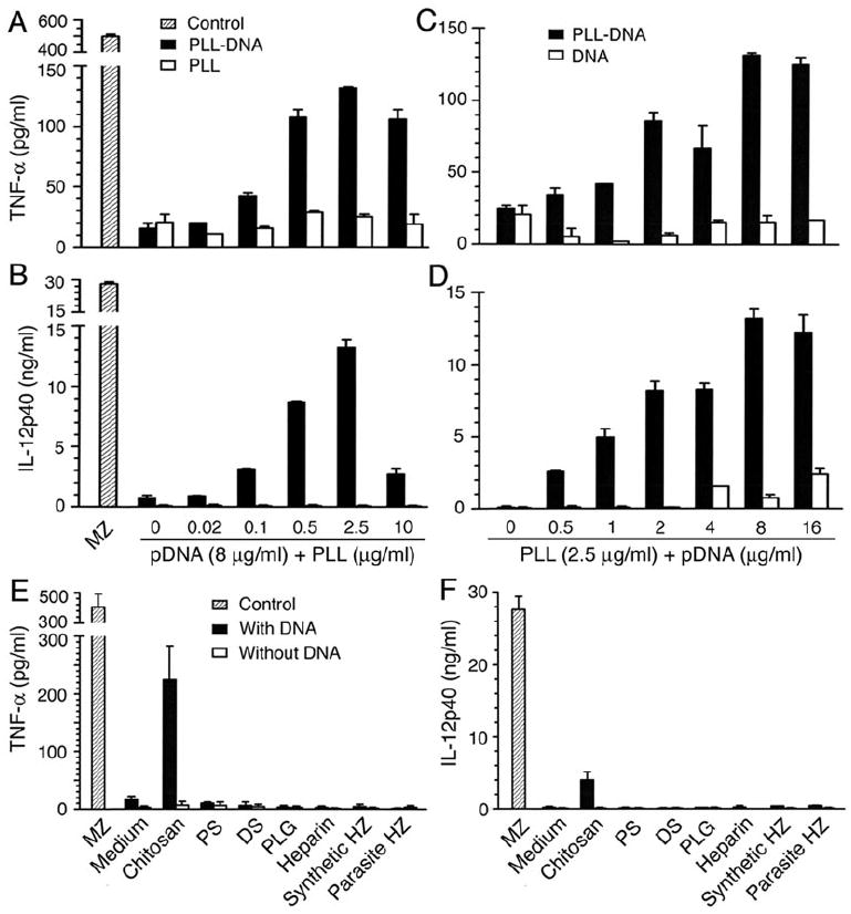
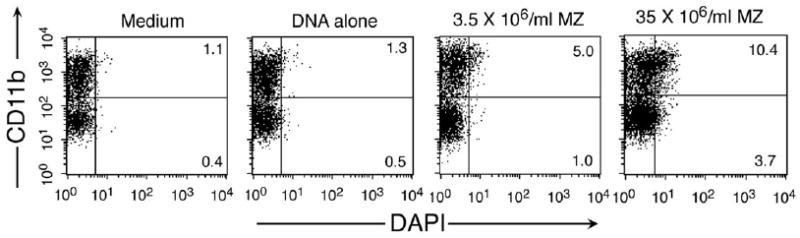
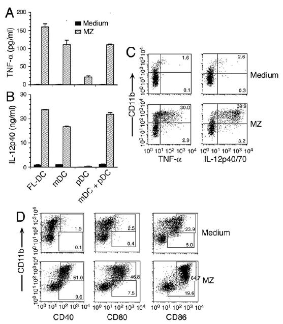
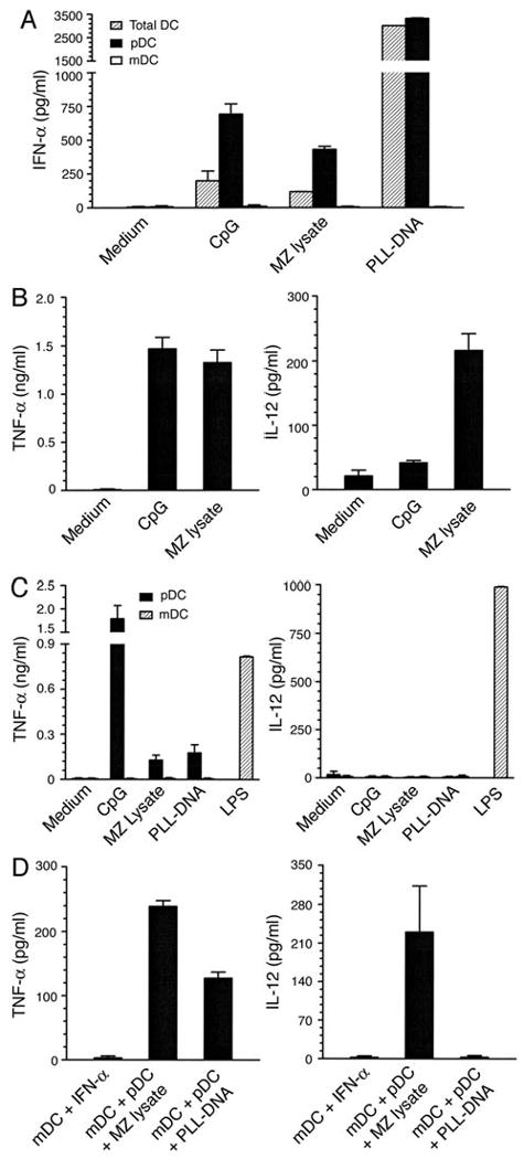
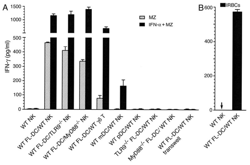
Dendritic cells (DCs) play a crucial role in the development of protective immunity to malaria. However, it remains unclear how malaria parasites trigger immune responses in DCs. In this study, we purified merozoites, food vacuoles, and parasite membrane fragments released during the Plasmodium falciparum schizont burst to homogeneity and tested for the activation of bone marrow-derived DCs from wild-type and TLR2(-/-), TLR4(-/-), TLR9(-/-), and MyD88(-/-) C57BL/6J mice. The results demonstrate that a protein-DNA complex is the exclusive parasite component that activates DCs by a TLR9-dependent pathway to produce inflammatory cytokines. Complex formation with proteins is essential for the entry of parasite DNA into DCs for TLR9 recognition and, thus, proteins convert inactive DNA into a potent immunostimulatory molecule. Exogenous cationic polymers, polylysine and chitosan, can impart stimulatory activity to parasite DNA, indicating that complex formation involves ionic interactions. Merozoites and DNA-protein complex could also induce inflammatory cytokine responses in human blood DCs. Hemozoin is neither a TLR9 ligand for DCs nor functions as a carrier of DNA into cells. Additionally, although TLR9 is critical for DCs to induce the production of IFN-gamma by NK cells, this receptor is not required for NK cells to secret IFN-gamma, and cell-cell contact among myeloid DCs, plasmacytoid DCs, and NK cells is required for IFN-gamma production. Together, these results contribute substantially toward the understanding of malaria parasite-recognition mechanisms. More importantly, our finding that proteins and carbohydrate polymers are able to confer stimulatory activity to an otherwise inactive parasite DNA have important implications for the development of a vaccine against malaria.


Figures

FIGURE 1
FIGURE 1
Analysis of P. falciparum MZs, FVs, and HZ for purity. (Panel 1) Light micrographs of Giemsa-stained MZ, FV and HZ. (Panels 2-4) Immunofluorescent micrographs after staining with anti-merozoite surface protein-1 monoclonal antibody (2), rabbit anti-Pgh antibodies (3) or DAPI (4). (Panel 5) Merge of micrographs 2, 3 and 4. (Inset) Enlarged immunofluorescent micrographs of a single MZ and FV stained as above; phase-contrast micrographs of MZ and FV (5).
FIGURE 2
FIGURE 2
MZs are the exclusive components of P. falciparum schizont rupture that induce the production of inflammatory cytokines in DCs. (A and B) TNF-α and IL-12 produced by FL-DCs (1 × 10⁵ cells/well in 200 μl of culture medium) stimulated with MZs, FVs (each at the indicated doses), parasite membrane fragments (2 × 10⁷ IRBC equivalent/well), or parasite cytoplasmic materials (2 × 10⁷ IRBC equivalent/well). CpG-1826 (2 μg/ml) was used as a control stimulant in all experiments. Cytokines secreted into culture media were measured by ELISA. Data are representative of six independent experiments, each performed in duplicates. In all figures, error bars represent mean values ± SEM. (C and D), TNF-α and IL-12 produced by unprimed and IFN-γ -primed mouse splenic DCs (1 × 10⁵ cells/well in 200 μl of culture medium) stimulated with MZs (3 × 10⁶/ml; 6 × 10⁵/well). Data are representative of three independent experiments. (E and F) Surface expression of costimulatory molecules (CD40, CD80, and CD86) by FL-DCs stimulated with the MZs (3 × 10⁶/ml/well, E), and schizont ruptured membrane fragments and cytoplasmic material (equivalent of 1 × 10⁸ IRBCs in 1 ml of culture medium, F) in 24 well plates. (E), Shaded area, thin line, and thick line represent, respectively, the IgG isotype controls, unstimulated cells, and cells stimulated with MZs (3 × 10⁶/ml/well). (F), Shaded area, thin line, thick line, and dashed line represent, respectively, unstimulated cells, stimulated with membrane fragments, cytoplasmic material and CpG control.
FIGURE 3
FIGURE 3
P. falciparum IRBCs activate DCs in a manner similar to that by MZs. ELISA measurement of TNF-α and IL-12 produced by FL-DCs (1 × 10⁵/well in 200 μl of culture medium) stimulated with the indicated doses of the late stage (trophozoites and schizonts) IRBCs. Cells stimulated with RBCs and CpG-1826 (2 μg/ml) were analyzed as controls. Data are representative of four independent experiments.
FIGURE 4
FIGURE 4
Activation of DCs by MZs and IRBCs is mediated by TLR9. (A and B) Analysis of TNF-α and IL-12 by WT, and TLR2, TLR4, TLR9 and MyD88 deficient FL-DCs (1 × 10⁵/well in 200 μl of culture medium) stimulated with MZs (3 × 10⁶/ml; 6 × 10⁵/well) or IRBCs (1.5 × 10⁶/ml; 3 × 10⁵/well). Pam3CSK4 (TLR2 ligand, 10 ng/ml), LPS (TLR4 ligand, 100 ng/ml), and CpG (TLR9 ligand, 2 μg/ml) were used as control stimulants. Data are representative of six independent experiments. (C) Co-stimulatory molecules expressed by WT (thick lines), TLR9-/- (shaded areas) and MyD88-/- (dashed line) FL-DCs stimulated with MZs (3 × 10⁶/ml/well) and unstimulated WT FL-DCs (thin lines).
FIGURE 5
FIGURE 5
The activation of DCs by P. falciparum merozoites involves recognition of a parasite protein-DNA complex. TNF-α and IL-12 produced by FL-DCs (1 × 10⁵/well in 200 μl of culture medium) stimulated with untreated, DNase-treated MZs (DNase-), trypsin-treated MZs (Trypsin-), mixture of DNase- and trypsin-treated MZs. The indicated exogenous proteins were added to trypsin-treated MZs at 2.5 μg/ml; in each case, 3 × 10⁶/ml of MZs (6 × 10⁵/well) were used. Parasite DNA was tested at 8 μg/ml. Data are representative of four independent experiments. DCs stimulated with CpG (2 μg/ml) were analyzed as a control. MZs incubated at 37 °C for 1 h were tested as controls.
FIGURE 6
FIGURE 6
PLL converts P. falciparum inactive DNA into a potent stimulatory molecule. (A and B) TNF-α and IL-12 produced by FL-DCs (1 × 10⁵/well in 200 μl of culture medium) stimulated with 8 μg/ml of parasite DNA containing the indicated concentrations of PLL. (C and D), TNF-α and IL-12 produced by FL-DCs stimulated with DNA containing in each case 2.5 μg/ml of PLL. (E and F) TNF-α and IL-12 produced by FL-DCs stimulated with parasite DNA (8 μg/ml) containing PLG, PS, DS, chitosan, heparin, parasite hemozoin or synthetic hemozoin (5 μg/ml each). All these reagents were also tested at different doses (see Fig. S5). Data are representative of five independent experiments. MZs (3 × 10⁶/ml; 6 × 10⁵/well) were analyzed as control stimuli.
FIGURE 7
FIGURE 7
The mDC subset of FL-DCs can uptake P. falciparum MZs and DNA complex more efficiently than the pDC subset. Parasite DNA and MZs were stained with DAPI (0.1 μg/ml in PBS) and FL-DCs (1 × 10⁶/well in 24-well plates) were stimulated with stained DNA (8 μg/ml) or MZs (at the indicated amounts) in 1 ml of culture medium. The uptake of parasite DNA and MZs by DCs was measured by flow cytometry. Data are representative of two independent experiments.
FIGURE 8
FIGURE 8
P. falciparum MZs can activate both myeloid DCs and plasmacytoid DCs to express costimulatory molecules, but only former DC population produce TNF-α and IL-12. (A and B) TNF-α and IL-12 production by sorted mDC and pDC populations (each 5 × 10⁴ cells/well in 200 μl of culture medium) of FL-DCs and by the reconstituted mixture of sorted mDCs and pDCs (mDC + pDC) stimulated with MZs (3 × 10⁶/ml; 6 × 10⁵/well). (C) TNF-α and IL-12 produced by FL-DCs (1 × 10⁶/well in 24-well plates) stimulated with MZs (3 × 10⁶/ml/well) were analyzed by intracellular staining. Data shown in each (A), (B) and (C) are representative of three independent experiments. (D) Costimulatory molecules expressed by FL-DCs (1 × 10⁶/well in 24-well plates) stimulated with MZs (3 × 10⁶/ml/well). Unstimulated FL-DCs were analyzed as controls. Experiments were done two times.
FIGURE 9
FIGURE 9
Human DCs efficiently produce IFN-α, TNF-α, and IL-12 in response to malaria parasite. DCs isolated from human blood were sorted by FACS into mDC and pDC. (A) Unsorted DCs (1 × 10⁵/well in 200 μl of culture medium) and sorted DCs (each 1 × 10⁵/well in 200 μl of culture medium) were stimulated with CpG-2006 (2 μg/ml), MZ lysates (MZ containing 2 μg DNA/ml) or parasite DNA (8 μg/ml) + PLL (2.5 μg/ml). IFN-α released into the cultured medium was measured by ELISA. (B) In separate experiments, human blood total DCs (1 × 10⁵ cells/well in 200 μl of culture medium) were stimulated with CpG-2006 (2 μg/ml) control or MZ lysates (MZ, 5 μg DNA/ml) (C) Production of cytokines produced by sorted human pDCs and mDCs stimulated with MZ lysate (equivalent of 2 μg/ml DNA), parasite DNA (8 μg/ml) + PLL (2.5 μg/ml), and CpG-2006 (2 μg/ml) and LPS (100 ng/ml) controls each in 200 μl culture medium. (D) Sorted human mDCs or mixtures of sorted pDCs and mDCs were stimulated with IFN-α (1000 pg/ml), MZ lysate or parasite DNA-PLL as indicated at concentrations outlined in panel C. In all cases, TNF-α and IL-12 secreted into culture medium were measured by ELISA.
FIGURE 10
FIGURE 10
Merozoite- and IRBC-stimulated DCs can activate NK and γδ T cells to produce IFN-γ. (A) WT FL-DCs (1 × 10⁵ cell/well) cocultured with NK cells (5 × 10⁴ cells/well) from WT, TLR9-/- or MyD88-/- mice or WT γδ T cells (5 × 10⁴/well) all in 96 well plates using 200 μl of culture medium. In each case, cells were stimulated with MZs (3 × 10⁶/ml; 6 × 10⁵/well) or MZs (3 × 10⁶/ml; 6 × 10⁵/well) plus IFN-α (500 units/ml). Similarly, TLR9-/- or MyD88-/- FL-DCs (1 × 10⁵/well in 200 μl of culture medium) cocultured with WT NK cells (5 × 10⁴/well) were stimulated with MZs (3 × 10⁶/ml; 6 × 10⁵/well) or MZs (3 × 10⁶/ml; 6 × 10⁵/well) plus IFN-α (500 units/ml). Sorted mDC and pDC populations (each 5 × 10⁴/well in 200 μl of culture medium) of WT FL-DCs cocultured with WT NK cells were also stimulated as above. To examine the cell-cell contact requirement for IFN-γ production, FL-DCs (1 × 10⁶/well) and NK cells (4 × 10⁵/well) were cultured in different compartments of 24-well transwells, and both DCs and NK cells were stimulated with MZs (3 × 10⁶/ml/well) or MZs (3 × 10⁶/ml/well) plus IFN-α (500 units/ml). Data are representative of five independent experiments. (B) IFN-γ production after stimulation with IRBCs; arrow indicates that IFN-γ was not detectable. Data are representative of three independent experiments.

Comment in

Similar articles

Cited by

References

    1. Augustine AD, Hall BF, Leitner WW, Mo AX, Wali TM, Fauci AS. NIAID workshop on immunity to malaria: Addressing immunological challenges. Nat Immunol. 2009;10:673–678. - PubMed
    1. Snow RW, Guerra CA, Noor AM, Myint HY, Hay SI. The global distribution of clinical episodes of Plasmodium falciparum malaria. Nature. 2005;434:214–217. - PMC - PubMed
    1. Cox-Singh J, Singh B. Knowlesi malaria: newly emergent and of public health importance. Trends Parasitol. 2008;24:406–410. - PMC - PubMed
    1. Pierce SK, Miller LH. World malaria day 2009: what malaria knows and the immune system that immunologists still do not. J Immunol. 2009;182:5171–5177. - PMC - PubMed
    1. Stevenson MM, Riley EM. Innate Immunity to malaria. Nat Rev Immunol. 2004;4:169–180. - PubMed

Publication types

MeSH terms