. 2010 Mar 9;4(3):e622.
doi: 10.1371/journal.pntd.0000622.

Diversity of Micrurus snake species related to their venom toxic effects and the prospective of antivenom neutralization

Affiliations

Diversity of Micrurus snake species related to their venom toxic effects and the prospective of antivenom neutralization

Gabriela D Tanaka et al. PLoS Negl Trop Dis. .

Abstract

Background: Micrurus snake bites can cause death by muscle paralysis and respiratory arrest, few hours after envenomation. The specific treatment for coral snake envenomation is the intravenous application of heterologous antivenom and, in Brazil, it is produced by horse immunization with a mixture of M. corallinus and M. frontalis venoms, snakes that inhabit the South and Southeastern regions of the country. However, this antivenom might be inefficient, considering the existence of intra- and inter-specific variations in the composition of the venoms. Therefore, the aim of the present study was to investigate the toxic properties of venoms from nine species of Micrurus: eight present in different geographic regions of Brazil (M. frontalis, M. corallinus, M. hemprichii, M. spixii, M. altirostris, M. surinamensis, M. ibiboboca, M. lemniscatus) and one (M. fulvius) with large distribution in Southeastern United States and Mexico. This study also analyzed the antigenic cross-reactivity and the neutralizing potential of the Brazilian coral snake antivenom against these Micrurus venoms.

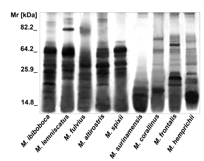
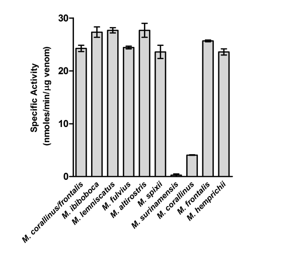
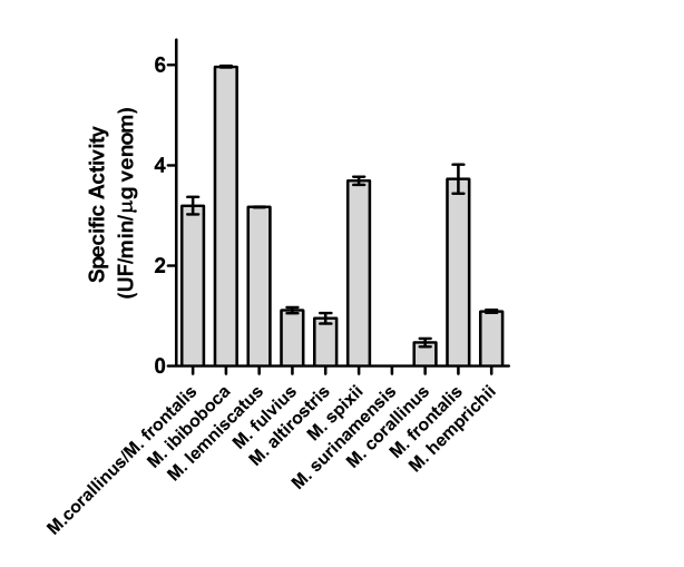
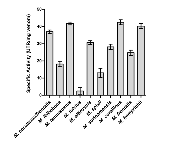
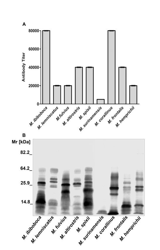
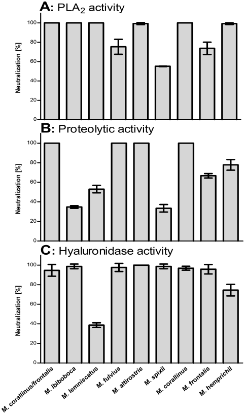
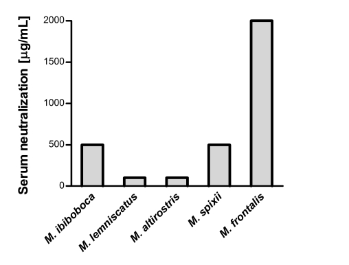
Methodology/principal findings: Analysis of protein composition and toxicity revealed a large diversity of venoms from the nine Micrurus species. ELISA and Western blot assays showed a varied capability of the therapeutic antivenom to recognize the diverse species venom components. In vivo and in vitro neutralization assays indicated that the antivenom is not able to fully neutralize the toxic activities of all venoms.

Conclusion: These results indicate the existence of a large range of both qualitative and quantitative variations in Micrurus venoms, probably reflecting the adaptation of the snakes from this genus to vastly dissimilar habitats. The data also show that the antivenom used for human therapy in Brazil is not fully able to neutralize the main toxic activities present in the venoms from all Micrurus species occurring in the country. It suggests that modifications in the immunization scheme, with the inclusion of other venoms in the antigenic mixture, should occur in order to generate effective therapeutic coral snake antivenom.


Conflict of interest statement

The authors have declared that no competing interests exist.

Figures

Figure 1
Figure 1. SDS-polyacrylamide gel eletrophoresis.
Samples (20 µg) of M. ibiboboca, M. lemniscatus, M. fulvius, M. altirostris, M. spixii, M. surinamensis, M. corallinus, M. frontalis and M. hemprichii venoms were analyzed by SDS-PAGE in a gradient gel (7.5% to 15%) and silver stained.
Figure 2
Figure 2. Determination of the phospholipase A2 activity.
Samples of individual Micrurus venoms (4 µg), or a mixture of M. frontalis (2 µg) and M. corallinus (2 µg), venoms were incubated for 20 min at 37°C with 180 µL of a mixture containing 5 mM Triton X-100, 5 mM phosphatidylcholine, 2 mM HEPES, 10 mM calcium chloride and 0.124% (wt./vol) bromothymol blue dye in water. Results are representative for three separate experiments and expressed as nanomoles acid per minute per µg of venom.
Figure 3
Figure 3. Determination of the proteolytic activity.
Samples of Micrurus venoms (50 µg), or a mixture of M. frontalis (25 µg) and M. corallinus (25 µg), were incubated at 37°C with the FRET substrate, Abz-FEPFRQ-EDnp, and the hydrolysis measured in a spectrofluorimeter. Results are representative for three separate experiments and expressed as units of free fluorescence per minute per µg of venom (UF/min/µg).
Figure 4
Figure 4. Determination of the hyaluronidase activity.
Samples of Micrurus venoms (30 µg), or a mixture of M. frontalis (15 µg) and M. corallinus (15 µg), were incubated for 15 min at 37°C with the hyaluronic acid as substrate. After this period, it was added cetyltrimethylammonium bromide, to develop the turbidity in the mixtures, and the absorbance measured in a spectrophotometer at λem 405 nm. Results are representative for three separate experiments and expressed in units of turbidity reduction (UTR) per mg of venom.
Figure 5
Figure 5. Cross-reactivity of coral snake antivenom.
[A] ELISA: plates were coated with 10 µg of Micrurus venoms and incubated with different dilutions of coral snake antivenom, followed by GAH/IgG-HRPO, diluted 1∶3,000. The absorbance of the samples was determined at 492 nm. The data presented correspond to the mean OD492 value +/− SD of experiments carried out in duplicate. [B] Western blot: Samples (20 µg) of M. ibiboboca, M. lemniscatus, M. fulvius, M. altirostris, M. spixii, M. surinamensis, M. corallinus, M. frontalis and M. hemprichii venoms were separated by SDS-PAGE in gradient gel (7.5% to 15%), electrotransfered to a nitrocellulose membrane and incubated with the coral snake antivenom diluted 1∶2,000 followed by GAH/IgG-AP. The reaction was revealed with NBT and BCIP.
Figure 6
Figure 6. In vitro serum neutralization assays.
[A] Phospholipase A2 activity: Samples of the venoms (4 µg) were incubated with the coral snake antivenom (1∶10) for 20 min at room temperature. As positive control, the mixture of M. corallinus (2 µg) and M. frontalis (2 µg) venoms, used for the production of the antivenom, was used. [B] Proteolytic activity: Samples of Micrurus venoms (50 µg) and the coral snake antivenom (1∶4) were incubated for 10 min at room temperature. As positive control, the mixture of the M. corallinus (25 µg) and M. frontalis (25 µg) venoms was used. [C] Hyaluronidase activity: Samples of Micrurus venoms (30 µg) and the coral snake antivenom (1∶20) were incubated for 15 min at room temperature. As positive control, the mixture of the M. corallinus (15 µg) and M. frontalis (15 µg) venoms was used. Venoms residual toxic activities were measured as described in materials and methods. Results are representative for three separate experiments and expressed as percentage of neutralization of the venoms enzymatic activities.
Figure 7
Figure 7. Antivenom neutralization of Micrurus venoms lethal toxicity.
Samples corresponding to 2 LD50, of each Micrurus venom, were mixed with serial dilutions of the coral snake horse antivenom. The mixtures were incubated for 30 min at 37°C and the animals were i.p. inoculated. The effective dose (ED50) was calculated, from the number of deaths within 48 h of injection of the venom/antivenom mixture, using probit analysis and expressed as mL of antivenom per µg of venom.

References

    1. Campbell JA, Lamar WW. 2004. The venomous reptiles of the western hemisphere. 2. ed., Ithaca, New York, Cornell University Press.
    1. Roze JA. A checklist of the New World venomous coral snakes (Elapidae), with description of new forms. Am Mus Novitates. 1967;2287:1–60.
    1. Roze JA. New World coral snake (Elapidae): a toxonomic and biological summary. Mem Inst Butantan. 1983;46:305–338.
    1. Roze JA, Bernal-Carlo A. Las serpientes corales venenosas del genero Leptomicrurus (Serpentes, Elapidae) de Sudamérica con descripción de una nueva subespecie. Boll Mus Sci Nat Torino. 1987;5:573–608.
    1. Roze JA. Coral snakes of the Americans. Biology, identification, and venoms. Krieger Publishing Co Florida 262. 1996.

Publication types