Phage display peptide probes for imaging early response to bevacizumab treatment
- PMID: 20232090
- PMCID: PMC3233971
- DOI: 10.1007/s00726-010-0548-9
Phage display peptide probes for imaging early response to bevacizumab treatment
Abstract
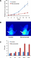
Early evaluation of cancer response to a therapeutic regimen can help increase the effectiveness of treatment schemes and, by enabling early termination of ineffective treatments, minimize toxicity, and reduce expenses. Biomarkers that provide early indication of tumor therapy response are urgently needed. Solid tumors require blood vessels for growth, and new anti-angiogenic agents can act by preventing the development of a suitable blood supply to sustain tumor growth. The purpose of this study is to develop a class of novel molecular imaging probes that will predict tumor early response to an anti-angiogenic regimen with the humanized vascular endothelial growth factor antibody bevacizumab. Using a bevacizumab-sensitive LS174T colorectal cancer model and a 12-mer bacteriophage (phage) display peptide library, a bevacizumab-responsive peptide (BRP) was identified after six rounds of biopanning and tested in vitro and in vivo. This 12-mer peptide was metabolically stable and had low toxicity to both endothelial cells and tumor cells. Near-infrared dye IRDye800-labeled BRP phage showed strong binding to bevacizumab-treated tumors, but not to untreated control LS174T tumors. In addition, both IRDye800- and (18)F-labeled BRP peptide had significantly higher uptake in tumors treated with bevacizumab than in controls treated with phosphate-buffered saline. Ex vivo histopathology confirmed the specificity of the BRP peptide to bevacizumab-treated tumor vasculature. In summary, a novel 12-mer peptide BRP selected using phage display techniques allowed non-invasive visualization of early responses to anti-angiogenic treatment. Suitably labeled BRP peptide may be potentially useful pre-clinically and clinically for monitoring treatment response.
Figures


Similar articles
-
Quantifying antivascular effects of monoclonal antibodies to vascular endothelial growth factor: insights from imaging.Clin Cancer Res. 2009 Nov 1;15(21):6674-82. doi: 10.1158/1078-0432.CCR-09-0731. Epub 2009 Oct 27. Clin Cancer Res. 2009. PMID: 19861458 Free PMC article.
-
Evaluation of 64Cu labeled GX1: a phage display peptide probe for PET imaging of tumor vasculature.Mol Imaging Biol. 2012 Feb;14(1):96-105. doi: 10.1007/s11307-011-0479-1. Mol Imaging Biol. 2012. PMID: 21360213 Free PMC article.
-
A Cy5.5-labeled phage-displayed peptide probe for near-infrared fluorescence imaging of tumor vasculature in living mice.Amino Acids. 2012 Apr;42(4):1329-37. doi: 10.1007/s00726-010-0827-5. Epub 2011 Jan 7. Amino Acids. 2012. PMID: 21212998
-
Use of bevacizumab in metastatic colorectal cancer: report from the Mexican opinion and analysis forum on colorectal cancer treatment with bevacizumab (September 2009).Drugs R D. 2011;11(2):101-11. doi: 10.2165/11590440-000000000-00000. Drugs R D. 2011. PMID: 21679003 Free PMC article. Review.
-
Biomarkers of anti-angiogenic therapy in metastatic colorectal cancer (mCRC): original data and review of the literature.Z Gastroenterol. 2011 Oct;49(10):1398-406. doi: 10.1055/s-0031-1281752. Epub 2011 Sep 30. Z Gastroenterol. 2011. PMID: 21964893 Review.
Cited by
-
High-Throughput Approaches to the Development of Molecular Imaging Agents.Mol Imaging Biol. 2017 Apr;19(2):163-182. doi: 10.1007/s11307-016-1016-z. Mol Imaging Biol. 2017. PMID: 27812924 Review.
-
Evaluation of Near-infrared Fluorescence-conjugated Peptides for Visualization of Human Epidermal Receptor 2-overexpressed Gastric Cancer.J Gastric Cancer. 2021 Jun;21(2):191-202. doi: 10.5230/jgc.2021.21.e18. Epub 2021 Jun 28. J Gastric Cancer. 2021. PMID: 34234980 Free PMC article.
-
PEGylated and Non-PEGylated TCP-1 Probes for Imaging of Colorectal Cancer.Mol Imaging Biol. 2023 Feb;25(1):133-143. doi: 10.1007/s11307-021-01684-z. Epub 2021 Nov 29. Mol Imaging Biol. 2023. PMID: 34845659 Free PMC article.
-
Combinatorial peptide libraries: mining for cell-binding peptides.Chem Rev. 2014 Jan 22;114(2):1020-81. doi: 10.1021/cr400166n. Epub 2013 Dec 3. Chem Rev. 2014. PMID: 24299061 Free PMC article. Review. No abstract available.
-
Protein and peptide probes for molecular imaging.Amino Acids. 2011 Nov;41(5):1009-12. doi: 10.1007/s00726-011-0945-8. Amino Acids. 2011. PMID: 21643775 Free PMC article. No abstract available.
References
-
- Browder T, Butterfield CE, Kraling BM, Shi B, Marshall B, O'Reilly MS, Folkman J. Antiangiogenic scheduling of chemotherapy improves efficacy against experimental drug-resistant cancer. Cancer Res. 2000;60:1878–1886. - PubMed
-
- Holash J, Maisonpierre PC, Compton D, Boland P, Alexander CR, Zagzag D, Yancopoulos GD, Wiegand SJ. Vessel cooption, regression, and growth in tumors mediated by angiopoietins and VEGF. Science. 1999;284:1994–1998. - PubMed
-
- Jain RK. The next frontier of molecular medicine: delivery of therapeutics. Nat Med. 1998;4:655–657. - PubMed
-
- Ferrara N. Vascular endothelial growth factor: basic science and clinical progress. Endocr Rev. 2004;25:581–611. - PubMed
-
- Hurwitz H, Fehrenbacher L, Novotny W, Cartwright T, Hainsworth J, Heim W, Berlin J, Baron A, Griffing S, Holmgren E, Ferrara N, Fyfe G, Rogers B, Ross R, Kabbinavar F. Bevacizumab plus irinotecan, fluorouracil, and leucovorin for metastatic colorectal cancer. N Engl J Med. 2004;350:2335–2342. - PubMed
Publication types
MeSH terms
Substances
Grants and funding
LinkOut - more resources
Full Text Sources
Other Literature Sources
Medical
Miscellaneous

