. 2010 Jul;224(1):262-72.
doi: 10.1002/jcp.22129.

Prolonged blockade of VEGF receptors does not damage retinal photoreceptors or ganglion cells

Affiliations

Prolonged blockade of VEGF receptors does not damage retinal photoreceptors or ganglion cells

Akiko Miki et al. J Cell Physiol. 2010 Jul.

Abstract

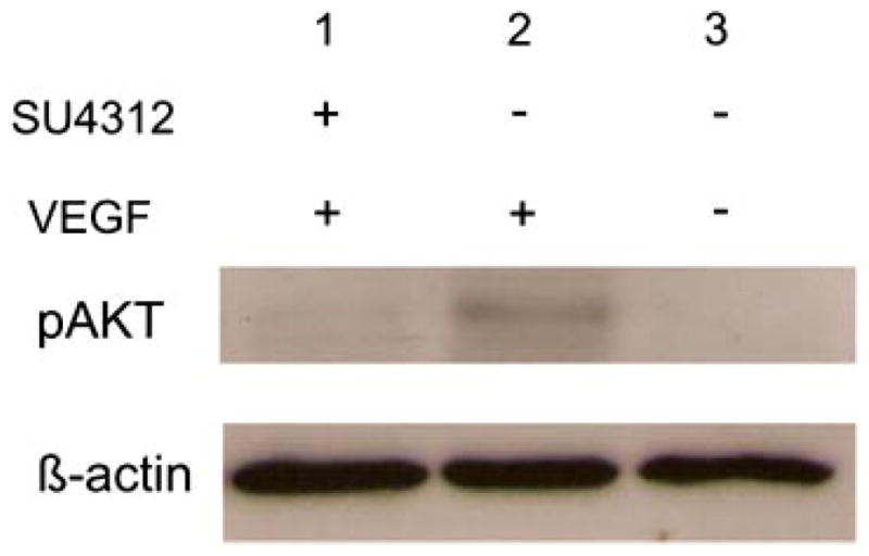
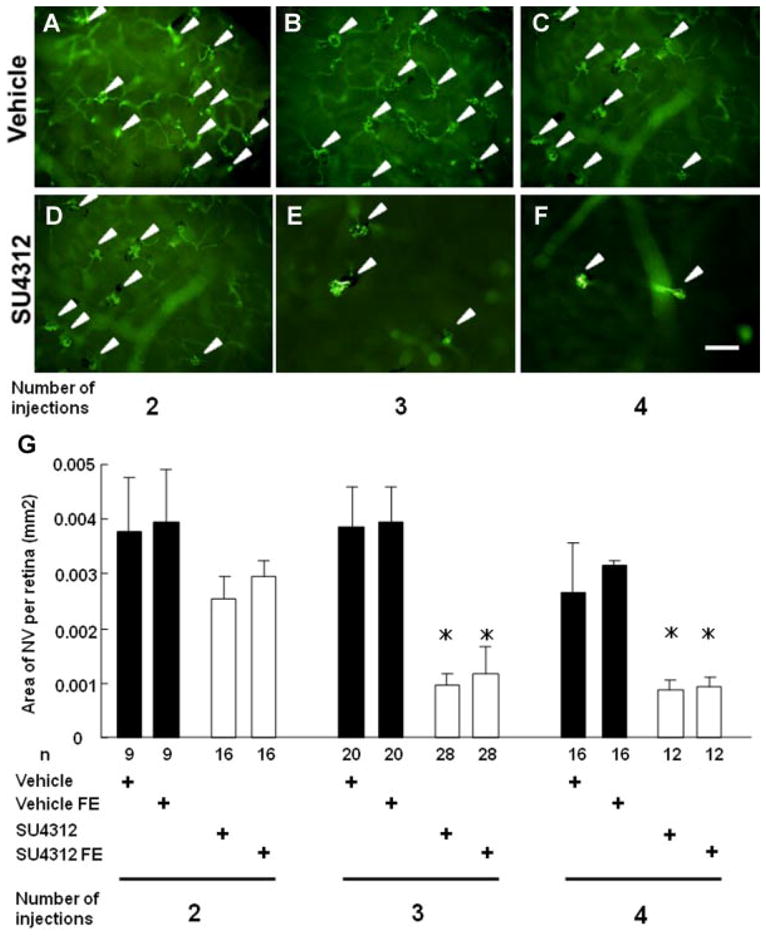
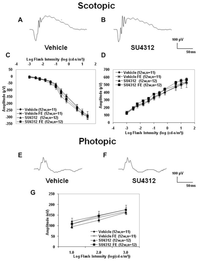
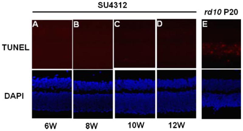
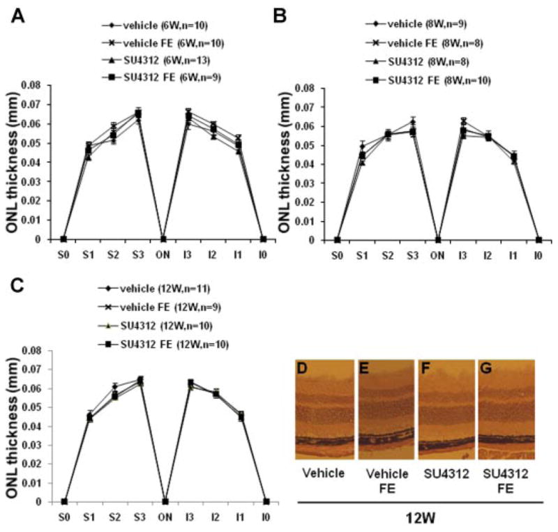
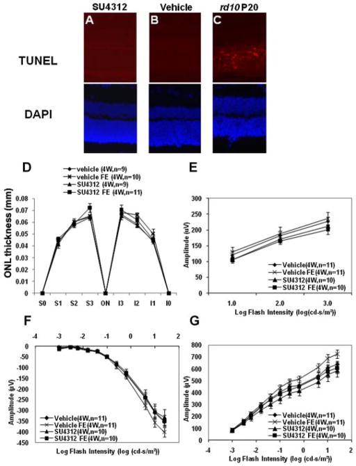
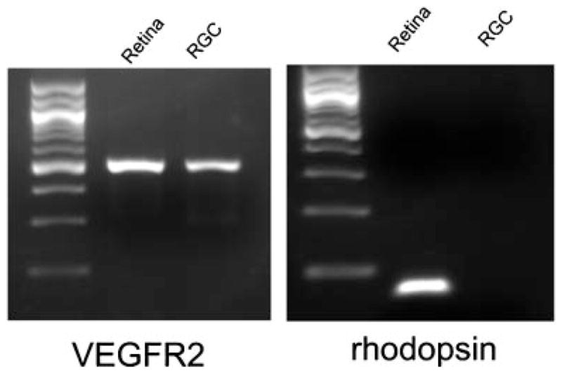
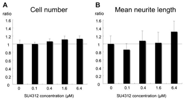
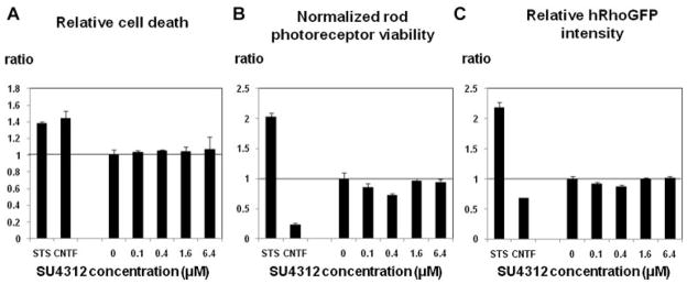
It has recently been reported that relatively short-term inhibition of vascular endothelial growth factor (VEGF) signaling can cause photoreceptor cell death, a potentially clinically important finding since VEGF blockade has become an important modality of treatment of ocular neovascularization and macular edema. However, in a set of studies in which we achieved extended and complete blockage of VEGF-induced vascular leakage through retinal expression of a VEGF binding protein, we did not observe any toxicity to retinal neurons. To follow-up on these apparently discrepant findings, we designed a set of experiments with the kinase inhibitor SU4312, which blocks phosphorylation of VEGF receptors, to look directly for evidence of VEGF inhibition-related retinal toxicity. Using transgenic mice with sustained expression of VEGF in photoreceptors, we determined that periocular injection of 3 microg of SU4312 every 5 days markedly suppressed subretinal neovascularization, indicating effective blockade of VEGF signaling. Wild-type mice given periocular injections of 5 microg of SU4312 every 5 days for up to 12 weeks showed normal scotopic and photopic electroretinograms (ERGs), no TUNEL stained cells in the retina, and no reduction in outer nuclear layer thickness. Incubation of cultured ganglion cells or retinal cultures containing photoreceptors with high doses of SU4312 did not reduce cell viability. These data suggest that blocking VEGF signaling in the retina for up to 12 weeks does not damage photoreceptors nor alter ERG function and should reassure patients who are receiving frequent injections of VEGF antagonists for choroidal and retinal vascular diseases.


Figures

Fig. 1
Fig. 1
SU4312 blocks vascular endothelial growth factor (VEGF)-induced phosphorylation of Akt in mouse retina. Retinas were dissected from C57BL/6 mice that were uninjected or 6 h after intraocular injection of 1 μl of 10−6 M μg of VEGF or coinjection of 1 μl containing 10−6 M VEGF and 3 × 10⁻⁶ M SU4312. Retinal homogenates (80 μg of protein) were run in immunoblots using an antibody specific for phosphorylated AKT, stripped and reblotted with an antibody for β-actin. Retinas from eyes injected with VEGF showed a signal for phosphorylated Akt (lane 2) which was not present in the retinas of uninjected eyes (lane 3) and was blocked by co-injection of SU4312 with the VEGF (lane 1).
Fig. 2
Fig. 2
Effect of periocular injections of SU4312 in transgenic mice in which the rhodopsin promoter drives expression of VEGF in photoreceptors (rho/VEGF mice). Starting at postnatal day (P) 14, hemizygous rho/VEGF mice were given a periocular injection of 3 μl of vehicle or 3 μl containing 3 μg of SU4312 every 4, 5, or 7 days. At P28, these three groups of mice which had had a total of 4, 3, or 2 injections were perfused with fluorescein-labeled dextran and retinal flat mounts were examined by fluorescence microscopy. The eyes of all mice injected with vehicle, regardless of the frequency of injections, showed numerous tufts of neovascularization (NV) on the outer surface of the retina (A–C). Mice given two injections of SU4312 every 7 days a part also showed many tufts of NV on the outer surface of the retina (D), but mice given injections of SU4312 every 5 days (E) or every 4 days (F) showed few tufts of NV. Image analysis with the investigator masked with respect to treatment group showed that eyes of mice given two injections of SU4312 over the span of 2 weeks had no significant difference in mean (±SEM) area of NV on the outer surface of the retina compared to eyes of mice that received two injections of vehicle (G, first column). However, eyes given injections of SU4312 every 5 days (second column) or 4 days (third column) had a significant reduction in the area of NV on the outer surface of the retina than their corresponding vehicle-injected control group. Fellow eyes (FE) of mice given injections of SU4312 every 5 or 4 days also showed significant reductions in the area of NV compared to eyes from vehicle-injected controls indicating that the amount of SU4312 getting into the systemic circulation was sufficient to block VEGF signaling in the fellow eye. *P < 0.001 by ANOVA with Bonferroni/Dunn’s correction for multiple comparisons. Scale bar =100 μm.
Fig. 3
Fig. 3
Prolonged blockade of VEGF signaling in the retina did not reduce electroretinogram (ERG) a- or b-wave amplitudes. Adult (4–6weeks old) C57BL/6 mice were given a periocular injection of 5 μl of vehicle or 5 μl of vehicle containing 5 μg of SU4312 every 5 days. Scotopic and photopic ERGs were done 4, 6, 8, 10, and 12 weeks after initiating injections. The scotopic ERG wave forms at 12 weeks (12w) after initiating injections appeared very similar in vehicle-treated mice (A) and SU4312-treated mice (B). The mean (±SEM) a-wave (C) and b-wave amplitudes (D) are shown for the 12-week time point. There was no significant difference in amplitude at any flash intensity between SU4312-treated eyes and vehicle-treated eyes and the curves for the fellow eyes (FE) for each group were superimposable on the curves for SU4312-treated and vehicle-treated eyes. Photopic ERG waveforms also appeared similar in eyes of mice treated for 12 weeks with vehicle (E) or SU4312 (F). The mean (±SEM) photopic b-wave amplitudes (G) showed no significant difference between eyes treated with SU4312 or vehicle for 12 weeks, or for FE for each group at three different flash intensities. The data from earlier time points are identical to those shown here for the 12-week time point.
Fig. 4
Fig. 4
Apoptosis was not induced in the retinas of mice treated with SU4312. Adult C57BL/6 mice were given a periocular injection of 5 μg of SU4312 every5 days and were euthanized after 4,6, 8, 10, or12 weeks. TUNEL staining of ocular frozen sections alsostained with DAPI to visualize nuclei showed no labeled cells in the retinas of mice treated with SU4312 for 6 (A), 8 (B), 10 (C), or 12 (D) weeks, but there were numerous labeled cells in the retina of postnatal day (P) 20 rd10 mice (E).
Fig. 5
Fig. 5
Treatment with SU4312 for upto 12 weeks did not cause reduction in thickness of the outer nuclear layer (ONL). Adult C57BL/6 mice were given periocular injections of 5 μl of vehicle or vehicle containing 5 μg of SU4312 every 5 days in one eye and after 4, 6, 8, 10, or 12 weeks, ONL thickness was measured by image analysis at six locations in each eye; 25% (S1), 50% (S2), and 75% (S3) of the distance between the superior pole and the optic nerve and 25% (I1), 50% (I2), and 75% (I3) of the distance between the inferior pole and the optic nerve. Each point represents the mean (±SEM) for the designated number of mice in each group. Compared to eyes injected with vehicle, eyes injected with SU4312 for 6 weeks (A), 8 weeks (B), or 12 weeks (C) showed no difference in mean ONL thickness at each of the corresponding six locations. The mean ONL thickness values for both of these groups were also not different from values in contralateral fellow eyes (FE) of eyes injected with SU4312 or FE of eyes injected with vehicle. Representative images are shown only for the 12-week time point—the retinas from SU4312-injected eyes, their contralateral fellow eyes (FE), vehicle-injected eyes from separate mice and their FE all looked similar.
Fig. 6
Fig. 6
SU4312 did not cause apoptosis, reduction of outer nuclear layer thickness, or reduction of ERG amplitudes in young mice. Starting at postnatal day (P) 14, C57BL/6 mice were given periocular injections of 3 μl ofvehicleor 3 μl of vehicle containing 3 μg of SU4312 every 5 days. After 4 weeks (4w) of injections, there were no TUNEL-positive cells in the retinas of mice treated with SU4312 (A) or vehicle (B), but numerous TUNEL-positive cells in the retinas of P20 rd10 mice (C). The sections were also stained with DAPI to visualize the nuclei (shown beneath each fluorescent image). There were no significant differences in mean (±SEM) outer nuclear layer (ONL) thickness at six corresponding locations in eyes injected with SU4312 for 4 weeks, their contralateral fellow eyes (FE), eyes of mice injected with vehicle for 4 weeks or their FE (D). There was no reduction in mean photopic b-wave amplitudes (E), or scotopic a- (F) and b-wave amplitudes (G) in eyes of mice treated with SU4312 for 4 weeks compared to vehicle-treated mice, or FE of each type of mice.
Fig. 7
Fig. 7
Expression of mRNA for vascular endothelial cell growth factor receptor 2 (VEGFR2) in retinal ganglion cell (RGC) cultures. Total RNA was prepared from immunopurified murine RGCs and from total mouse retina (as positive control), cDNA was synthesized, and 1 μg was used for RT-PCR using primers specific for VEGFR2 or rhodopsin. Both RGC cultures and total retina showed clear expression of VEGFR2 mRNA. The mRNA for rhodopsin was present in total retina, but not in the immunopurified RGCs.
Fig. 8
Fig. 8
Blockade of vascular endothelial growth factor (VEGF) signaling does not alter viability or mean neurite length of cultured retinal ganglion cells. Rat RGC cultures were maintained in normal growth media or growth media supplemented with 0.1, 0.4, 1.6, or 6.4 μM SU4312 (A,B). After 72 h cell number (A) and mean neurite length (B) were analyzed by image analysis. The bars represent the mean (±SEM) cell number or neurite length value for each concentration of SU4312 normalized to control cultures without SU4312, which are defined to have a value of 1. There were no significant differences between any of the treatment groups and the controls in cell number or mean neurite length.
Fig. 9
Fig. 9
Blockade of vascular endothelial growth factor (VEGF) signaling does not alter viability or rhodopsin expression of dissociated mixed retinal cell cultures. Mixed retinal murine cell cultures were maintained in normal growth media or growth media supplemented with 0.1, 0.4, 1.6, or 6.4 μM SU4312 (all cultures contained 0.1% DMSO). Staurosporine (20 nM) was used as a positive control, and CNTF (1 ng/ml) was used as a negative control. After 6 days in vitro, cultures were imaged on an ArrayScan VTI and cell number and fluorescence intensity (relative rhodopsin level) per cell were quantified by image analysis. Each bar represents the mean value (±SEM) of the indicated parameter for the given SU4312 concentration normalized to the mean of the negative control cultures (three cultures for each experimental group). One-way ANOVA analysis did not show any significant effect of SU4312 treatment on the measured cell parameters.

Similar articles

Cited by

References

    1. Barres BA, Chun Linda LY. Purification of rat retinal ganglion cells by immunopanning. Neuroprotocols. 1993;2:201–204.
    1. Bocker-Meffert S, Rowenstiel P, Rohl C, Warneke N, Held-Feindt J, Sievers J, Lucius R. Erythropoietin and VEGF promote neural outgrowth from retinal explants in postnatal rats. Invest Ophthalmol Vis Sci. 2002;43:2021–2026. - PubMed
    1. Bonnet D, Garcia M, Vecino E, Lorentz JG, Sahel J, Hicks D. Brain-derived neurotrophic factor signalling in adult pig retinal ganglion cell neurite regeneration in vitro. Brain Res. 2004;1007:142–151. - PubMed
    1. Bradford RL, Wang C, Zack DJ, Adler R. Roles of cell-intrinsic and microenvironmental factors in photoreceptor cell differentiation. Dev Biol. 2005;286:31–45. - PMC - PubMed
    1. Brown DM, Kaiser PK, Michels M, Soubrane G, Heier JS, Kim RY, Sy JP, Schneider S the Anchor Study Group. Ranibizumab versus verteporfin for neovascular age-related macular degeneration. N Engl J Med. 2006;355:1432–1444. - PubMed

Publication types

MeSH terms

Substances

LinkOut - more resources