. 2010 Jul;224(1):17-27.
doi: 10.1002/jcp.22071.

Transcriptional regulation of Rex1 (zfp42) in normal prostate epithelial cells and prostate cancer cells

Affiliations

Transcriptional regulation of Rex1 (zfp42) in normal prostate epithelial cells and prostate cancer cells

Mi-Young Lee et al. J Cell Physiol. 2010 Jul.

Abstract

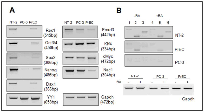
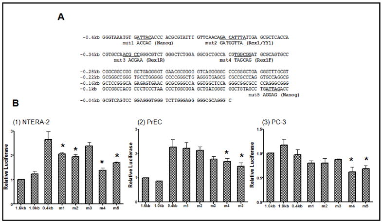
Rex1 (zfp42) was identified by our laboratory because of its reduced expression in F9 teratocarcinoma stem cells after retinoic acid (RA) treatment. The Rex1 (Zfp42) gene is currently widely used as a marker of embryonic stem cells. We compared the transcriptional regulation of the human Rex1 gene in NTera-2 (NT-2) human teratocarcinoma, normal human prostate epithelial cells (PrEC), and prostate cancer cells (PC-3) by promoter/luciferase analyses. Oct4, Sox2, Nanog, and Dax1 transcripts are expressed at higher levels in NT-2 and PrEC cells than in PC-3 cells. Co-transfection analyses showed that YY1 and Rex1 are positive regulators of hRex1 transcription in NT-2 and PrEC cells, whereas Nanog is not. Serial deletion constructs of the hRex1 promoter were created and analyzed, by which we identified a potential negative regulatory site that is located between -1 and -0.4 kb of the hRex1 promoter. We also delineated regions of the hRex1 promoter between -0.4 kb and the TSS that, when mutated, reduced transcriptional activation; these are putative Rex1 binding sites. Mutation of a putative Rex1 binding site in electrophoretic mobility shift assays (EMSA) resulted in reduced protein binding. Taken together, our results indicate that hRex1 binds to the hRex1 promoter region at -298 bp and positively regulates hRex1 transcription, but that this regulation is lost in PC-3 human prostate cancer cells. This lack of positive transcriptional regulation by the hRex1 protein may be responsible for the lack of Rex1 expression in PC-3 prostate cancer cells.


Figures

Fig. 1
Fig. 1
Analysis of Stem Cell Marker Gene Expression. Reverse transcription–PCR(RT-PCR). Total RNA was isolated from NT-2, PC-3 and PrEC cells and 2mg of RNA was used to make cDNA by reverse transcription. cDNA was diluted 1:5 and 2ml of the cDNA was used to perform PCR. 35 reaction cycles were used, except for GAPDH (30 cycles), and the PCR products were sequenced for confirmation of gene identity (Rex1 was amplified using E2F and E4R primers). Primers used are in Table 2. Each primer set is designed so that the set spans at least two different exons. (A) Various stem cell marker genes, (B) Rex1 transcripts (1, 4: E1F and E4R, size of product, 650 bp; 2, 5: E2F and E4R, size of product, 515 bp; and 3, 6: E3F and E4R primer, size of product, 396 bp). These experiments were performed at least three times, starting with cell culture, with very similar results. The products were sequenced to verify their identities.
Fig. 2
Fig. 2
(A) Maps of Promoter Deletion Constructs of the Human Rex1 Promoter. Genomic DNA was isolated from human mammary epithelial cells (HMEC) and used as a template to amplify the human Rex1 promoter region of 1.6 kb.A1.6 kb PCR product was cloned into the pGL3-Basic vector and named pGL3-hRex1-1.6 kb. The other five serial deletion constructs, hRex1-1.4 kb, hRex1-1.0 kb, hRex1-0.4 kb, hRex1-0.2 kb and hRex1-0.13 kb, were generated from hRex1-1.6 kb by performing PCR. The sequence of each construct was confirmed by DNA sequencing. (B) Control Level of hRex1 Promoter Activity. The luciferase activity of the hRex1-1.6 promoter construct was measured to compare the levels of the hRex1 promoter activity among three cell types. The hRex1-1.6 promoter construct was transfected into each cell line along with the Renilla luciferase (pTK-RL) as an internal control. After 24 hours, media were changed and cells were incubated for an additional 48 hr. Cells were harvested and extracts were used for the dual luciferase assay. The luciferase activities were normalized to that in NT-2 cells. (C) hRex1 Promoter Activity Analysis of Serial hRex1 Promoter Deletion Constructs. Six hRex1 promoter constructs were transfected into each cell line along with the Renilla luciferase (pTK-RL) as an internal control. The luciferase activities of the deletion constructs were normalized to the hRex1-1.6 control group and presented as a relative level of luciferase activity. Transfections were repeated at least three times starting with fresh cells and the data are presented as the mean WS.E.M. (MUp < 0.05, one way Anova, comparison among groups to hRex1-1.6).
Fig. 3
Fig. 3
(A) Maps of Mutant Constructs of The Human Rex1 Promoter. Mutant constructs were generated using deletion constructs as templates; DATTA was created by removing the -1.0 kb to _0.4 kb region of the hRex1-1.6 kb construct and DATTA 0.2 was created by deletion of the _0.4 kb to -0.2 kb of the hRex1-1.0 construct. Site-directed mutagenesis was performed to replace the putative Tcf1A/IRF1,2 and Cdx/Nanog binding sites to generate the Mut1 and Mut2 constructs. DTcfCdx1 was created by a deletion covering both binding sites. The sequence of each construct was confirmed by sequencing. (B) Promoter Activity Analysis of hRex1 Mutant Constructs. Human Rex1 promoter mutants were cloned into the pGL3-Basic vector. These constructs were transfected into each cell line with a Renilla luciferase (pTK-RL) as an internal control. Twenty four hr after transfection, media were changed and fresh media added. Cell extracts were harvested after 48 hours for the dual luciferase assays. The luciferase levels were normalized to the hRex1-1.6 sample and presented as the relative level of luciferase activity. Transfections were performed at least three times, starting with cells in culture, and the data are presented as mean WS.E.M. (MUp < 0.05, one way Anova, comparison among groups against hRex1-1.6). [Color figure can be viewed in the online issue, which is available at www.interscience.wiley.com.]
Fig. 4
Fig. 4
(A) Structures of Rex1, YY1 and, Nanog Proteins. Rex1 and YY1 are members of the zinc finger motif transcription factor family (Kim et al., 2007). Four repeated C2H2-type zinc finger motifs are located in the C-terminal halves of the Rex1 and YY1 proteins. The DNA sequences show approximately 70% sequence homology in their C-terminal zinc-finger motif regions (Mongan et al., 2006). Nanog is a member of the homeodomain transcription factor family and the homeobox is located in the proximal N-terminal portion of the protein (Mitsui et al., 2003). (B) Cotransfection Analysis of the hRex1 Promoter. The cDNAs for human Rex1, YY1 and Nanog were cloned into the pSG5 vector and used for cotransfections with the hRex1-1.6, hRex1-1.0 and hRex1-0.4 promoter constructs, with the Renilla Luciferase (pTK-RL) co-transfected as an internal control. The ratio between the promoter fragment and a transcription factor construct was 1:5. The total amount of DNA was equalized in all samples by addition of pSG5 vector DNA. The luciferase levels were normalized to each promoter construct without the pSG5-cDNA constructs and presented as a relative level of luciferase. Transfections were performed a minimum of three times, and the data are presented as the mean WS.E.M. (MUp < 0.05, one way Anova
Fig. 5
Fig. 5
(A) Sequences of The Human Rex1-0.4 kb Construct and Generation of Mutant Constructs. Five different hRex1-0.4 kb mutant constructs were created by site-directed mutagenesis using the pGL3-hRex1-0.4 kb as a template. Mut1 was created by mutation of a potential Nanog binding site (Mitsui et al., 2003; Loh et al., 2006) locating on -390 to -380, and mut2 for potential Rex1/YY1 binding site located between −370 to -355 (Shrivastava and Calame, 1994; Yant et al., 1995; Kim et al., 2007), [Zheng, unpublished] mut3 for a reverse Rex1 binding site located between −340 to -325 (Zhang et al., 2006), mut4 for a forward Rex1 binding site on located between −300 to -290 and mut5 for a Nanog binding site (Mitsui et al., 2003; Loh et al., 2006) located between −50 to -40 of hRex1 promoter region. All five mutant constructs were sequenced to confirm the base changes. (B) Promoter Activity Analysis of hRex1 0.4 kb Mutant Constructs The human Rex1 0.4 kb promoter mutants were cloned into the pGL3-Basic vector. These constructs were transfected into each cell line along with the Renilla luciferase (pTK-RL) as an internal control. Twenty four hr after transfection, media was changed and fresh media added. Cell extracts were harvested after 48 hours for dual luciferase assays. The luciferase levels were normalized to hRex1-1.6 and presented as a relative level of luciferase activity. Transfections were performed at least three times and the data are presented as the mean WS.E.M. (MUp < 0.05, one way Anova, samples compared to hRex1-1.6).
Fig. 6
Fig. 6
(A) Location and Sequences of Binding Probes hRex1-0.4 kb to 0.0 kb. The sequences of two binding probes are underlined and the specific binding sites are indicated by the boxes. (B) Electrophoretic Mobility Shift Assays. Putative Nanog, Rex1, or YY1 binding sites, (Nanog/YY1/Rex1A) or Rex1 binding site (Rex1B) are found between -0.4 kb to -0 kb in the hRex1 promoter region. The binding sites were labeled by -[32P]-dCTP.5mg of nuclear extract of each cell line was incubated with the radio-labeled probe and the reactions were separated on 5% acrylamide gel in 0.5X TBE buffer. For the competition assays, 100X unlabeled oligonucleotide was incubated before adding the labeled probeonice. These experiments were performed at least three times and the data were reproducible. One representative experiment is shown. (1), (2). 1, 4, 7: free probe, 2, 5, 8: probe R nuclear extract, 3, 6, 9: probe R nuclear extract R100X wt unlabeled competitor, (3). 1, 6, 11: freeprobe, 2, 7, 12: probe R nuclear extract, 3, 8, 13: probe R nuclear extract R100X wt unlabeled competitor, 4, 9, 14: probe R nuclear extract R100X mut1 unlabeled competitor, 5, 10, 15: probe R nuclear extract R100X mut2 unlabeled competitor

Similar articles

Cited by

References

    1. Ben-Shushan E, Thompson JR, Gudas LJ, Bergman Y. Rex-1, a gene encoding a transcription factor expressed in the early embryo, is regulated via Oct-3/4 and Oct-6 binding to an octamer site and a novel protein, Rox-1, binding to an adjacent site. Mol Cell Biol. 1998;18(4):1866–1878. - PMC - PubMed
    1. Boyer LA, Lee TI, Cole MF, Johnstone SE, Levine SS, Zucker JP, Guenther MG, Kumar RM, Murray HL, Jenner RG, Gifford DK, Melton DA, Jaenisch R, Young RA. Core transcriptional regulatory circuitry in human embryonic stem cells. Cell. 2005;122(6):947–956. - PMC - PubMed
    1. Boyer LA, Plath K, Zeitlinger J, Brambrink T, Medeiros LA, Lee TI, Levine SS, Wernig M, Tajonar A, Ray MK, Bell GW, Otte AP, Vidal M, Gifford DK, Young RA, Jaenisch R. Polycomb complexes repress developmental regulators in murine embryonic stem cells. Nature. 2006;441(7091):349–353. - PubMed
    1. Chambers I, Colby D, Robertson M, Nichols J, Lee S, Tweedie S, Smith A. Functional expression cloning of Nanog, a pluripotency sustaining factor in embryonic stem cells. Cell. 2003;113(5):643–655. - PubMed
    1. Chan EM, Ratanasirintrawoot S, Park IH, Manos PD, Loh YH, Huo H, Miller JD, Hartung O, Rho J, Ince TA, Daley GQ, Schlaeger TM. Live cell imaging distinguishes bona fide human iPS cells from partially reprogrammed cells. Nat Biotechnol 2009 - PubMed

Publication types

MeSH terms