. 2011 Jan 15;128(2):283-93.
doi: 10.1002/ijc.25326. Epub 2010 Mar 15.

Unimpeded skin carcinogenesis in K14-HPV16 transgenic mice deficient for plasminogen activator inhibitor

Affiliations

Unimpeded skin carcinogenesis in K14-HPV16 transgenic mice deficient for plasminogen activator inhibitor

Anne Masset et al. Int J Cancer. .

Abstract

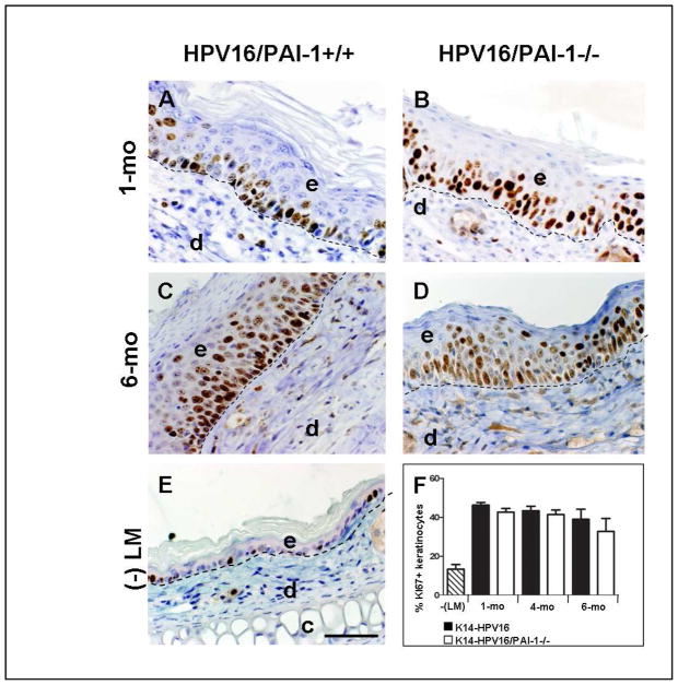
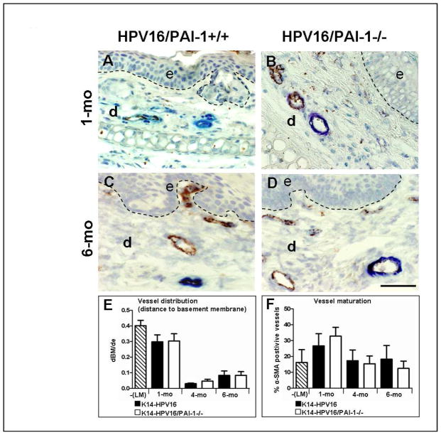
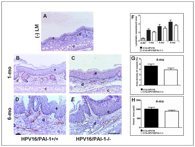
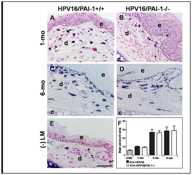
Angiogenesis, extracellular matrix remodeling and cell migration are associated with cancer progression and involve at least, the plasminogen activating system and its main physiological inhibitor, the plasminogen activator inhibitor-1 (PAI-1). Considering the recognized importance of PAI-1 in the regulation of tumor angiogenesis and invasion in murine models of skin tumor transplantation, we explored the functional significance of PAI-1 during early stages of neoplastic progression in the transgenic mouse model of multistage epithelial carcinogenesis (K14-HPV16 mice). We have studied the effect of genetic deletion of PAI-1 on inflammation, angiogenesis, lymphangiogenesis and tumor progression. In this model, PAI-1 deficiency neither impaired keratinocyte hyperproliferation or tumor development nor affected the infiltration of inflammatory cells and development of angiogenic or lymphangiogenic vasculature. We are reporting evidence for concomitant lymphangiogenic and angiogenic switches independent to PAI-1 status. Taken together, these data indicate that PAI-1 is not rate limiting for neoplastic progression and vascularization during premalignant progression, or that there is a functional redundancy between PAI-1 and other tumor regulators, masking the effect of PAI-1 deficiency in this long-term model of multistage epithelial carcinogenesis.


Figures

Figure 1
Figure 1. Keratinocyte proliferation is not affected in the absence of PAI-1
A–E: Immunohistochemical detection of Ki-67 positive cells on tissue sections of ears issued from K14-HPV16/PAI-1+/+ (A and C) and K14-HPV16/PAI-1−/− (B and D) mice with hyperplastic (A and B) and dysplastic (C and D) lesions; and on section of ears from negative littermate (-LM) (E). F: Quantitative analysis of the percentage of Ki-67-positive keratinocytes in K14-HPV16 and K14-HPV16/PAI-1−/− mice at 1, 4 and 6 months of age. Values represent keratinocyte proliferation which was determined as the percentage of Ki-67 positive keratinocytes as a fraction of total keratinocytes averaged from five high-power fields per mouse (n=5). Error bars represent SEM. No statistically significant difference was observed between age-matched neoplastic tissue issued from K14-HPV16 and K14-HPV16/PAI-1−/− (1 month P = 0.1143; 4 months P = 1; 6 months P = 0.62; Mann-Whitney unpaired t-test). Dashed line indicates epidermal-dermal interface. The epidermis (e), dermis (d), and cartilage (c) are indicated. Original magnification: 400x. Bars, 50μm.
Figure 2
Figure 2. Tumor vascularization is not affected by PAI-1 deficiency
A–D: Immunocolocalization of von Willebrand factor (endothelial cells in brown) and alpha-smooth muscle actin (pericytes in blue) in paraffin-embedded sections of age-matched neoplastic ear tissue of K14-HPV16 (A and C) and K14-HPV16/PAI-1−/− mice (B and D). E: Quantitative analysis of vessel distribution at distinct stages of neoplastic progression in ear tissue from K14-HPV16, K14-HPV16/PAI-1−/− and negative littermate (-LM). Values represent the average ratio between “dBM” and “de”. “dBM” corresponds to the distance separating epithelial basement membrane and the 5 closest blood vessels. “de” is the thickness of dermis. Errors bars represent SEM (1 month P = 0;8857; 4 months P = 0;547; 6 months P = 0.9166; Mann-Whitney unpaired t-test). F: Quantitative analysis of vessel maturity at distinct stages of neoplastic progression in ear tissue from K14-HPV16, K14-HPV16/PAI-1−/− and negative littermate (-LM). Values represent the percentage of vessels covered by pericytes (% α-SMA positive vessels) evaluated on five high-power fields per mouse and five mice per category. Errors bars represent SEM. No statistically significant differences were observed between age-matched neoplastic tissues between K14-HPV16 and K14-HPV16/PAI-1−/− (1 month P = 0.3429; 4 months P = 0.8413; 6 months P = 0.3457; Mann-Whitney unpaired t-test). Original magnification: 400x. Bars, 50μm.
Figure 3
Figure 3. The lymphangiogenic switch during squamous epithelial carcinogenesis is independent of PAI-1 status
A–F: Immunohistochemical detection of lymphatic vessels by Lyve-1 staining in normal skin of negative littermate (-LM) (A); hyperplastic lesions (B, C), dysplastic lesions (D, E) of ear skin issued from K14-HPV16/PAI-1+/+ mice (B, D) and K14-HPV16/PAI-1−/− mice (C, E). F-H: Quantification of ear vascularization performed by determining the vessel density (number of vessels per area) (1 month P = 0.5386; 4 months P = 0.3321; 6 months P = 0.2530; Mann-Whitney unpaired t-test) (F); the area density of vessels (percentage of tissue occupied by lymphatic vessels) (P = 0;1508; Mann-Whitney unpaired t-test) (G) and the mean surface of lymphatic vessels (P = 0.4206; Mann-Whitney unpaired t-test) (H). In all quantifications, values represent average values from five high-power fields per mouse and five mice per group. Errors bars represent SEM. Original magnification: 200x. Bars, 100μm.
Figure 4
Figure 4. The infiltration of neoplastic skin by mast cells is not affected by PAI-1-deficiency
A–E: Detection of mast cell infiltration by toluidin blue coloration on hyperplastic ear skin (A and B), in dysplastic ear skin (C and D) from K14-HPV16 (A and C) and K14-HPV16/PAI-1−/− (B and D) and in normal ear skin (E). F: Quantitative analysis of mast cells at distinct stages of neoplastic progression in ear tissue from K14-HPV16, K14-HPV16/PAI-1−/− and negative littermate (-LM). Values represent the number of mast cells averaged from five high-power fields per mouse and five mice per experimental group. Error bars represent SEM (1 month P = 0.3314; 4 months P = 0.9166; 6 months P = 0.92; Mann-Whitney unpaired t-test). Original magnification: 400x. Bars, 50μm.

References

    1. Binder BR, Mihaly J. The plasminogen activator inhibitor “paradox” in cancer. Immunology Letters. 2008;118:116–124. - PubMed
    1. Andreasen PA, Egelund R, Petersen HH. The plasminogen activation system in tumor growth, invasion, and metastasis. Cell Mol Life Sci. 2000;57:25–40. - PMC - PubMed
    1. Rakic JM, Maillard C, Jost M, Bajou K, Masson V, Devy L, Lambert V, Foidart JM, Noel A. Role of plasminogen activator-plasmin system in tumor angiogenesis. Cell Mol Life Sci. 2003;60:463–473. - PMC - PubMed
    1. Duffy MJ. Urokinase plasminogen activator and its inhibitor, PAI-1, as prognostic markers in breast cancer: From pilot to level I evidence studies. Clinical Chemistry. 2002;48:1194–1197. - PubMed
    1. Wilkins-Port CE, Higgins CE, Freytag J, Higgins SP, Carlson JA, Higgins PJ. PAI-1 is a critical upstream regulator of the TGF-beta 1/EGF-induced invasive phenotype in mutant p53 human cutaneous squamous cell carcinoma. Journal of Biomedicine and Biotechnology. 2007 - PMC - PubMed

Publication types