. 2010 Jun;27(6):843-59.
doi: 10.1080/07391102.2010.10508586.

Sequence-dependent Kink-and-Slide deformations of nucleosomal DNA facilitated by histone arginines bound in the minor groove

Affiliations

Sequence-dependent Kink-and-Slide deformations of nucleosomal DNA facilitated by histone arginines bound in the minor groove

Difei Wang et al. J Biomol Struct Dyn. 2010 Jun.

Abstract

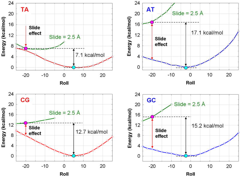
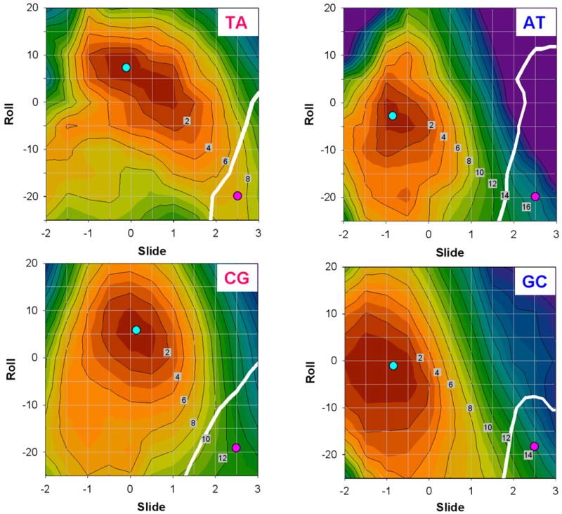
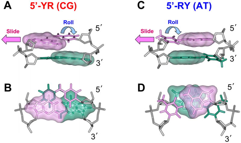
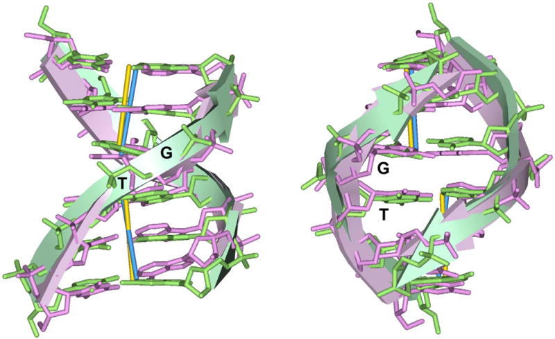
In addition to bending and twisting deformabilities, the lateral displacements of the DNA axis (Kink-and-Slide) play an important role in DNA wrapping around the histone core (M. Y. Tolstorukov, A. V. Colasanti, D. M. McCandlish, W. K. Olson, V. B. Zhurkin, J. Mol. Biol. 371, 725-738 (2007)). Here, we show that these Kink-and-Slide deformations are likely to be stabilized by the arginine residues of histones interacting with the minor groove of DNA. The arginines are positioned asymmetrically in the minor groove, being closer to one strand. The asymmetric arginine-DNA interactions facilitate lateral displacement of base pairs across the DNA grooves, thus leading to a stepwise accumulation of the superhelical pitch of nucleosomal DNA. To understand the sequence dependence of such Kink-and-Slide deformations, we performed all-atom calculations of DNA hexamers with the YR and RY steps in the center. We found that when the unrestrained DNA deformations are allowed, the YR steps tend to bend into the major groove, and RY steps bend into the minor groove. However, when the nucleosomal Kink-and-Slide deformation is considered, the YR steps prove to be more favorable for bending into the minor groove. Overall, the Kink-and-Slide deformation energy of DNA increases in the order TA < CA < CG < GC < AC < AT. We propose a simple stereochemical model accounting for this sequence dependence. Our results agree with experimental data indicating that the TA step most frequently occurs in the minor-groove kink positions in the most stable nucleosomes. Our computations demonstrate that the Kink-and-Slide distortion is accompanied by the BI to BII transition. This fact, together with irregularities in the two-dimensional (Roll, Slide) energy contour maps, suggest that the Kink-and-Slide deformations represent a nonharmonic behavior of the duplex. This explains the difference between the two estimates of the DNA deformation energy in nucleosome - the earlier one made using knowledge-based elastic energy functions, and the current one based on all-atom calculations. Our findings are useful for refining the score functions for the prediction of nucleosome positioning. In addition, the reverse bending behavior of the YR and RY steps revealed under the Kink-and-Slide constraint is important for understanding the molecular mechanisms of binding transcription factors (such as p53) to DNA exposed on the surface of nucleosome.


Figures

Figure 1
Figure 1
Variability of arginines penetrating into the DNA minor groove in a nucleosome. The 5′-end ‘sequence’ strand is shown in yellow and the 3′-end ‘non-sequence’ strand is in gray. A. Superhelix Locations (SHLs) in the ‘anterior’ half of nucleosomal crystal structure, PDB 1KX5 (5). The dyad axis is marked by 0 (in red). To distinguish between the two halves of the nucleosome related by twofold pseudo-symmetry, we call the first half ‘anterior’ and the second half ‘posterior.’ Seven arginines interacting with the DNA minor groove are shown in magenta. Blue circles show conformationally ‘rigid’ arginines, and magenta circles show ‘flexible’ arginines. H3, H4, H2A and H2B histones are shown in blue, green, yellow and orange, respectively. B. Conformationally ‘rigid’ R77 (H2A) penetrating into the minor groove at SHL −5.5. Images shown here are based on nine nucleosomal structures aligned through 300 Cα atoms of histone α-helices conserved in all nucleosomes. (The PDB entries are 1KX5, 1KX3, 1KX4, 1ID3, 1EQZ, 2CV5, 2PYO, 2NQB and 2NZD. These nine structures were selected to represent variability in the DNA sequence and length, as well as in the origin of histones – yeast, fly, frog, chicken and human.) For clarity, only four structures are shown (1KX5, 1KX3, 1ID3 and 1EQZ). Note that all nine arginines have nearly identical conformation – the distance between two positions of Cζ atom varies from 0.1 to 1.1 Å. C. Conformationally ‘flexible’ R63 (H3) interacting with the minor groove of DNA at SHL −1.5. Note that the side chain of R63 (H3) is more variable than that of R77 (H2A) shown in B – the distance between two positions of the Cζ atom varies from 0.3 to 7.7 Å.
Figure 2
Figure 2
Arginines in the minor groove of the 434 repressor-DNA complex and in the nucleosome. A. Symmetrically positioned arginine 43 in the minor groove of DNA in complex with the 434 repressor (21). The distances between the guanidinium NH2 groups and the closest O4′ atoms from nucleotides (i′) and (i+2) are 3.59 and 3.61 Å. The O4′ atoms are shown as red balls. (The distance between the left NH2 group and O4′(i+3) is 4.3 Å.) B. Asymmetrically positioned arginine 77 (H2A) in the minor groove of nucleosomal DNA (5) at the site SHL −5.5 with a strong Kink-and-Slide distortion. Arginine is closer to the gray strand than to the yellow ‘sequence’ strand – the distances between the guanidinium NH2 groups and the O4′ atoms from nucleotides (i′) and (i+3) are 3.38 Å and 4.36 Å, respectively. (The distance between the right NH2 group and O4′(i+2) is 6.7 Å.) C, D. Schematic representation of the DNA minor groove in the 434 repressor-DNA complex (canonical B-DNA), C, and in the nucleosome (at the site with the Kink-and-Slide distortion), D. In C, the closest opposite O4′ atoms belong to nucleotides (i′) and (i+2), with the O4′-O4′ distance 6.8 Å. Position of the guanidinium group of arginine (blue star) is close to the center of O4′-O4′ vector (see panel A). In D, geometry of the minor groove differs from that in canonical B-form because of the strong positive Slide and negative Roll (shown by blue arrows in panel C). As a result, the two closest O4′ atoms belong to nucleotides (i′) and (i+3), the O4′-O4′ distance being 7.1 Å. The guanidinium group of arginine (blue star) is closer to the non-sequence strand shown in gray (see panel B).
Figure 3
Figure 3
Arginines facilitate the lateral displacement (Slide) in the nucleosomal DNA (stereo image). The DNA fragment shown in gray is the same as in Figure 2B – arginine 77 of H2A (magenta surface) asymmetrically penetrates into the minor groove at SHL −5.5. The base pairs shown as blue sticks represent the Kink-and-Slide step (positions #16 and #17 in 147-bp long DNA fragment (4)). For comparison, the DNA conformation modeled with Slide = 0 at this step is shown in orange; in this case, there is unacceptable steric hindrance between arginine and sugar ring of the DNA backbone. Note that the arginine NH2 group is close to the line connecting O4′(#16′) and O4′(#19) atoms (red balls). That is, arginine 77 ‘bridges’ deoxyribose oxygens located at the 3′-ends of tetramer 16-19 (marked by green ribbon in the sequence strand). The line between O4′(#16′) and O4′(#18), which is distant from the arginine NH2 groups, corresponds to the line (i′, i+2) in the complex with the 434 repressor (Figures 2A, C).
Figure 4
Figure 4
Histone arginines facilitate the axial dislocation of DNA in the nucleosome. A. ‘Outside’ view. The zig-zag trajectory of DNA (yellow and gray ribbons) around the histone core (gray cylinder). For clarity, only arginines at SHLs −5.5, −4.5, −3.5 and 1.5, 2.5 and 3.5 are shown (magenta balls). The centers of base pairs corresponding to dimeric steps with Kink-and-Slide deformations are shown as blue balls. These balls are linked by sticks to emphasize the zig-zag trajectory of the DNA axis. The arrows indicate directions of the base pair movements facilitated by arginines: white arrows for the ‘anterior’ half and yellow arrows for the ‘posterior’ half of the nucleosome. B. Stereo ‘inside’ view of the nucleosomal DNA fragment. Arginines 77 and 42 at the ends of H2A α2 helix, interacting with the non-sequence DNA strand (gray ribbon), produce a cooperative effect leading to the lateral Slide displacements and the Roll angles (white arrows at SHLs −5.5 and −3.5).
Figure 5
Figure 5
Minor groove width, kinks and arginine positions in nucleosome 1KX5 (5). The minor groove width is measured as the distance between the O4′ atoms in nucleotides (i′) and (i+3), see Figure 2D; this value is assigned to the midpoint position (i+1.5). The arginine positions (pink circles) are defined as follows. For each arginine, the O4′ atom is selected, which is the closest to its guanidinium NH2 groups. The opposite O4′ atom is selected from the complementary DNA strand, according to the ‘i+3’ rule described above. Then, the arginine is assigned to the center of the tetramer (i, i+3), that is, to the position (i+1.5). For example, for arginine 77 of H2A in Figure 3, the closest oxygen is O4′(#16′) and the opposite one is O4′(#19); therefore, the arginine position is defined as 17.5. The dimeric steps with Kink-and-Slide distortions are shown as blue diamonds; their positions are also half-integer, in particular, the kink between base pairs #16 and #17 is assigned to position 16.5 (Figure 3). The SHL sites are shown at the top; see Table I for the description of arginines interacting with the minor groove at each SHL site.
Figure 6
Figure 6
Schematic representation of hexamers calculated in this study. Roll and Slide in the central step S3 served as ‘principal’ variables during minimization. The dimeric step parameters in the terminal steps S1 and S5, as well as Propeller twist and Buckle in the terminal base pairs (#1):(#12) and (#6):(#7) were fixed – see Methods for details.
Figure 7
Figure 7
Bending profiles of energy versus Roll angle for hexamers with the YR (TA and CG) and RY (AT and GC) central steps (see Methods for the hexamer sequences). Red and blue lines: the YR and RY deformation energies versus Roll without the Slide constraint. The energetically optimal structures are shown by blue dots. Green lines: the energy profiles for Roll scan with the Slide constraint of 2.5 Å. The Kink-and-Slide deformed structures with Roll = −20° and Slide = 2.5 Å are shown by red dots.
Figure 8
Figure 8
Two-dimensional (Roll versus Slide) energy contour plots for hexamers with the YR and RY central steps. The hexamer sequences are the same as in Figure 7. The contour line separation is 2 kcal/mol. Cyan circles: the optimal structures. Pink circles: the Kink-and-Slide deformed structures. White thick lines: the boundary between BI and BII forms, i.e. structures with equal backbone dihedral angles ε and ζ.
Figure 9
Figure 9
The Kink-and-Slide deformation is more favorable for the YR dimeric steps than for the RY steps. The Kink-and-Slide structures of the CG (A, B) and AT dimers (C, D) are shown. Note that in the CG dimers, the purine-purine repulsion (overlap between the pink and green surfaces) is relatively weak compared to that in AT.
Figure 10
Figure 10
Comparison of the hexamer at SHL −3.5 in 1KX5 (Roll = −18° and Slide = 2.7 Å) and the calculated structure with the Kink-and-Slide deformation (Roll = −20° and Slide = 2.5 Å), aligned together through the base-pair centers. In both hexamers, the central dimer is TG:CA. The hexamer at SHL −3.5 in the X-ray structure (5) is shown in pink stick rendering, and the calculated structure is shown in green sticks. The RMSD of six base pair centers is 0.71 Å.

Similar articles

Cited by

References

    1. Sekinger EA, Moqtaderi Z, Struhl K. Mol Cell. 2005;18:735–748. - PubMed
    1. Anderson JD, Widom J. Mol Cell Biol. 2001;21:3830–3839. - PMC - PubMed
    1. Thastrom A, Lowary PT, Widlund HR, Cao H, Kubista M, Widom J. J Mol Biol. 1999;288:213–229. - PubMed
    1. Gabdank I, Barash D, Trifonov EN. J Biomol Struct Dyn. 2009;26:403–411. - PubMed
    1. Davey CA, Sargent DF, Luger K, Maeder AW, Richmond TJ. J Mol Biol. 2002;319:1097–1113. - PubMed

Publication types