. 2010 Jun;27(6):861-6.
doi: 10.1080/07391102.2010.10508587.

Electrostatic interactions between arginines and the minor groove in the nucleosome

Affiliations

Electrostatic interactions between arginines and the minor groove in the nucleosome

Sean M West et al. J Biomol Struct Dyn. 2010 Jun.

Abstract

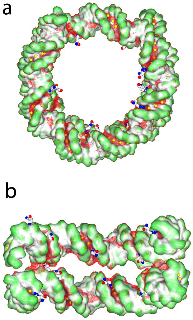
Proteins rely on a variety of readout mechanisms to preferentially bind specific DNA sequences. The nucleosome offers a prominent example of a shape readout mechanism where arginines insert into narrow minor groove regions that face the histone core. Here we compare DNA shape and arginine recognition of three nucleosome core particle structures, expanding on our previous study by characterizing two additional structures, one with a different protein sequence and one with a different DNA sequence. The electrostatic potential in the minor groove is shown to be largely independent of the underlying sequence but is, however, dominated by groove geometry. Our results extend and generalize our previous observation that the interaction of arginines with narrow minor grooves plays an important role in stabilizing the deformed DNA in the nucleosome.


Figures

Figure 1
Figure 1. Minor-groove shape recognition in the nucleosome
Plot of the minor groove width and electrostatic potential for (a) DNA from the α- satellite/X. laevis histone structure, (b) DNA from the α-satellite/D. melanogaster histone structure, and (c) DNA from the A16/X. laevis histone structure. Regions of the A16 DNA that vary in sequence from the α-satellite structures are underlined in blue. Arginines from each histone class are denoted with a marker as described in the legend. Arginine residues are placed at the base pair whose reference point is closest to their guanidinium Cζ atom. Arginines from the histone core are shown as filled markers; those in histone tails are shown as empty markers. Markers circled in green represent arginines located between 6.0 and 8.0 Å from the bottom of the minor groove. The vertical position of the markers is based on the most negative potential experienced by any atom of the guanidinum group.
Figure 2
Figure 2. Helical parameters of the α-satellite/X. laevis, α-satellite/D. Melanogaster and A16/X.laevis DNA structures
DNA structural parameters are plotted in red for the α-satellite/X. laevis structure, in black for the α-satellite/D. melanogaster structure and in blue for the A16/X. laevis structure. Local parameters as defined by Curves are shown for helical parameters. Regions where the sequences vary between α-satellite and A16 are underlined in blue. Horizontal black lines indicate helical parameters for canonical B-DNA modeled from fiber diffraction data.
Figure 3
Figure 3. Electrostatic isosurfaces for nucleosomal DNA
GRASP2 surface representations (22) of the DNA taken from the α-satellite/X. laevis structures with (a) a view along the superhelical axis and (b) a view of a portion of the DNA facing the histone core are colored-coded for shape, with convex surfaces in green and concave surfaces in dark gray. Arginines are displayed as sticks and are located in the groove where the DNA is compressed. The yellow spheres represent the reference points inside of the minor groove where the electrostatic potential was measured. Mesh representations of the -6.0 kT/e isosurface are displayed in red. All arginines that contact the minor groove within a distance of 8.0 Å from the bases are shown.
Figure 4
Figure 4. Effect of salt concentration on electrostatic potentials
Electrostatic potentials in the minor groove of the DNA from the α-satellite/X. laevis structure were obtained from solutions to the nonlinear Poisson-Boltzmann equation at varying salt concentrations. Electrostatic potentials are plotted at the center of the minor groove as a function of the base sequence.

Similar articles

Cited by

References

    1. Rohs R, West SM, Jin X, Joshi R, Honig B, Mann RS. Annu Rev Biochem. 2010;79 - PMC - PubMed
    1. Tolstorukov MY, Colasanti AV, McCandlish DM, Olson WK, Zhurkin VB. J Mol Biol. 2007;371:725–38. - PMC - PubMed
    1. Rohs R, Sklenar H, Shakked Z. Structure. 2005;13:1499–509. - PubMed
    1. Rohs R, West SM, Liu P, Honig B. Curr Opin Struct Biol. 2009;19:171–7. - PMC - PubMed
    1. Joshi R, Passner JM, Rohs R, Jain R, Sosinsky A, Crickmore MA, Jacob V, Aggarwal AK, Honig B, Mann RS. Cell. 2007;131:530–43. - PMC - PubMed

Publication types

LinkOut - more resources