. 2010 Apr;159(7):1418-28.
doi: 10.1111/j.1476-5381.2009.00630.x. Epub 2010 Mar 3.

Atherosclerosis induced by chronic inhibition of the synthesis of nitric oxide in moderately hypercholesterolaemic rabbits is suppressed by pitavastatin

Affiliations

Atherosclerosis induced by chronic inhibition of the synthesis of nitric oxide in moderately hypercholesterolaemic rabbits is suppressed by pitavastatin

Masaki Kitahara et al. Br J Pharmacol. 2010 Apr.

Abstract

Background and purpose: It is not clear if the new 3-hydroxyl-3-methylglutaryl coenzyme A (HMG-CoA) reductase inhibitor pitavastatin prevents atherogenesis by a direct effect. Statins have a cholesterol-lowering effect, so an accessible animal model of atherosclerosis showing only moderate hypercholesterolaemia as in humans, is needed. The effects of pitavastatin were evaluated on atherosclerotic lesions accumulating foam cells derived from macrophages, produced in rabbits with moderate hypercholesterolaemia by chronic inhibition of nitric oxide synthase (NOS).

Experimental approach: White New Zealand rabbits were fed a 0.2% cholesterol diet with the NOS inhibitor N(omega)-nitro-L-arginine methyl ester (L-NAME) in the same diet. Pitavastatin (0.1 and 0.3 mg x kg(-1)) was given orally once a day for 8 weeks. The aortic arch and thoracic aorta were analysed by histochemistry and atherosclerotic lesions were quantified. The effect of pitavastatin on adhesion of THP-1 cells to endothelial cells, and cholesterol content in RAW264.7 cells incubated with oxidized or acetylated LDL were also investigated.

Key results: Atherosclerotic lesions containing foam cells were induced in a model of atherosclerosis in rabbits with moderate hypercholesterolaemia by chronic inhibition of NOS. The area of atherosclerotic lesions was diminished by pitavastatin administration. The adhesion of THP-1 cells and cholesteryl ester content in RAW macrophages were decreased by pitavastatin treatment.

Conclusion: Atherosclerosis induced by chronic inhibition of NOS in moderately hypercholesterolaemic rabbits was suppressed by pitavastatin via inhibition of macrophage accumulation and macrophage foam cell formation.


Figures

Figure 1
Figure 1
Experimental schedule of cholesterol feeding and administration of L-NAME and pitavastatin. The feeding regimen of cholesterol chow and administration of L-NAME and pitavastatin is summarized. Rabbit were fed a cholesterol diet containing L-NAME and pitavastatin was given orally once a day. L-NAME, Nω-nitro-L-arginine methyl ester.
Figure 4
Figure 4
Quantification of the atherosclerotic areas of cross-sections in the aortic arch and thoracic aorta. The ratio of intima/media was calculated by measurement of each area of H&E-stained cross-section using LUZEX. Quantitative analysis of areas of lipid deposition and macrophage content was performed in Oil Red O-stained cross-sections and RAM11-immunostained cross-sections by LUZEX respectively. Graphs A–C show the data from the aortic arch samples, and graphs D–F that from the thoracic aorta samples. A and D, ratio of intima/media; B and E, percentage of Oil Red O-stained area; C and F, percentage of RAM 11-stained area. Each column represents mean ± SE. Asterisk (*) indicates significant difference from control at P < 0.05 by Dunnett's test.
Figure 2
Figure 2
Effect of pitavastatin on the area of atherosclerotic surface lesions in the aortic arch (A) and thoracic aorta (B). Rabbits were treated with 0.1 and 0.3 mg·kg−1 pitavastatin once a day during the experimental period. The aortic arch and thoracic aorta were stained with Sudan III. Each column represents mean ± SE. Asterisk (*) indicates significant difference from control at P < 0.05 by Dunnett's test.
Figure 3
Figure 3
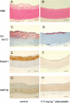
Cross-sections from a typical aortic arch. Rabbits were treated as described in Figure 1. Panels A and B, H&E staining; panels C and D, Oil Red O staining; panels E and F, immunostaining using anti-macrophage antibody (RAM11); panels G and H, immunostaining using anti-smooth muscle actin antibody (HHF35). Panels A, C, E and G were control rabbits and panels B, D, F and H were pitavastatin (0.3 mg·kg−1)-treated rabbits. Bar denotes 200 µm in each photograph. H&E, haematoxylin–eosin.
Figure 5
Figure 5
Effect of pitavastatin on THP-1 monocyte adhesion to human umbilical vein endothelial cells (HUVECs). (A) Dose-dependent manner of pitavastatin treatment. At the confluency of HUVEC culture, THP-1 cells or HUVECs were treated with pitavastatin for 48 h. HUVECs were treated with TNF-α (10 ng·mL−1) for 8 h and THP-1 cells were labelled with BCECF-AM. THP-1 cells (2 × 10⁵cells per well) were loaded on HUVECs and incubated for 2 h. The square at the lower left of the Figure represents the number of untreated THP-1 cells adherent to untreated HUVEC. (B) Effect of pitavastatin on cell surface expression of ICAM-1 and VCAM-1 in HUVECs. HUVECs were treated with pitavastatin alone or combined with mevalonic acid lactone (MV, 100 µM), geranylgeraniol (GG, 15 µM) or farnesol (Fa, 15 µM) for 48 h. (C) Effect of MV, GG and Fa on THP-1 cell adhesion. HUVECs were treated with pitavastatin (300 nM) alone or combined with MV (100 µM), GG (15 µM) or Fa (15 µM) for 48 h. (D) Adhesion of THP-1 cells to HUVECs treated with Y-27632. HUVECs were treated with Y-27632 for 1 h before TNF-α treatment. (E) Adhesion of THP-1 cells treated with Y-27632 to HUVECs. THP-1 cells were treated with Y-27632 for 6 h before adhesion assays. Basal, number of THP-1 cells adherent to non-treated HUVEC; cont, control; At, atorvastatin (600 nM); TB, toxin B (0.15 µg·mL−1). Each column or point represents mean ± SD from a triplicate set of culture wells. *P < 0.05; **P < 0.01, significant difference from control (Student's t-test). HUVECs, human umbilical vein endothelial cells.
Figure 6
Figure 6
Effect of pitavastatin on cholesteryl ester content of RAW264.7 macrophages. RAW264.7 cells were treated with pitavastatin or atorvastatin for 48 h, then cells were treated with oxidized human plasma LDL (A) or acetylated human plasma LDL (B) for 48 h. Preparation of oxidized LDL and acetylated LDL is described in the Methods. The squares on the left of the Figures represent the levels of cholesteryl ester in RAW264.7 macrophages, without statin, on incubation with oxidized LDL (A) or acetylated LDL (B). Each point represents mean ± SD from a triplicate set of culture wells. *P < 0.05; **P < 0.01, significant difference from control [or pitavastatin (−) (Dunnett's test)]. LDL, low-density lipoprotein.
Figure 7
Figure 7
The mevalonate pathway and the pleiotropic effects of statins. Statins exert pleiotropic effects, via the inhibition of HMG-CoA reductase. This figure was summarized from previous reports (Endres, 2005; Alegret and Silvestre, 2006; Konstantinopoulos et al., 2007). Mevalonate, a product of HMG-CoA reductase, produces several isoprenoids in the cholesterol synthesis pathway. These isoprenoids associate with proteins and modify their functions. Geranylgeranylpyrophosphate (geranylgeranyl-pp), an isoprenoid derived from cholesterol synthesis, is formed from isopentenylpyrophosphate (isopentenyl-pp) and farnesylpyrophosphate (farnesyl-pp). Geranylgeranyl-pp and farnesyl-pp are associated with the C-terminal motif of several small G proteins such as those in the Rho, Rac, Rab and Ras families, and regulate their functions. This pathway is called the ‘mevalonate pathway’ and plays significant roles in the biological functions of cells in several organs. Depletion of mevalonic acid via inhibition of HMG-CoA reductase by statins leads to depletion of geranyl-pp and farnesyl-pp. Depletion of geranyl-pp in turn decreases the synthesis of gernylgeranyl-pp. Finally, depletion of farnesyl-pp and geranylgeranyl-pp decrease the isoprenylation of small G proteins and thus decrease their activation and their functions. HMG-CoA, developed 3-hydroxyl-3methylglutaryl coenzyme A; GGTase, geranylgeranyltransferase I; FTase, farnesyltransferase.

References

    1. Aikawa M, Rabkin E, Okada Y, Voglic SJ, Clinton SK, Brinckerhoff CE, et al. Lipid lowering by diet reduces matrix metalloproteinase activity and increases collagen content of rabbit atheroma: a potential mechanism of lesion stabilization. Circulation. 1998;97:2433–2444. - PubMed
    1. Alegret M, Silvestre JS. Pleiotropic effects of statins and related pharmacological experimental approaches. Methods Find Exp Clin Pharmacol. 2006;28:627–656. - PubMed
    1. Aoki T, Nishimura H, Nakagawa S, Kojima J, Suzuki H, Tamaki T, et al. Pharmacological profile of a novel synthetic inhibitor of 3-hydroxy-3-methylglutaryl-coenzyme A reductase. Arzneimittelforschung. 1997;47:904–909. - PubMed
    1. Aoki T, Yoshinaka Y, Yamazaki H, Suzuki H, Tamaki T, Sato F, et al. Triglyceride-lowering effect of pitavastatin [corrected] in a rat model of postprandial lipemia. Eur J Pharmacol. 2002;444:107–113. - PubMed
    1. Basu SK, Goldstein JL, Anderson GW, Brown MS. Degradation of cationized low density lipoprotein and regulation of cholesterol metabolism in homozygous familial hypercholesterolemia fibroblasts. Proc Natl Acad Sci USA. 1976;73:3178–3182. - PMC - PubMed