

Diminished cardiac fibrosis in heart failure is associated with altered ventricular arrhythmia phenotype
- PMID: 20233273
- PMCID: PMC3703442
- DOI: 10.1111/j.1540-8167.2010.01736.x
Diminished cardiac fibrosis in heart failure is associated with altered ventricular arrhythmia phenotype
Abstract
Objectives: We sought to define the role of interstitial fibrosis in the proarrhythmic phenotype of failing ventricular myocardium.
Background: Multiple cellular events that occur during pathological remodeling of the failing ventricle are implicated in the genesis of ventricular tachycardia (VT), including interstitial fibrosis. Recent studies suggest that ventricular fibrosis is reversible, and current anti-remodeling therapies attenuate ventricular fibrosis. However, the role of interstitial fibrosis in the proarrhythmic phenotype of failing ventricular myocardium is currently not well defined.
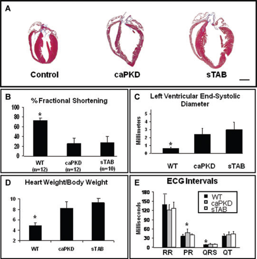
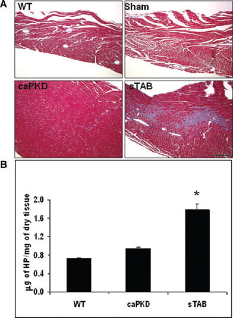
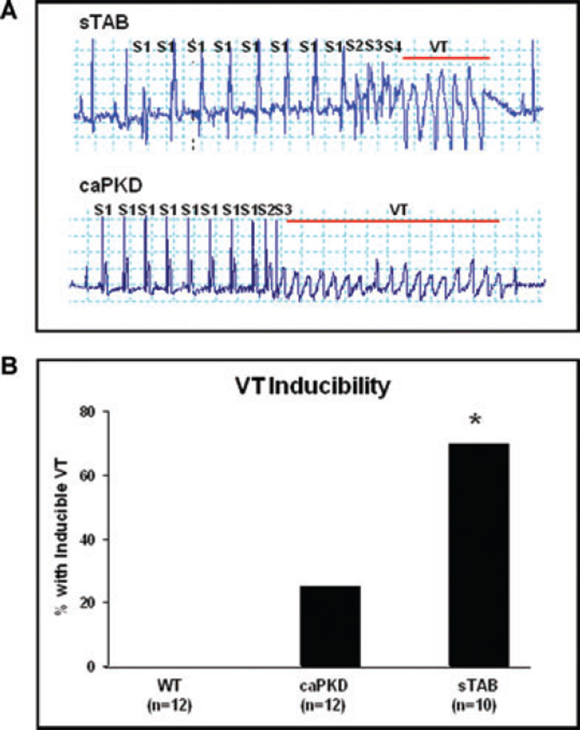
Methods: Class II histone deacetylases (HDACs) have been implicated in promoting collagen biosynthesis. As these enzymes are inhibited by protein kinase D1 (PKD1), we studied mice with cardiomyocyte-specific transgenic over-expression of a constitutively active mutant of PKD1 (caPKD). caPKD mice were compared with animals in which cardiomyopathy was induced by severe thoracic aortic banding (sTAB). Hearts were analyzed by echocardiographic and electrocardiographic means. Interstitial fibrosis was assessed by histology and quantified biochemically. Ventricular arrhythmias were induced by closed-chest, intracardiac pacing.
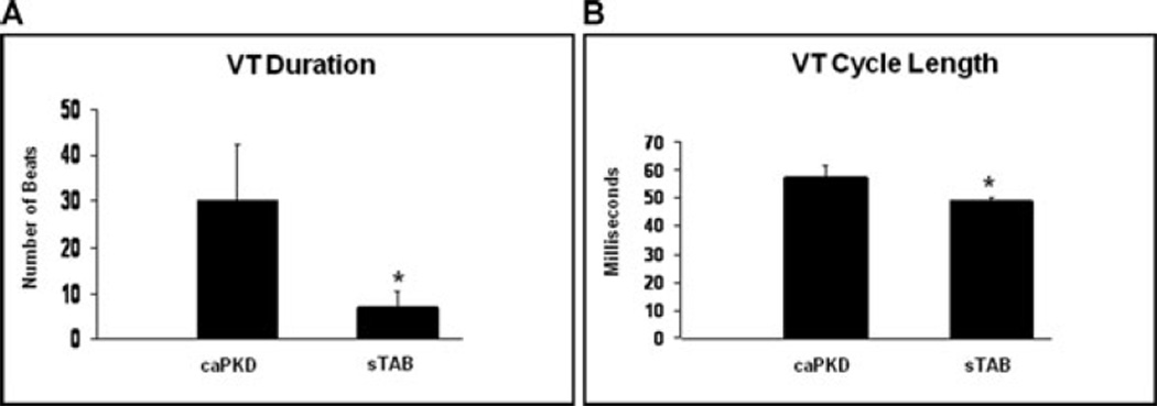
Results: Similar degrees of hypertrophic growth, systolic dysfunction and mortality were observed in the two models. In sTAB mice, robust ventricular fibrosis was readily detected, but myocardial collagen content was significantly reduced in caPKD mice. As expected, VT was readily inducible by programmed stimulation in sTAB mice and VT was less inducible in caPKD mice. Surprisingly, episodes of VT manifested longer cycle lengths and longer duration in caPKD mice.
Conclusion: Attenuated ventricular fibrosis is associated with reduced VT inducibility, increased VT duration, and significantly longer arrhythmia cycle length.
© 2010 Wiley Periodicals, Inc.
Figures
Similar articles
-
Phosphoinositide 3-kinase p110α is a master regulator of exercise-induced cardioprotection and PI3K gene therapy rescues cardiac dysfunction.Circ Heart Fail. 2012 Jul 1;5(4):523-34. doi: 10.1161/CIRCHEARTFAILURE.112.966622. Epub 2012 Jun 15. Circ Heart Fail. 2012. PMID: 22705768
-
Inducible NO synthase is constitutively expressed in porcine myocardium and its level decreases along with tachycardia-induced heart failure.Cardiovasc Pathol. 2016 Jan-Feb;25(1):3-11. doi: 10.1016/j.carpath.2015.08.003. Epub 2015 Aug 12. Cardiovasc Pathol. 2016. PMID: 26361649
-
Determinants of ventricular arrhythmias in human explanted hearts with dilated cardiomyopathy.Eur J Clin Invest. 2015 Dec;45(12):1286-96. doi: 10.1111/eci.12549. Epub 2015 Nov 18. Eur J Clin Invest. 2015. PMID: 26444674
-
Why Clinicians Should Care About the Cardiac Interstitium.JACC Cardiovasc Imaging. 2019 Nov;12(11 Pt 2):2305-2318. doi: 10.1016/j.jcmg.2019.04.025. Epub 2019 Aug 14. JACC Cardiovasc Imaging. 2019. PMID: 31422140 Review.
-
Electrical Ventricular Remodeling in Dilated Cardiomyopathy.Cells. 2021 Oct 15;10(10):2767. doi: 10.3390/cells10102767. Cells. 2021. PMID: 34685747 Free PMC article. Review.
Cited by
-
Oxidative stress, fibrosis, and early afterdepolarization-mediated cardiac arrhythmias.Front Physiol. 2013 Feb 15;4:19. doi: 10.3389/fphys.2013.00019. eCollection 2013. Front Physiol. 2013. PMID: 23423152 Free PMC article.
-
Pathological ventricular remodeling: mechanisms: part 1 of 2.Circulation. 2013 Jul 23;128(4):388-400. doi: 10.1161/CIRCULATIONAHA.113.001878. Circulation. 2013. PMID: 23877061 Free PMC article. Review.
-
Effect of angiotensin-converting enzyme inhibitors and receptor blockers on appropriate implantable cardiac defibrillator shock in patients with severe systolic heart failure (from the GRADE Multicenter Study).Am J Cardiol. 2015 Apr 1;115(7):924-31. doi: 10.1016/j.amjcard.2015.01.020. Epub 2015 Jan 15. Am J Cardiol. 2015. PMID: 25682436 Free PMC article. Clinical Trial.
-
Effect of spironolactone on patients with atrial fibrillation and structural heart disease.Clin Cardiol. 2011 Jul;34(7):415-9. doi: 10.1002/clc.20914. Epub 2011 Jun 14. Clin Cardiol. 2011. PMID: 21674535 Free PMC article.
-
Assessment of Remote Myocardium Heterogeneity in Patients with Ventricular Tachycardia Using Texture Analysis of Late Iodine Enhancement (LIE) Cardiac Computed Tomography (cCT) Images.Mol Imaging Biol. 2018 Oct;20(5):816-825. doi: 10.1007/s11307-018-1175-1. Mol Imaging Biol. 2018. PMID: 29536321 Free PMC article.
References
-
- Hill JA, Olson EN. Cardiac plasticity. N Engl J Med. 2008;358:1370–1380. - PubMed
-
- Frey N, Olson EN. Cardiac hypertrophy: The good, the bad, and the ugly. Annu Rev Physiol. 2003;65:45–79. - PubMed
-
- Nass RD, Aiba T, Tomaselli GF, Akar FG. Mechanisms of disease: Ion channel remodeling in the failing ventricle. Nat Clin Pract Cardiovas Med. 2008;5:196–207. - PubMed
-
- Spinale FG. Myocardial matrix remodeling and the matrix metal-loproteinases: Influence on cardiac form and function. Physiol Rev. 2007;87:1285–1342. - PubMed
-
- Tomaselli GF, Zipes DP. What causes sudden death in heart failure? Circ Res. 2004;95:754–763. - PubMed
Publication types
MeSH terms
Substances
Grants and funding
LinkOut - more resources
Full Text Sources
Medical
Research Materials
