Comparative Study
. 2010 Jul;16(5):453-64.
doi: 10.1111/j.1601-0825.2010.01656.x. Epub 2010 Mar 9.

Expression and distribution of SIBLING proteins in the predentin/dentin and mandible of hyp mice

Affiliations
Comparative Study

Expression and distribution of SIBLING proteins in the predentin/dentin and mandible of hyp mice

B Zhang et al. Oral Dis. 2010 Jul.

Abstract

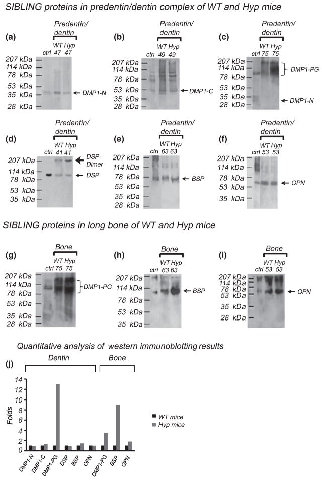
Objectives: Human X-linked hypophosphatemia (XLH) and its murine homologue, Hyp are caused by inactivating mutations in PHEX gene. The protein encoded by PHEX gene is an endopeptidase whose physiological substrate(s) has not been identified. Dentin matrix protein 1 (DMP1) and dentin sialophosphoprotein (DSPP), two members of the Small Integrin-Binding LIgand, N-linked Glycoprotein (SIBLING) family are proteolytically processed. It has been speculated that PHEX endopeptidase may be responsible for the proteolytic cleavage of DMP1 and DSPP. To test this hypothesis and to analyse the distribution of SIBLING proteins in the predentin/dentin complex and mandible of Hyp mice, we compared the expression of four SIBLING proteins, DMP1, DSPP, bone sialoprotein (BSP) and osteopontin (OPN) between Hyp and wild-type mice.

Methods: These SIBLING proteins were analysed by protein chemistry and immunohistochemistry.

Results: (1) Dentin matrix protein 1 and DSPP fragments are present in the extracts of Hyp predentin/dentin and bone; (2) the level of DMP1 proteoglycan form, BSP and OPN is elevated in the Hyp bone.

Conclusions: The PHEX protein is not the enzyme responsible for the proteolytic processing of DMP1 and DSPP. The altered distribution of SIBLING proteins may be involved in the pathogenesis of bone and dentin defects in Hyp and XLH.


Figures

Figure 1
Figure 1
Chromatographic separation of non-collagenous proteins (NCPs) extracted from the predentin/dentin complex of WT (a) and Hyp (b) mice. NCPs in the predentin/dentin complex of the WT and Hyp mice were extracted by 4 M Gdm-HCl/0.5 M EDTA. The Q-Sepharose column separated NCPs from the predentin/dentin complex of the WT (a) and Hyp mice (b) into 120 fractions. Each fraction contained 0.5 ml of 6 M urea solution. Note that the separation profile for the NCPs from the WT mice was similar, but not identical to that from the Hyp mice. The slight differences in the elution profile between the WT and Hyp mice might be due to the fact that the Hyp mice had more predentin, and thus might contain greater amounts of proteoglycans including DMP1-PG which eluted in later fractions (after fraction 65)
Figure 2
Figure 2
Stains-All staining for chromatographic fractions 41–85 from the predentin/dentin complex of the WT (a) and Hyp (b) mice. Digits at the top of each figure represent fraction numbers of the Q-Sepharose chromatography. MW refers to molecular weight markers. The blue protein bands around 57 and 37 kDa in fractions 45–57 (more prominent in fraction 49) represent DMP1 C- and DMP1 N-terminal fragments respectively. The blue-stained, broad protein bands in fractions 51–57 migrating between the 78 and 114 kDa molecular weight markers represent DPP. The light-blue protein bands in fractions 41–47, migrating at ~100 kDa (close to the 114 kDa molecular weight marker), represent DSP. BSP, migrating just above the 78 kDa molecular weight marker, eluted in fractions 61–85. Minor amounts of DPP co-eluted with BSP in the fractions 61–69; the protein bands between the 78 and 114 kDa molecular weight markers in these fractions represent a mixture of DPP and BSP. The identity of DMP1 fragments, DSP and BSP was confirmed by Western immunoblotting analyses; Western immunoblotting results for a representative chromatographic fraction for each of these molecules are shown in Figure 3
Figure 3
Figure 3
Western immunoblotting for the SIBLING family members in the extracts from the predentin/dentin complex and the long bone of WT and Hyp mice. A representative fraction is shown, to illustrate the Western immunoblotting results for the SIBLING members. As appropriate antibodies against DPP are not available, we could not carry out Western immunoblotting for DPP. (af) Results for the SIBLING members extracted from the predentin/dentin complex. (gi) Results for the SIBLING members extracted from the long bone. (a, c) Western immunoblotting using the anti-DMP1-N-terminal-9B6.3 monoclonal Ab. Positive control (Ctrl): 1 μg of the N-terminal fragment of DMP1. The core protein of the DMP1-N terminal fragment (37 kDa) (a) and DMP1-PG (c) were detected by the anti-DMP1-N-terminal-9B6.3 Ab. Although there was no difference for the core protein of the DMP1 N-terminal fragment (37 kDa) between the WT and Hyp mice, DMP1-PG was more abundant in the predentin/dentin of the Hyp mice. (b) Western immunoblotting using the anti-DMP1-C-terminal-857 polyclonal Ab. Ctrl: 1 μg of COOH-terminal fragment of DMP1. Note significant difference in the quantity was observed for the DMP1 C-terminal fragment between the WT and Hyp mice. (d) Western immunoblotting using the anti-DSP polyclonal Ab. Ctrl: 0.5 μg of DSP isolated from rat dentin. The expression level of DSP in the dentin of the Hyp mice was similar to WT mice. (e) Western immunoblotting using the anti-BSP-10D9.3 monoclonal Ab. Ctrl: 0.5 μg of BSP isolated from rat long bone. The quantity of BSP in the predentin/dentin of Hyp mice was similar to WT mice. (f) Western immunoblotting using the anti-OPN monoclonal Ab. Ctrl: 1 μg of OPN isolated from rat long bone. The quantity of OPN in the predentin/dentin of Hyp mice was similar to WT mice. (g) Western immunoblotting using the anti-DMP1-N-terminal-9B6.3 monoclonal Ab. Ctrl: 1 μg of DMP1 isolated from the rat long bone. The extracellular matrix (ECM) of the long bone of the Hyp mice had more DMP1-PG than in the WT mice. (h) Western immunoblotting using the anti-BSP-10D9.3 monoclonal Ab. Positive control (Ctrl): 0.5 μg of BSP isolated from the rat long bone. The ECM of the long bone of the Hyp mice had more BSP than the WT mice. (i) Western immunoblotting using the anti-OPN monoclonal Ab. Ctrl: 1 μg of OPN isolated from the rat long bone. The ECM of the long bone of the Hyp mice had more OPN than the WT mice. (j) Quantitative analyses of Western immunoblotting results for SIBLING proteins extracted from the dentin and bone of Hyp and WT mice. For normalization, the quantity of any of the SIBLING proteins from the WT was set as 1, while the quantity of each protein from the Hyp mice was expressed as relative folds to that from the WT mice. The amount of DMP1-PG in the Hyp mouse dentin was more than 12-fold greater than the WT mice. Also note that there was more than eight times of BSP in the Hyp mouse bone than in the WT. The data represent calculations from three separate Western immunoblots that agreed closely
Figure 4
Figure 4
H&E staining for the predentin/dentin complex and the mandible from 5-week-old WT mice and 5, 10, 15-week-old Hyp mice. Column 1, H&E staining for the mandible at a lower magnification; column 2, H&E staining for the predentin/dentin complex; column 3, H&E staining for the mandible at a higher magnification. ac, 5-week-old WT mice; df, 5-week-old Hyp mice; gi, 10-week-old Hyp mice; jl, 15-week-old Hyp mice. The two arrows (two bars) in figure b, e, h, and k were used to indicate the width of predentin. In comparison with the WT mice, the predentin of Hyp mice was wider (compare b with e, h, and k). The mandible of Hyp mice contained greater number of osteocytes and more osteoid (arrowheads) than the WT mice (compare c with f, i and l). (m) Quantity analyses to compare the width of predentin (H&E staining) between the WT and Hyp mice. For normalization, the width of predentin of the WT mice was set as 1; the predentin width of the Hyp mice was expressed as relative folds to that of the WT mice. Note that the predentin of the Hyp mouse was approximately three times wider than the WT mice. The results were obtained from three specimens of three animals, and for each specimen, the predentin width was measured at five points and an average was calculated
Figure 5
Figure 5
Immunohistochemical (IHC) staining for the predentin/dentin complex and the mandible of 5-week-old WT and Hyp mice. Column 1: predentin/dentin of 5-week-old WT mice; column 2, predentin/dentin of 5-week-old Hyp mice; column 3, mandible of 5-week-old WT mice; column 4, mandible of 5-week-old Hyp mice. (ad) IHC for the DMP1 N-terminal; eh, IHC for the DMP1 C-terminal; il, IHC for DSP; mp, IHC for BSP; qt, IHC for OPN. Red arrows were used to indicate a relatively weak IHC staining and black arrows were used for a relatively strong IHC staining. In the predentin/dentin complex of both the WT and Hyp mice, the signal for the DMP1 N-terminal was mainly observed in the predentin (PD in a, b) whereas the DMP1 C-terminal was primarily located in the dentin (e, f). In the mandible, a relatively strong signal for the DMP1 N-terminal was observed in the lower border region of the mandible in the Hyp mice while the signal for the N-terminal was generally weak in the mandible of the WT mice (compare c with d). In the Hyp mice, the signal for the N-terminal was also strong in the osteoid or osteoid-like tissues in the sponge bone regions of the alveolar bone. In the WT mice, the signal for the DMP1 C-terminal was strong in the lower border region (cortical bone) of the mandible, but was weak in the spongy bone area (g). In the Hyp mice, the staining for the DMP1 C-terminal was weak in the lower border region of the mandible (h) in which the signal for the N-terminal fragment was strong. The distribution of DSP was similar in the predentin and dentin between the WT and Hyp mice (i, j). In the alveolar bone of the Hyp mice, DSP was more abundant in the osteoid or osteoid-like regions surrounding osteocytes (k, l), and appeared more abundant in the Hyp than in the WT mice. In the tooth, a relatively strong signal for BSP was observed in the cementum (c) in the WT and Hyp mice (m, n). The IHC staining for BSP was stronger in the mandible of the Hyp mice than in the WT mice, especially in the lower border region of the mandible of the Hyp mice (o, p) in which a strong signal for the DMP1 N-terminal was seen. In the tooth, the signal for OPN was strong in the cementum of both the WT and Hyp mice (q, r). The IHC staining for OPN in the alveolar bone of Hyp mice was stronger than in the WT mice. OPN was mainly observed in the areas containing the newly formed bone in the WT mice, but in the Hyp mice, the whole areas of the alveolar bone, especially the osteoid/osteoid-like regions surrounding the osteocytes, displayed strong IHC staining (s, t)

Similar articles

Cited by

References

    1. Abe K, Ooshima T, Tong SML, Yasufuku Y, Sobue S. Structural deformities of deciduous dentin in patients with hypophosphatemic vitamin D-resistant rickets. Oral Surg Oral Med Oral Pathol. 1988;65:191–198. - PubMed
    1. Abe K, Ooshima T, Masatomi Y, Sobue S, Moriwaki Y. Microscopic and crystallographic examinations of the dentin of the X-linked hypophosphatemic mouse. J Dent Res. 1989;68:1519–1524. - PubMed
    1. Baba O, Qin C, Brunn JC, et al. Detection of dentin sialoprotein in rat periodontium. Eur J Oral Sci. 2004a;112:163–170. - PubMed
    1. Baba O, Qin C, Brunn JC, Wygant JN, Mcintyre BW, Butler WT. Colocalization of dentin matrix protein 1 and dentin sialoprotein at late stages of rat molar development. Matrix Biol. 2004b;23:371–379. - PubMed
    1. Baroncelli GI, Angiolini M, Ninni E, Galli V, Saggese R, Giuca MR. Prevalence and pathogenesis of dental and periodontal lesions in children with X-linked hypophosphatemic rickets. Eur J Paediatr Dent. 2006;7:61–66. - PubMed

Publication types

MeSH terms