. 2004 Nov 15;2(4):161-73.
doi: 10.1186/1897-4287-2-4-161.

Multiple osteochondromas: clinicopathological and genetic spectrum and suggestions for clinical management

Affiliations

Multiple osteochondromas: clinicopathological and genetic spectrum and suggestions for clinical management

Liesbeth Hameetman et al. Hered Cancer Clin Pract. .

Abstract

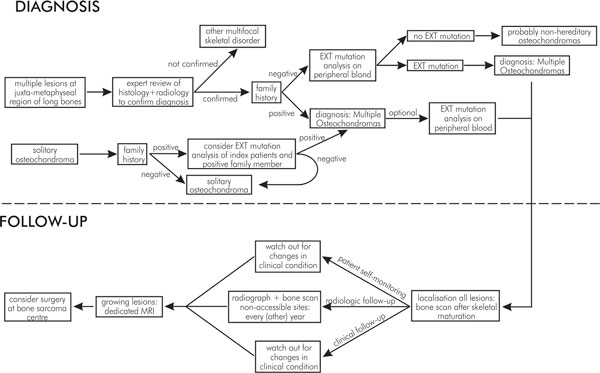
Multiple Osteochondromas is an autosomal dominant disorder characterised by the presence of multiple osteochondromas and a variety of orthopaedic deformities. Two genes causative of Multiple Osteochondromas, Exostosin-1 (EXT1) and Exostosin-2 (EXT2), have been identified, which act as tumour suppressor genes. Osteochondroma can progress towards its malignant counterpart, secondary peripheral chondrosarcoma and therefore adequate follow-up of Multiple Osteochondroma patients is important in order to detect malignant transformation early.This review summarizes the considerable recent basic scientific and clinical understanding resulting in a multi-step genetic model for peripheral cartilaginous tumorigenesis. This enabled us to suggest guidelines for clinical management of Multiple Osteochondroma patients. When a patient is suspected to have Multiple Osteochondroma, the radiologic documentation, histology and patient history have to be carefully reviewed, preferably by experts and if indicated for Multiple Osteochondromas, peripheral blood of the patient can be screened for germline mutations in either EXT1 or EXT2. After the Multiple Osteochondroma diagnosis is established and all tumours are identified, a regular follow-up including plain radiographs and base-line bone scan are recommended.


Figures

Figure 2
Figure 2
Mutation spectrum of the EXT1 and EXT2 genes in MO patients described so far [106].
Figure 3
Figure 3
Genomic DNA structures of the EXT1 and EXT2 genes.
Figure 4
Figure 4
The mode of action of the EXT-proteins in heparan sulphate biosynthesis. After a tetrasaccharide linker is synthesised on conserved serine residues of the core protein, EXTL2 and/or EXTL3 initiate the polymerisation of the heparan sulphate chain by the addition of N-acetylglucosamine [60,107]. The EXT1/EXT2 complex subsequently catalyses further elongation of the heparan sulphate chain by adding alternating units of N-acetylglucosamine and glucuronic acid [59-62]. Subsequent deacetylation and sulphation of most N-acetylglucosamines, epimerisation of the glucoronic into iduronic acid and further sulphation result in a large spectrum of structural heterogenic heparan sulphate chains [58,108]. Adapted from Couchman et al [109] and Nybakken et al [110].
Figure 5
Figure 5
Growth plate signalling. EXT1 and EXT2 are expressed in the proliferative and transition zone [77] (Fig. 5). The HSPGs, expressed in all zones of the growth plate [67-72]. A. In the embryonic growth plate chondrocytes in the transition zone secrete IHh protein, which diffuses to its receptor Patched (Ptc) in the lateral perichondrium. Subsequently, via a yet incompletely understood mechanism, increased secretion of ParaThyroid Hormone related Protein (PTHrP, PTHlP) is induced at the apical perichondrium, which diffuses to its receptor expressed in the late proliferating chondrocytes [80]. Terminal differentiation is inhibited by direct or indirect upregulation of Bcl-2, prolonging cell survival [78]. In this way, PTHrP regulates chondrocyte differentiation by delaying the progression of chondrocytes towards the hypertrophic zone and allowing longitudinal bone growth. B. In the post-natal growth plate the signalling is confined to the growth plate [79].
Figure 6
Figure 6
Peripheral Cartilaginous Tumorigenesis.
Figure 7
Figure 7
Overview of systematic steps to screen and follow-up (suspected) Multiple Osteochondroma patients.
Figure 1
Figure 1
Specimen radiographs and histology. A pedunculated osteochondroma shown in a macroscopic whole mount section (A) and specimen radiograph (B); C, whole mount section of a sessile osteochondroma. Note the presence of a small cartilage cap in both osteochondromas (< 0.5 cm); D, radiograph of the forearm of a Multiple Osteochondroma patient. Several osteochondromas can be seen at the ends of the ulna and radius. Note that the ulna is shortened, which caused subsequent bowing of the radius; E and F, gross specimen and whole mount section of secondary peripheral chondrosarcoma. The cartilage cap is thicker than 2 cm and in the whole mount section the lobules are clearly visible.

Similar articles

Cited by

References

    1. Mulder JD, Schütte HE, Kroon HM, Taconis WK. Radiologic Atlas of Bone Tumors. 2. Elsevier, Amsterdam; 1993.
    1. Bovee JVMG, Hogendoorn PCW. In: World Health Organization Classification of Tumours. Pathology and Genetics of Tumours of Soft Tissue and Bone. Fletcher CDM, Unni KK, Mertens F, editor. IARC Press, Lyon; 2002. Multiple osteochondromas.
    1. Dahlin's Bone Tumors General Aspects and Data on 11,087 Cases. 5. Lippincott-Raven Publishers, Philadelphia; 1996.
    1. Legeai-Mallet L, Munnich A, Maroteaux P, Le Merrer M. Incomplete penetrance and expressivity skewing in hereditary multiple exostoses. Clin Genet. 1997;52:12–16. - PubMed
    1. Schmale GA, Conrad EU, Raskind WH. The natural history of hereditary multiple exostoses. J Bone Joint Surg [Am] 1994;76A:986–992. - PubMed

LinkOut - more resources