. 2010 Jun;88(3):341-52.
doi: 10.1016/j.yexmp.2010.03.006. Epub 2010 Mar 15.

Northwestern profiling of potential translation-regulatory proteins in human breast epithelial cells and malignant breast tissues: evidence for pathological activation of the IGF1R IRES

Affiliations

Northwestern profiling of potential translation-regulatory proteins in human breast epithelial cells and malignant breast tissues: evidence for pathological activation of the IGF1R IRES

Scott W Blume et al. Exp Mol Pathol. 2010 Jun.

Abstract

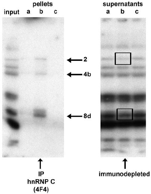
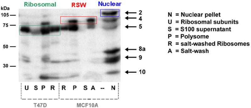
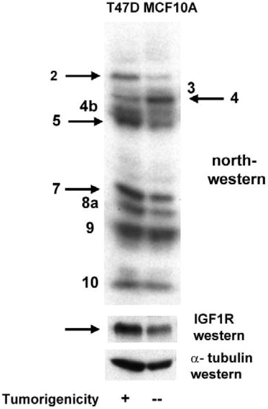
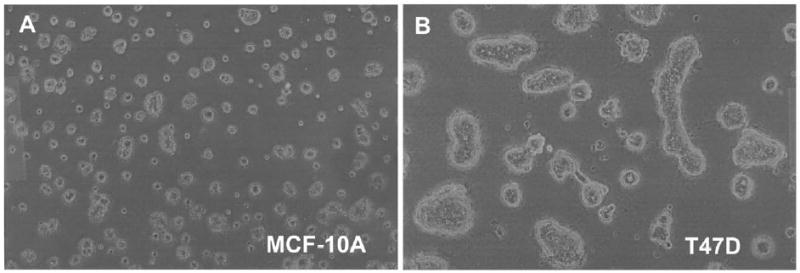
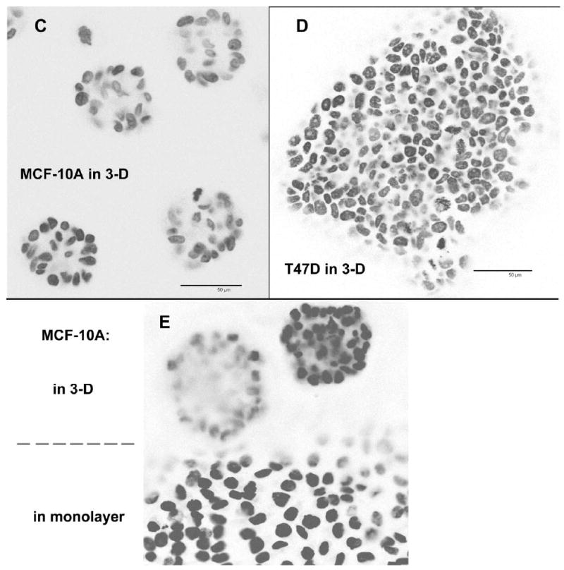
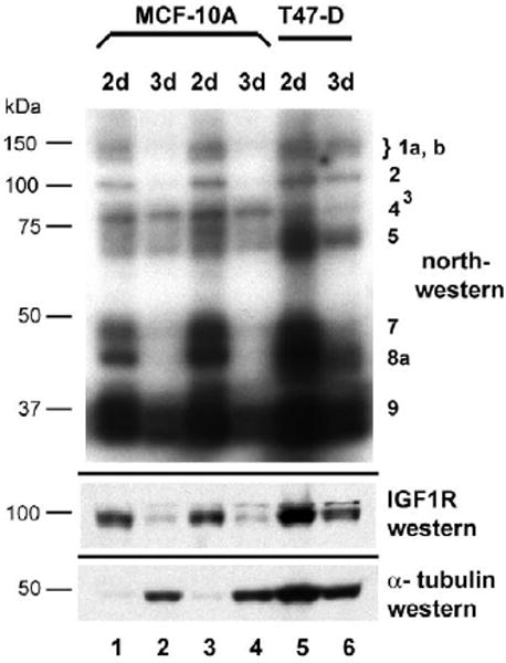
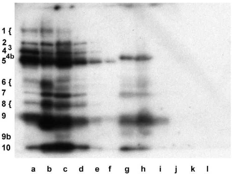
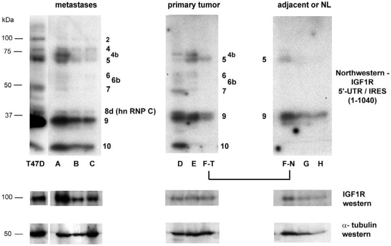
Genes involved in the control of cell proliferation and survival (those genes most important to cancer pathogenesis) are often specifically regulated at the translational level, through RNA-protein interactions involving the 5'-untranslated region of the mRNA. IGF1R is a proto-oncogene strongly implicated in human breast cancer, promoting survival and proliferation of tumor cells, as well as metastasis and chemoresistance. Our lab has focused on the molecular mechanisms regulating IGF1R expression at the translational level. We previously discovered an internal ribosome entry site (IRES) within the 5'-untranslated region of the human IGF1R mRNA, and identified and functionally characterized two individual RNA-binding proteins, HuR and hnRNP C, which bind the IGF1R 5'-UTR and differentially regulate IRES activity. Here we have developed and implemented a high-resolution northwestern profiling strategy to characterize, as a group, the full spectrum of sequence-specific RNA-binding proteins potentially regulating IGF1R translational efficiency through interaction with the 5'-untranslated sequence. The putative IGF1R IRES trans-activating factors (ITAFs) are a heterogeneous group of RNA-binding proteins including hnRNPs originating in the nucleus as well as factors tightly associated with ribosomes in the cytoplasm. The IGF1R ITAFs can be categorized into three distinct groups: (a) high molecular weight external ITAFs, which likely modulate the overall conformation of the 5'-untranslated region of the IGF1R mRNA and thereby the accessibility of the core functional IRES; (b) low molecular weight external ITAFs, which may function as general chaperones to unwind the RNA, and (c) internal ITAFs which may directly facilitate or inhibit the fundamental process of ribosome recruitment to the IRES. We observe dramatic changes in the northwestern profile of non-malignant breast cells downregulating IGF1R expression in association with acinar differentiation in 3-D culture. Most importantly, we are able to assess the RNA-binding activities of these putative translation-regulatory proteins in primary human breast surgical specimens, and begin to discern positive correlations between individual ITAFs and the malignant phenotype. Together with our previous findings, these new data provide further evidence that pathological dysregulation of IGF1R translational control may contribute to development and progression of human breast cancer, and breast metastasis in particular.


Conflict of interest statement

Conflict of interest statement The authors declare that there are no conflicts of interest.

Figures

Fig 1
Fig 1. IGF1R translational regulation: Architecture of the 5’-untranslated region of the human IGF1R mRNA
The structural and regulatory features contained within the 5’-untranslated region of the human IGF1R mRNA are indicated (drawn to scale, coordinates refer to distance in nucleotides). The conventional mechanism for translation initiation in eukaryotes involves a process of scanning by the 40S ribosomal subunit from the beginning of the mRNA (blue arrow) to the initiation codon. The complex IGF1R 5’-UTR presents multiple obstacles to ribosomal scanning. Domains 2 and 3 are highly stable secondary structures with individual free energies of -98.4 and -159.4 kCal/mol and a combined free energy of -257.8 kCal/mol. (For reference, a structure with dG > -60 kCal / mol is considered a significant obstacle to ribosomal scanning.) The upstream open reading frame (uORF) encodes a 28 amino acid peptide with no relationship to IGF1R. The uORF initiation codon is in good Kozak consensus, thus scanning ribosomes which reach this point will tend to initiate translation (upon joining of the 60S ribosomal subunit), and then dissociate from the mRNA upon reaching the uORF termination codon (red arrow). Ribosome dissociation is facilitated by the G-C rich element which immediately follows the uORF termination codon. We have referred to this G-C rich segment (56 out of 59 residues are G or C) as the “third obstacle”. Finally, however, there exists an internal ribosomal entry site (IRES, nucleotides 951 – 1040) positioned just upstream of the authentic IGF1R initiation codon. The IRES provides an alternative mechanism for IGF1R translation initiation, recruiting the 40S ribosomal subunit directly to the vicinity of the AUG (green arrow), allowing it to bypass the obstacles presented by the remainder of the 5’-UTR. Importantly, the IRES is not constitutively active, but rather is specifically regulated through competitive and cooperative interactions with multiple RNA-binding proteins recognizing specific sequence elements within or outside of the IGF1R IRES. The RNA-binding proteins regulating IGF1R translation are the subject of this investigation.
Fig. 2
Fig. 2. Characterization of Sequence-specific RNA-binding proteins by the Northwestern assay: Development of a protocol for optimal recovery of functional RNA-binding proteins from cells
A: Northwestern assay evaluating recovery of sequence-specific RNA-binding proteins from the nucleus. Following hypotonic lysis of T47D breast tumor cells in the presence of 0.25% NP-40, pelleted nuclei were resuspended in an extraction buffer containing various concentrations of NaCl (lanes a through c). For lanes d through f, nuclei were incubated with DNase I and / or micrococcal nuclease prior to 500 mM NaCl. For lanes g through i, nuclear remnants following extraction with 150, 500, or 1000 mM NaCl respectively were resuspended and extracted with additional detergents: T-PER (Pierce), 1% Triton X-100/ 0.5% deoxycholate/ 0.1% SDS, or 50% T-PER/ 1% SDS respectively. Numerical designations for the proteins interacting specifically with the IGF1R 5’-untranslated RNA are indicated to the left. Note that the majority of bands detected by IGF1R northwestern required at minimum 500 mM NaCl for efficient extraction from the nucleus, while several individual RNA-binding proteins (particularly Bands 4b and 8a, see arrows) required additional NaCl, nuclease digestion, and/or supplemental detergent for efficient recovery. B: Cytoplasmic lysates obtained by hypotonic lysis of T47D cells were incubated with 500 mM NaCl and / or micrococcal nuclease, and then analyzed by northwestern. Note that the same manipulations which enhance recovery of RNA-binding proteins from the nucleus result in loss of RNA-binding activity in the cytoplasm. This phenomenon, which we refer to as “cytoplasmic susceptibility”, may be explained by targeted inactivation of RNA-binding translation-regulatory proteins once they become dissociated from their intended target sequences in the cytoplasm.
Fig. 3
Fig. 3. Sequence-specificity of RNA-binding proteins regulating IGF1R translation: internal and external ITAFs
Northwestern assays were performed to assess the sequence-specificity of the RNA-protein interactions taking place on the 5’-untranslated region of the IGF1R mRNA. Three different RNA probes were synthesized: the full length 5’-UTR (nucleotides 1 – 1040, panels A and B), the isolated 90 nucleotide sequence (nucleotides 951-1040) to which the core functional IRES has been delimited (panel C), and the 5’-untranslated sequence from which the core functional IRES sequence has been deleted (nucleotides 1-959, panel D). Distinct patterns of protein binding are observed for the core IRES and the remainder of the 5’-UTR, while the pattern of protein binding to the full-length 5’-UTR (including the IRES) is a composite of those two series of bands. Pottential translation-regulatory proteins binding within the IGF1R core functional IRES are designated “internal” ITAFs (IRES trans-acting factors), while those binding 5’-untranslated sequence outside the core functional IRES are referred to as “external” ITAFs. The relative affinity or accessibility for the internal versus the external ITAFs can be modulated by varying the magnesium concentration (and thereby the level of secondary structure of the RNA). Molecular weight markers are shown on the left, and the positions of the IGF1R ITAFs are indicated on the right. Whole cell extracts: lanes i, MCF-10A, Protocol A; lanes ii, T47D, Protocol A; lanes iii, T47D, Protocol B.
Fig. 4
Fig. 4. Characterization of IGF1R ITAFs by intermolecular association
A: T47D whole cell extract (Protocol B) was subjected to immunoprecipitation using the 4F4 monoclonal antibody (mouse IgG1) to hnRNP C, which immunoprecipitates the hnRNP complex (composed of >20 proteins). Aliquots of the input, pellet, and supernatant were separated by SDS / PAGE and analyzed by northwestern using the full-length IGF1R 5’-UTR / IRES probe. Three of the IGF1R northwestern bands are selectively recovered by 4F4 immunoprecipitation (lane b): Band 8d, which is hnRNP C itself (as confirmed by western analysis performed sequentially on the same blot), as well as Bands 2 and 4b. Significant immunodepletion of hnRNP C (8d) and Band 2 is observed in the corresponding supernatant (lane b of right panel). Lanes a and c of each panel are negative control immunoprecipitations and supernatants (no immunodepletion) obtained using antibodies to two other RNA-binding proteins (3A2 mouse monoclonal IgG1 to HuR and goat polyclonal C-18 to TIAR). The experiment was repeated with identical results B: MCF-10A and T47D cells were subjected to conventional fractionation of the cytoplasm by differential ultracentrifugation to yield an S100 supernatant (lanes S) and polysomal pellet (lanes P). Aliquots of polysomal pellets were incubated in 500 mM KCl, and centrifuged again to yield a supernatant (ribosomal salt wash, lane A) and pellet (salt-washed ribosomes, lanes R). Finally, an aliquot of salt-washed ribosomes was incubated with puromycin and 500 mM KCl, and centrifuged again to yield free ribosomal subunits (lane U). Each of these fractions was analyzed by northwestern using the IGF1R IRES probe. Note that Bands 2 and 8a are exclusively nuclear, Band 5 is tightly associated with ribosomes, while Band 4 is quantitatively recovered in the ribosomal salt wash. A proportion of Bands 9 and 10 are also tightly associated with the ribosomes.
Fig. 5
Fig. 5. Distinctions in the pattern of protein binding to the IGF1R 5’-UTR / IRES between non-malignant and malignant cultured breast cells
Whole cell extracts (Protocol B) of T47D (malignant) or MCF-10A (non-malignant) breast epithelial cells were prepared and subjected to northwestern analysis (4 × 10⁵ cell equivalents per lane) using the IGF1R 5’-UTR / IRES probe. Though the patterns are generally quite similar, certain bands exhibit significant differences in relative intensity (RNA-binding activity). In particular, Bands 2, 5, and 7 are considerably more intense in T47D, correlating with the substantially higher IRES activity (Meng et al, 2005) and IGF1R protein levels in these cells. Band 4 is considerably more intense in MCF-10A cells, suggesting this may represent an IRES inhibitor.
Fig. 6
Fig. 6. Alterations in RNA-binding activities of IGF1R translation-regulatory proteins accompanying differentiation of non-malignant breast epithelial cells in 3-D culture
A, B: Single-cell suspensions of MCF-10A and T47D cells were embedded in Matrigel in 35 mm dishes and allowed to proliferate or differentiate for 15 days. Representative phase contrast micrographs reveal the distinction between the small, relatively uniform acinar structures formed by the non-transformed MCF-10A breast epithelial cells versus the large, amorphous clusters formed by the T47D breast tumor cells. C, D: MCF-10A and T47D cells were also plated on the surface of a thin layer of Matrigel on 8-well chamber slides to facilitate examination by confocal microscopy. Following fixation, nuclei were stained with DAPI, and a series of confocal images was captured demonstrating the organized morphology of the MCF-10A acinar structures versus the disorganized clusters of T47D cells. The immunofluorescent micrographs are shown in reverse image (nuclei are dark). Bar = 50 μm. E: MCF-10A cells were seeded on an 8-well chamber slide in which Matrigel had been intentionally spread so as to cover only a portion of the surface of the well, forming a “beach” with the inorganic growth surface. This allowed us to capture, in a single image, acinar structures on the surface of the Matrigel and a continuously proliferating monolayer on the adjacent uncoated portion of the slide. This illustrates the capacity of the non-transformed MCF-10A cells to differentiate and cease dividing when provided the appropriate environmental context, versus indefinite proliferation in standard monolayer culture. F: Northwestern analysis revealing changes in the pattern of protein binding to the IGF1R 5’-UTR / IRES accompanying differentiation of non-malignant MCF-10A breast epithelial cells in 3-D culture. MCF-10A and T47D cells were recovered from 3-D culture following dissolution of the matrix (see Materials and Methods) and whole cell extracts prepared (Protocol A). Extracts were also prepared in parallel from cells harvested from standard monolayer culture (2-D). Equal numbers of cell equivalents of these extracts were analyzed by northwestern using the IGF1R 5’-UTR / IRES probe. Results of western analyses for IGF1R (C-20 rabbit polyclonal, Santa Cruz) and alpha tubulin (B-5-1-2, Sigma) performed sequentially on the northwestern blot are shown below.
Fig. 7
Fig. 7. Northwestern analysis of sequence-specific translation-regulatory proteins binding to the IGF1R 5’-UTR in primary human breast tumors, metastases, and normal tissues
A: A primary human breast tumor specimen (poorly differentiated ductal carcinoma, specimen D, Table 1) was cryosectioned and subjected to varying degrees of processing to enable laser microdissection. Processed materials were then recovered from microdissection cap or slides and subjected to whole cell extraction (Protocol H) before performing northwestern analysis. Lane g: cryosections washed in water and immediately recovered from slides and transferred to lysis buffer (containing protease and phosphatase inhibitors). Lane h: cryosections (still in OCT) directly transferred to lysis buffer with no histological processing. Lane i: Sections stained with hematoxylin (standard protocol, but omitting final xylene rinse), then immediately recovered from slides and transferred to lysis buffer. Lane j: Sections stained with hematoxylin including final xylene rinse. Lane k: Sections stained with both hematoxylin and eosin. Lane l: Section stained with H&E and subjected to laser microdissection, with captured malignant epithelial cells selectively transferred to lysis buffer. Reference samples: Lane a: HeLa nuclear extract; Lane b: K-1 (T47D derivative) Protocol H whole cell extract; Lane c: T47D Protocol H whole cell extract (2.5 × 10⁵ cell equivalents); Lane d: same as Lane c, one-fourth sample loaded; Lane e: same as Lane c, one-tenth of sample loaded; Lane f: same as Lane c, one-twenty-fifth of sample loaded. B: Human breast surgical specimens (snap frozen or embedded in OCT) were examined by H&E staining to assess distribution of epithelial cells and guide gross microdissection as appropriate. Relative proportion of malignant breast epithelial cells within each tumor sample ranged from ~85 to near 100%. Consecutive cryosections (unprocessed) were then transferred directly to lysis buffer and used for preparation of whole cell extract (Protocol H). Equivalent aliquots were subjected to northwestern analysis using the full-length IGF1R 5’-UTR / IRES probe. Specimens A through C are metastatic lesions of breast tumor origin. Specimens D through F(T) are primary invasive breast tumors. Specimen F(N) is adjacent histologically uninvolved tissue obtained from the same block as primary tumor F(T). Specimens G and H are normal breast tissue obtained from reduction mammoplasty. The results are representative of four replicate northwestern analyses performed on the same series of specimens. Molecular weight markers are indicated on the left, while the positions of IGF1R ITAFs are indicated adjacent to each panel. Results of western analyses for IGF1R (N-20 rabbit polyclonal, Santa Cruz) and alpha tubulin (B-5-1-2, Sigma) performed sequentially on the northwestern blot are shown below. C: Representative H&E stained sections of each of the human breast surgical specimens. Bar = 50 μm.

Similar articles

Cited by

References

    1. Abdelmohsen K, Pullmann R, Jr, Lal A, Kim HH, Galban S, Yang X, Blethrow JD, Walker M, Shubert J, Gillespie DA, Furneaux H, Gorospe M. Phosphorylation of HuR by Chk2 regulates SIRT1 expression. Mol Cell. 2007;25(4):543–557. - PMC - PubMed
    1. Baird SD, Turcotte M, Korneluk RG, Holcik M. Searching for IRES. RNA. 2006;12(10):1755–1785. - PMC - PubMed
    1. Belfiore A, Frasca F. IGF and insulin receptor signaling in breast cancer. J Mammary Gland Biol Neoplasia. 2008;13(4):381–406. - PubMed
    1. Chappell SA, LeQuesne JP, Paulin FE, deSchoolmeester ML, Stoneley M, Soutar RL, Ralston SH, Helfrich MH, Willis AE. A mutation in the c-myc-IRES leads to enhanced internal ribosome entry in multiple myeloma: a novel mechanism of oncogene de-regulation. Oncogene. 2000;19(38):4437–4440. - PubMed
    1. Chiocchetti A, Gibello L, Carando A, Aspesi A, Secco P, Garelli E, Loreni F, Angelini M, Biava A, Dahl N, Dianzani U, Ramenghi U, Santoro C, Dianzani I. Interactions between RPS19, mutated in Diamond-Blackfan anemia, and the PIM-1 oncoprotein. Haematologica. 2005 Nov;90(11):1453–62. - PubMed

Publication types

MeSH terms