

Review
doi: 10.1102/1470-7330.2010.0009.
Imaging of leiomyosarcoma of the inferior vena cava: comparison of 2 cases and review of the literature
Affiliations
- PMID: 20233678
- PMCID: PMC2842181
- DOI: 10.1102/1470-7330.2010.0009
Item in Clipboard
Review
Imaging of leiomyosarcoma of the inferior vena cava: comparison of 2 cases and review of the literature
Cancer Imaging.
.
Abstract
Leiomyosarcoma of the inferior vena cava is a rare tumour arising from the smooth muscle fibres of the media with a mean size at diagnosis generally around 12 cm (range 2-38 cm). This study compares a 4-cm leiomyosarcoma of the inferior vena cava discovered incidentally with a symptomatic late stage leiomyosarcoma.
Figures
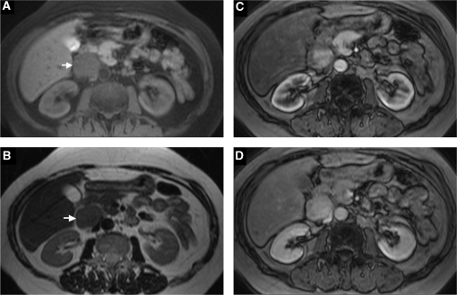
MR axial images: GRE FLASH T1w fs (A) and TSE T2w (B). GRE VIBE T1w post-Gd in arterial (C) and venous phase (D). The lesion appears homogenous and hypointense (arrow) on T1w fs scan (A) and shows an intermediate signal intensity (arrow) on the T2w sequence (B). An intense, non-homogeneous enhancement in arterial phase (C), with greater filling in venous phase (D) is detected. The IVC appears compressed but well opacified. No plane of cleavage with the duodenal wall is visible.
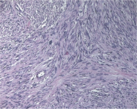
Leiomyosarcoma with typical intersecting clusters of spindle cells (haematoxylin and eosin stain, magnification×20).
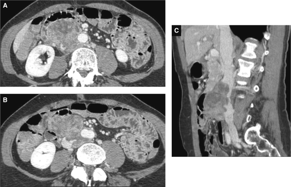
Contrast-enhanced CT axial images in venous phase A large heterogeneously enhanced retroperitoneal mass, arising from the lower IVC, involving more than 50% of the aortic wall and displacing the intestinal loops anteriorly (A) is depicted. The tumour extends to the confluence of the iliac vessels (B). A sagittal multi-planar reconstruction shows a narrowed and irregular IVC for throughout the longitudinal spread of the mass (C).
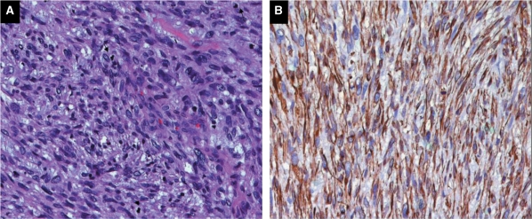
Leiomyosarcoma composed of elongated cells some with larger and hyperchromatic nuclei (haematoxylin and eosin stain, ×20) (A). Neoplastic cells showed cytoplasmic staining with desmin (immunohistochemistry, ×20) (B).
References
-
- Hilliard NJ, Heslin MJ, Castro CY. Leiomyosarcoma of the inferior vena cava. Three case reports and review of the literature. Ann Diagn Pathol. 2005;9:259–66. - PubMed
-
- Abisi S, Morris-Stiff GJ, Scott-Coombes D, et al. Leiomyosarcoma of the inferior vena cava: clinical experience with four cases. World J Surg Oncol. 2006;4:1. doi:10.1186/1477-7819-4-1. PMid:16393338. - DOI - PMC - PubMed
-
- Mingoli A, Cavallaro A, Sapienza P, et al. International registry of inferior vena cava leiomyosarcoma: analysis of a world series on 218 patients. Anticancer Res. 1996;16:3201–5. - PubMed
-
- Hemant D, Krantikumar R, Amita J, Chawla A, Ranjeet N. Primary leiomyosarcoma of inferior vena cava, a rare entity: imaging features. Aust Radiol. 2001;45:448–51. doi:10.1046/j.1440-1673.2001.00955.x. PMid:11903177. - DOI - PubMed
Publication types
MeSH terms
Substances
LinkOut - more resources
Full Text Sources