. 2010 May 28;285(22):16562-71.
doi: 10.1074/jbc.M110.110304. Epub 2010 Mar 15.

Tipin-replication protein A interaction mediates Chk1 phosphorylation by ATR in response to genotoxic stress

Affiliations

Tipin-replication protein A interaction mediates Chk1 phosphorylation by ATR in response to genotoxic stress

Michael G Kemp et al. J Biol Chem. .

Abstract

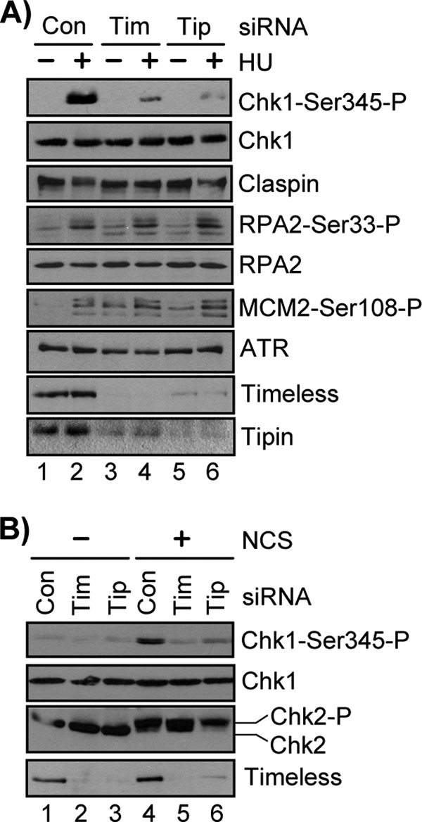
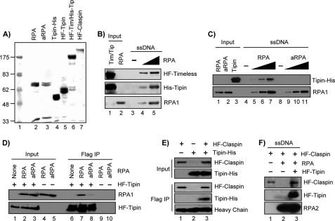
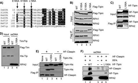
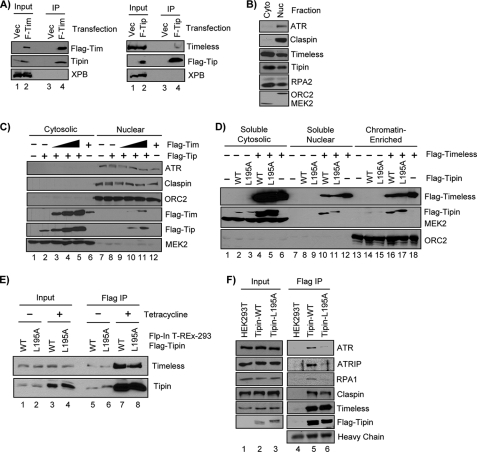
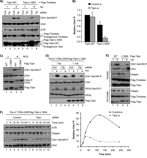
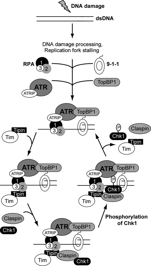
Mammalian Timeless is a multifunctional protein that performs essential roles in the circadian clock, chromosome cohesion, DNA replication fork protection, and DNA replication/DNA damage checkpoint pathways. The human Timeless exists in a tight complex with a smaller protein called Tipin (Timeless-interacting protein). Here we investigated the mechanism by which the Timeless-Tipin complex functions as a mediator in the ATR-Chk1 DNA damage checkpoint pathway. We find that the Timeless-Tipin complex specifically mediates Chk1 phosphorylation by ATR in response to DNA damage and replication stress through interaction of Tipin with the 34-kDa subunit of replication protein A (RPA). The Tipin-RPA interaction stabilizes Timeless-Tipin and Tipin-Claspin complexes on RPA-coated ssDNA and in doing so promotes Claspin-mediated phosphorylation of Chk1 by ATR. Our results therefore indicate that RPA-covered ssDNA not only supports recruitment and activation of ATR but also, through Tipin and Claspin, it plays an important role in the action of ATR on its critical downstream target Chk1.


Figures

FIGURE 1.
FIGURE 1.
Timeless and Tipin are required to specifically mediate Chk1 phosphorylation in response to DNA damage and replication stress. A, 48 h after transfection of HeLa cells with either Control (Con), Timeless (Tim), or Tipin (Tip) siRNA, the cells were treated with 1 mm HU for 30 min. The lysates were prepared, separated by SDS-PAGE, and analyzed by immunoblotting with antibodies against the indicated proteins. B, cells were transfected and analyzed as in A, but cells were instead treated with 100 ng/ml NCS for 1 h.
FIGURE 2.
FIGURE 2.
Interaction between RPA2 and Tipin stabilizes Timeless-Tipin and Claspin on RPA-coated ssDNA. A, Coomassie Blue-stained gel of purified proteins. RPA, aRPA, and Tipin-His were purified from E. coli, and His-FLAG-Tipin (HF-Tipin), His-FLAG-Timeless/His-Tipin (HF-Tim/His-Tip), and His-FLAG-Claspin (HF-Claspin) were purified from baculovirus-infected insect cells. Note that the gel displays only the 70-kDa (RPA1) and 34-kDa (RPA2/RPA4) subunits of RPA and aRPA, because the 17-kDa (RPA3) subunit was electrophoresed off of the gel. Lane 1 contains a molecular weight ladder, where numbers indicate molecular mass in kDa. B, His-FLAG-Timeless/His-Tipin complex (Tim/Tip; 10 pmol) was incubated with 5 pmol of immobilized 80-mer ssDNA preincubated for 30 min with 0, 2.5, or 5 pmol RPA in binding buffer. Input represents 5 pmol of HF-Timeless/His-Tipin complex and 5 pmol RPA. C, ssDNA (1 pmol) was preincubated with 0, 0.2, 0.5, or 1.25 pmol of RPA or aRPA in 50 μl of binding buffer before washing and addition of 20 pmol of Tipin. Input shows 0.5 pmol of RPA or aRPA and 1 pmol of Tipin. D, His-FLAG-tagged Tipin from baculovirus-infected insect cells was immobilized on anti-FLAG-agarose and then incubated with 1 μg of either RPA or aRPA overnight at 4 °C in 100 μl of binding buffer. Input represents 5% of the binding reactions. E, Tipin-His purified from E. coli (1 μg) was incubated with anti-FLAG resin alone or resin containing His-FLAG-tagged Claspin from baculovirus-infected insect cells. The reactions were in 100 μl of binding buffer overnight at 4 °C. Input shows 5% of the indicated reactions. Heavy chain of anti-FLAG IgG is displayed as a loading control. F, immobilized ssDNA (5 pmol) was incubated with RPA (10 pmol), His-FLAG-Tipin (10 pmol), and/or His-FLAG-Claspin (10 pmol), as indicated, and bound proteins were analyzed as in B and C. IP, immunoprecipitation.
FIGURE 3.
FIGURE 3.
Identification and characterization of a Tipin RPA-binding mutant. A, alignment of XPA and Tipin amino acid sequences. Black shading shows amino acid identity, and gray shading highlight indicates similarity. B, FLAG-tagged forms of Tipin were transiently expressed in HEK293T cells, immunoprecipitated with anti-FLAG-agarose, and analyzed by SDS-PAGE and immunoblotting with antibodies against FLAG, RPA1, and RPA2. Note that mutation of these amino acids alters Tipin mobility on SDS-PAGE. C, His-FLAG-tagged Tipin (HF-Tipin; WT and L195A) from baculovirus-infected insect cells was immobilized on anti-FLAG-agarose and then incubated with RPA. Resin was washed, and bound proteins were analyzed by SDS-PAGE and immunoblotting. D, FLAG-Timeless/His-Tipin (Tim/Tip) complexes prepared by baculoviral co-infection and anti-FLAG-agarose purification were incubated with immobilized ssDNA containing saturating amounts of RPA. Bound proteins were analyzed by SDS-PAGE and immunoblotting. Input represents 50% of the Timeless-Tipin complex used in the binding reaction. E, His-FLAG-tagged Claspin (HF-Claspin) from baculovirus-infected insect cells and immobilized on anti-FLAG resin (lanes 1–3) was incubated overnight at 4 °C with 1.5 μg of either Tipin-His-WT or Tipin-His-L195A purified from E. coli in 100 μl of binding buffer. Anti-FLAG resin lacking Claspin was used as a negative control (lanes 4–5). Input represents 5% of the Tipin used in the binding reaction. F, immobilized 80-mer ssDNA lacking or containing saturating amounts of RPA was incubated in reactions with HF-Claspin alone or together with HF-Tipin-WT or HF-Tipin-L195A. The beads were washed, and bound proteins were analyzed by SDS-PAGE and immunoblotting. IP, immunoprecipitation.
FIGURE 4.
FIGURE 4.
Characterization of Tipin protein expression and nuclear localization in human cells. A, an empty vector (Vec) and vectors encoding FLAG-Timeless (F-Tim; left panel) or FLAG-Tipin (F-Tip; right panel) were transiently transfected into HEK293T cells. The lysates were prepared and then immunoprecipitated (IP) with anti-FLAG resin. Input lanes show 5% of the lysate used for immunoprecipitation. B, HEK293T cells fractionated to yield a hypotonic cytosolic (Cyto) extract and a high salt nuclear (Nuc) fraction were separated by SDS-PAGE and immunoblotted with antibodies against the indicated proteins. C, HEK293T cells were transfected with an empty vector (lanes 1 and 7), vectors expressing FLAG-Tipin and increasing amounts of FLAG-Timeless (lanes 2–5 and 8–11), or a vector expressing FLAG-Timeless alone (lane 6 and 12). The total amount of plasmid DNA used per transfection (15 μg) was identical and was normalized with an empty vector plasmid DNA. The cells were fractionated as in B to yield cytosolic and nuclear fractions. D, HEK293T cells were transfected with empty vector or vectors expressing FLAG-Tipin and/or FLAG-Timeless, as indicated. The cells were fractionated to yield Triton-soluble (soluble cytosolic), soluble nuclear, or chromatin-enriched extracts. E, Flp-InTM T-RExTM-293 cell lines were generated to express FLAG-Tipin-WT or FLAG-Tipin-L195A under control of a tetracycline-inducible promoter. Uninduced (− Tetracycline) or cells induced with tetracycline (+ Tetracycline) for 3 days were lysed and immunoprecipitated with anti-FLAG-agarose, and the immunoprecipitates were probed for Timeless and Tipin. Input represents 5% of the lysate used for the immunoprecipitation. F, nuclear extracts from HEK293T or induced Flp-InTM T-RExTM-293-FLAG-Tipin (WT and L195A) were immunoprecipitated with anti-FLAG resin, separated by SDS-PAGE, and immunoblotted with antibodies against the indicated proteins.
FIGURE 5.
FIGURE 5.
Tipin-L195A does not support maintenance of Chk1 phosphorylation in response to DNA damage and replication stress. A, HeLa cells transiently transfected with vectors encoding FLAG-Timeless and siRNA-resistant FLAG-Tipin (WT or L195A) and either a nontargeting control (Con) siRNA or an siRNA targeting Tipin (Tip) were treated with 1 mm HU for 30 min. B, quantitation of phospho-Chk1 signals from experiments performed as in A. Phospho-Chk1 signals for each HU-treated sample were normalized to cells transfected with control siRNA and FLAG-Tipin-WT. The data show the averages and standard deviation from three independent experiments. C, HeLa cells transiently transfected with Tipin siRNA and vectors encoding FLAG-Timeless and siRNA-resistant FLAG-Tipin (WT or L195A) were treated with 100 ng/ml NCS for 1 h. D, uninduced (− Tet) and induced (+ Tet) Flp-In T-REx-293-FLAG-Tipin-L195A (siRNA-resistant) cells were transfected with either a control siRNA or an siRNA targeting endogenous Tipin and then treated with 1 mm HU for 2 h. E, HEK293T cells transfected with vectors expressing FLAG-Timeless and either FLAG-Tipin-WT or FLAG-Tipin-L195A were treated with 1 mm HU for 6 h or left untreated before fractionation to enrich for chromatin-associated proteins. Lysate from an equivalent number of cells was separated by SDS-PAGE and immunoblotted with antibodies against the indicated proteins. F, Flp-In T-REx-293-FLAG-Tipin-L195A cells induced with tetracycline to express FLAG-Tipin-L195A (siRNA-resistant) were transfected with either control or Tipin siRNA and then treated with 1 mm HU for the indicated lengths of time. The graph shows average Chk1 phosphorylation at each time point, relative to untreated control, from two independent experiments.
FIGURE 6.
FIGURE 6.
Model for Tipin function in ATR-Chk1 signaling. In response to DNA damage or replication stress, the generation of ssDNA and presence of primer-template junctions leads to the association of RPA and 9-1-1 at sites of DNA damage. Through an interaction of the RPA1 subunit of RPA with ATRIP, the ATR kinase is recruited to these sites. Through the C-terminal domain of Rad9, TopBP1 also stably associates with damage sites, resulting in activation of ATR kinase activity. Through an interaction of the RPA2 subunit of RPA with Tipin, the Timeless-Tipin complex and then Claspin are able to associate with ATR at sites of DNA damage. The presence of Claspin allows binding of Chk1 and then phosphorylation by ATR. The data showing that Timeless-Tipin, Claspin, and Chk1 do not appear to stably associate with sites of damage in human cells indicate that these proteins may be released to allow phosphorylation of additional Chk1 molecules.

Similar articles

Cited by

References

    1. Sancar A., Lindsey-Boltz L. A., Unsal-Kaçmaz K., Linn S. (2004) Annu. Rev. Biochem. 73, 39–85 - PubMed
    1. Kerzendorfer C., O'Driscoll M. (2009) DNA Repair 8, 1139–1152 - PubMed
    1. Lee J. H., Paull T. T. (2007) Oncogene 26, 7741–7748 - PubMed
    1. Palm W., de Lange T. (2008) Annu. Rev. Genet. 42, 301–334 - PubMed
    1. Cimprich K. A., Cortez D. (2008) Nat. Rev. Mol. Cell Biol. 9, 616–627 - PMC - PubMed

Publication types

MeSH terms