. 2010 Jun;185(2):671-84.
doi: 10.1534/genetics.109.113670. Epub 2010 Mar 16.

Identification of genes affecting wing patterning through a loss-of-function mutagenesis screen and characterization of med15 function during wing development

Affiliations

Identification of genes affecting wing patterning through a loss-of-function mutagenesis screen and characterization of med15 function during wing development

Ana Terriente-Félix et al. Genetics. 2010 Jun.

Abstract

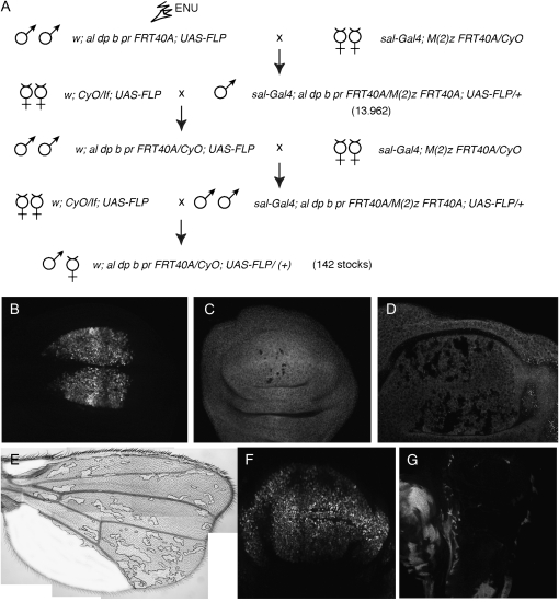
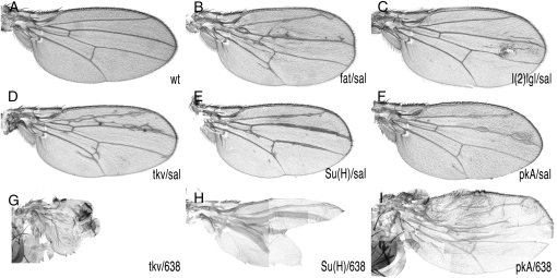
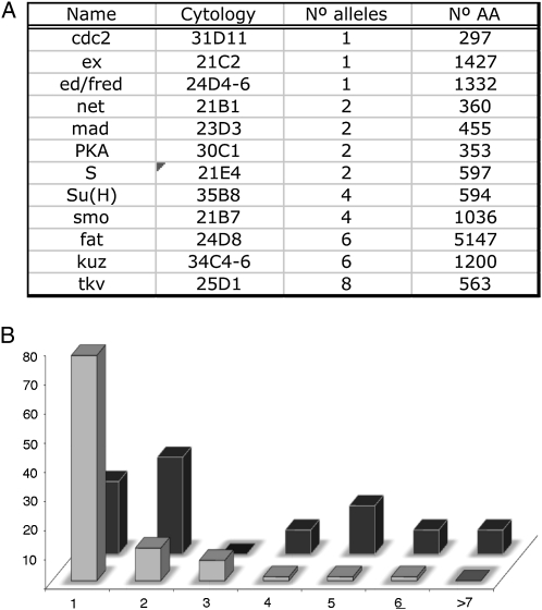
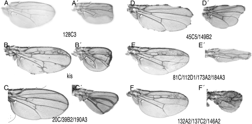
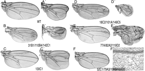
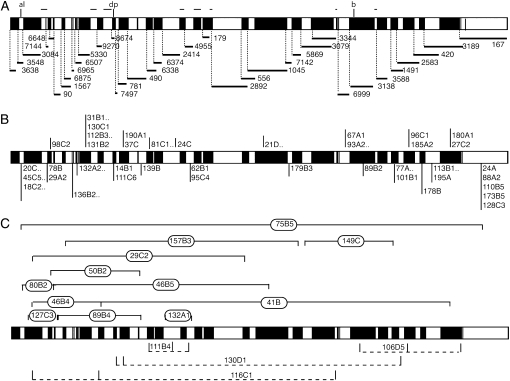
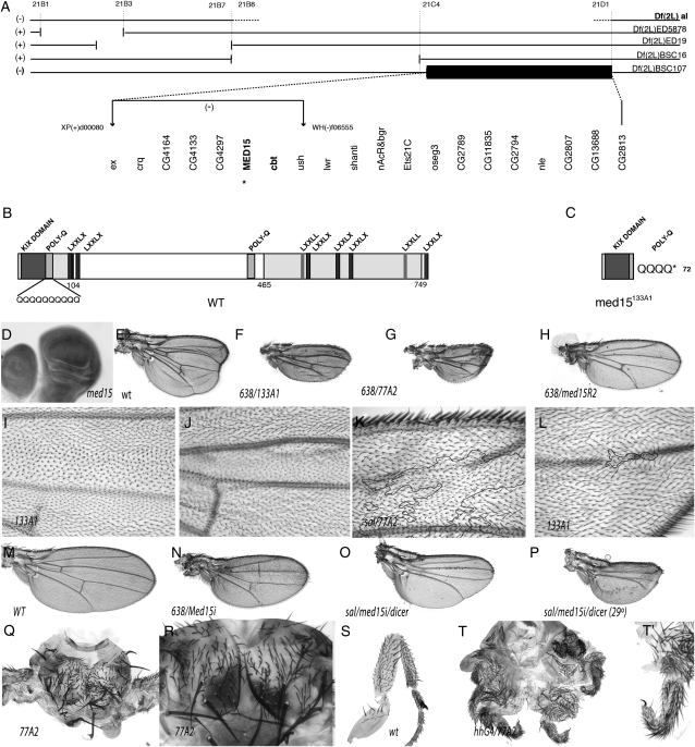
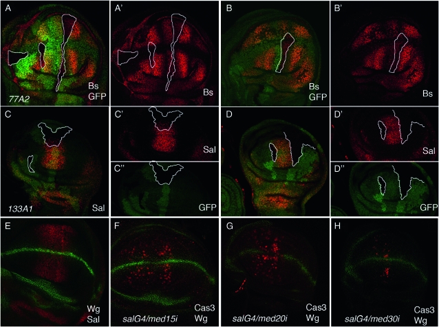
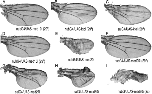
The development of the Drosophila melanogaster wing depends on the correct regulation of cell survival, growth, proliferation, differentiation, and pattern formation. These processes, and the genes controlling then, are common to the development of epithelia in many different organisms. To identify additional genes contributing to wing development we have carried out a genetic screen in mosaic wings carrying clones of homozygous mutant cells. We obtained 12 complementation groups corresponding to genes with a proven role in wing formation such as smoothened, thick veins, mothers against dpp, expanded, and fat and 71 new complementation groups affecting the pattern of veins and the size of wing. We mapped one of these groups to the mediator15 gene (med15), a component of the Mediator complex. We show that Med15 and other members of the Mediator complex are required, among other processes, for the transcription of decapentaplegic target genes.


Figures

F<sc>igure</sc> 1.—
Figure 1.—
Crosses and Gal4 lines used to generate homozygous mutant wings in heterozygous flies. (A) Chromosomes and genetic crosses used to generate mosaic flies. Males of w; al dp b pr FRT40A; UAS-FLP genotype were treated with ENU and crossed in groups of 50 with salEPv-Gal4; M(2)Z FRT40A/CyO females (first row). The sal-Gal4; M(2)Z FRT40A/al dp b pr FRT40A; UAS-FLP/+ male progeny (13,962 males, second row) was screened for wing phenotypes. Selected males were crossed with w; CyO/If; UAS-FLP females, and the male progeny of w; al dp b pr FRT40A/CyO; UAS-FLP genotype were crossed with salEPv-Gal4; M(2)Z FRT40A/CyO females. We established stable w; al dp b pr FRT40A;UAS-FLP/+ stocks when the original phenotype was found in the progeny of this last cross (142 cases). (B) Expression of GFP in the wing blade region of the salEPv-Gal4/UAS-GFP wing disc. (C and D) Early (C) and late (D) third instar wing discs of salEPv-Gal4; M(2)Z FRT40A tubGFP/al dp b pr FRT40A; UAS-FLP/+ genotype, showing the clones as black spots. (E) Adult wing of f36a salEPv-Gal4; M(2)Z FRT40A Pf+30C/al dp b pr FRT40A; UAS-FLP/+ genotype, showing the area not covered by forked clones in white. (F) Third instar wing disc showing the expression of the 638-Gal4 line (638-Gal4/UAS-GFP). (G) Third instar wing disc of 638-Gal4; M(2)Z FRT40A tubGFP/al dp b pr FRT40A; UAS-FLP/+ genotype. Most of the wing blade is composed of al dp b pr FRT40A homozygous cells (shown in black).
F<sc>igure</sc> 2.—
Figure 2.—
Mosaic wings of known mutants affecting wing development. (A) Wild-type wing. (B) salEPv-Gal4; M(2)Z FRT40A/ fat18 FRT40A; UAS-FLP/+ (fat/sal). (C) salEPv-Gal4; M(2)Z FRT40A/ l(2)gl4 FRT40A; UAS-FLP/+ [l(2)gl/sal]. (D) salEPv-Gal4; M(2)Z FRT40A/tkva12 FRT40A; UAS-FLP/+ (tkv/sal). (E) salEPv-Gal4; M(2)Z FRT40A/ TE35BC-GW24 FRT40A; UAS-FLP/+ [Su(H)/sal]. (F) salEPv-Gal4; M(2)Z FRT40A/ PKAB3 FRT40A; UAS-FLP/+ (PKA/sal). (G) 638-Gal4; M(2)Z FRT40A/tkva12 FRT40A; UAS-FLP/+ (tkv/638). (H) 638-Gal4; M(2)Z FRT40A/ TE35BC-GW24 FRT40A; UAS-FLP/+ [Su(H)/638]. (I) 638-Gal4; M(2)Z FRT40A/ PKAB3 FRT40A; UAS-FLP/+ (PKA/638). Note that the phenotypes involving the Gal4 driver 638-Gal4 are much stronger than those generated with sal-Gal4.
F<sc>igure</sc> 3.—
Figure 3.—
General results of the screen. (A) Known genes identified in the screen, indicating their names (“Name”), cytological position (“Cytology”), number of alleles found (“No. alleles”), and the number of amino acids of the corresponding proteins (“No. AA”). (B) Relationship between the numbers of complementation groups (vertical values) and the number of alleles included in each complementation group (horizontal values). Data with light shading correspond to the new complementation groups identified in this screen and data with solid shading to genes that were known to play a role in wing development.
F<sc>igure</sc> 4.—
Figure 4.—
Representative wings of the phenotypic classes affecting the formation of veins. (A and A′) Loss of veins and reduced wing size in salEPv-Gal4; M(2)Z FRT40A/al dp b pr 128C3 FRT40A; UAS-FLP/+ (A) and 638-Gal4; M(2)Z FRT40A/al dp b pr 128C3 FRT40A; UAS-FLP/+ (A′). (B and B′) Loss and gain of veins in salEPv-Gal4; M(2)Z FRT40A/al dp b pr 61C FRT40A; UAS-FLP/+ (B) and 638-Gal4; M(2)Z FRT40A/al dp b pr 61C FRT40A; UAS-FLP/+ (B′). The mutant 61C is an allele of kismet (kis). (C and C′) Thick vein phenotype in salEPv-Gal4; M(2)Z FRT40A/al dp b pr 20C FRT40A; UAS-FLP/+ (C) and 638-Gal4; M(2)Z FRT40A/al dp b pr 20C FRT40A; UAS-FLP/+ (C′). (D and D′) Wing margin phenotype of salEPv-Gal4; M(2)Z FRT40A/al dp b pr 45C5 FRT40A; UAS-FLP/+ (D) and 638-Gal4; M(2)Z FRT40A/al dp b pr 45C5 FRT40A; UAS-FLP/+ (D′). (E and E′) Ectopic veins and loss of wing margin in salEPv-Gal4; M(2)Z FRT40A/al dp b pr 81C FRT40A; UAS-FLP/+ (E) and 638-Gal4; M(2)Z FRT40A/al dp b pr 81C FRT40A; UAS-FLP/+ (E′). (F and F′) Ectopic veins and reduction in wing size in salEPv-Gal4; M(2)Z FRT40A/al dp b pr 132A2 FRT40A; UAS-FLP/+ (F) and 638-Gal4; M(2)Z FRT40A/al dp b pr 132A2 FRT40A; UAS-FLP/+ (F′). The phenotypes of other mutations affecting the veins or the wing margin are shown in Figure S1 and Figure S3.
F<sc>igure</sc> 5.—
Figure 5.—
Representative wings of the phenotypic classes affecting wing size, cell adhesion, and cell differentiation. (A and A′) Wild-type wing of salEPv-Gal4; M(2)Z FRT40A/al dp b pr FRT40A; UAS-FLP/+ (A) and 638-Gal4; M(2)Z FRT40A/al dp b pr FRT40A; UAS-FLP/+ (A′) genotypes. (B and B′) Reduced wing size in salEPv-Gal4; M(2)Z FRT40A/al dp b pr 31B1 FRT40A; UAS-FLP/+ (B) and 638-Gal4; M(2)Z FRT40A/al dp b pr 31B1 FRT40A; UAS-FLP/+ (B′). (C and C′) Increased wing size in salEPv-Gal4; M(2)Z FRT40A/al dp b pr 130C1 FRT40A; UAS-FLP/+ (C) and 638-Gal4; M(2)Z FRT40A/al dp b pr 130C1 FRT40A; UAS-FLP/+ (C′). (D and D′) Failures in dorso-ventral adhesion in salEPv-Gal4; M(2)Z FRT40A/al dp b pr 18C2 FRT40A; UAS-FLP/+ (D) and 638-Gal4; M(2)Z FRT40A/al dp b pr 18C2 FRT40A; UAS-FLP/+ (D′). (E and E′) Extra veins and increased wing size with loss of wing margin in salEPv-Gal4; M(2)Z FRT40A/al dp b pr 77A FRT40A; UAS-FLP/+ (E) and 638G4-Gal4; M(2)Z FRT40A/al dp b pr 77A FRT40A; UAS-FLP/+ (E′). (F and F′) Formation of several trichomes per cell in salEPv-Gal4; M(2)Z FRT40A/al dp b pr 52C1 FRT40A; UAS-FLP/+. F′ is a higher magnification of the L3/L4 dorsal intervein. Other phenotypes affecting wing size, cell adhesion, and differentiation are shown in Figure S2 (size) and Figure S4 (adhesion and cell differentiation).
F<sc>igure</sc> 6.—
Figure 6.—
Schematic representation of the complementation data with 2L deficiencies for all the mutants isolated. (A–C) The horizontal bars represent the chromosomal arm 2L subdivided in solid and open areas that exemplified the subdivision of the arm by the overlapping of the deficiencies used for the mapping. (A) Extent of the different deletions used for the complementation tests (see material and methods). The thin lines represent the main gaps left by the deficiencies. (B) Localization of mutants (single numbers) and complementation groups (single numbers and dots) that fail to complement with only one deficiency. (C) Localization of mutants that fail to complement with more than one deficiency.
F<sc>igure</sc> 7.—
Figure 7.—
The complementation group formed by 77A2 and 133A1 corresponds to med15. (A) Chromosomal interval 21B3–21C4 and extent of the deficiencies used (named to the right) shown as open spaces between horizontal bars. The region included in Df(2L)BSC107, which defines the localization of the 77A2/133A1 complementation group, is shown as a solid bar, and the genes included in this deficiency are shown at the bottom. The position of two PiggyBac insertions used to generate the Df(2L)d0080-f06555 chromosome is shown by vertical arrows. (B) Protein sequence and domains of Med15. (C) Expected protein produced by the med15133A1 allele. (D) Generalized expression of med15 in the wing disc. (E) Control wing of 638-Gal4/+; FRT40A al dp b pr/FRT40A M(2)z; UAS-FLP/+ genotype showing the characteristic dp phenotype. (F and G) Homozygous wings for the med15 alleles med15133A1 (133A1) (E) and med1577A2 (77A2) (F). The genotypes of these wings are 638-Gal4/+; FRT40A al med15133A1 dp b pr/FRT40A M(2)z; UAS-FLP/+ (F) and 638-Gal4/+; FRT40A al med1577A2 dp b pr/FRT40A M(2)z; UAS-FLP/+ (G). (H) Phenotype of med15f04180 homozygous wing in 638-Gal4/+; FRT40A al med15f04180 dp b pr/FRT40A M(2)z; UAS-FLP/+ female flies (compare the wing size with its control shown in M). (I and J) Mitotic recombination clones generated in hsFLP1.22 f36a; ck P[f+]30C FRT40A/al med15133A1 dp b pr FRT40A flies. The clone in I is labeled with ck and is wild type for the med15 gene (twin spot). The clone in J is labeled with forked and is homozygous for the med15133A1 allele. Both clones are located in the L3/L4 intervein, occupying a large fraction of this dorsal (I) and ventral (J) intervein. (K) med1577A2 clone labeled with forked generated in f36a salEPv-Gal4/+; FRT40A al med1577A2 dp b pr/FRT40A P[f+]30C M(2)z; UAS-FLP/+ flies. The forked territory is enclosed by a solid line and is associated with the loss of the ventral L2 vein. (L) Small med15133A1 clone in the distal dorsal L4 vein causing the loss of this vein. (M–P) Phenotypes resulting from the expression of med15 interference RNA (med15i) in the genotypes 638-Gal4/+; UAS-med15i (N), salEPv-Gal4/UAS-med15i; UAS-dicer/+ (O), and salEPv-Gal4/UAS-med15i; UAS-dicer/+ grown at 29° (P). The wild-type control wing is shown in M. (Q and R) Two examples of thoraxes taken at different magnification showing the failure in the fusion between the left and the right hemithorax when the central region is occupied by med1577A mutant cells (labeled with forked in hsFLP1.22 f36a; P[f+]30C M(2)z FRT40A/al med1577A2 dp b pr FRT40A flies). (S) Wild-type male first leg. (T and T′) Two examples of legs taken at different magnifications (T′ is ×4 T) showing the defects in leg morphogenesis and tarsal segmentation in w; M(2)z P[f+]30C FRT40A/al med1577A dp b pr FRT40A; hh-Gal4/UAS-FLP flies.
F<sc>igure</sc> 8.—
Figure 8.—
Developmental analysis of med15 function in the wing disc. (A and B) Expression of Bs (red) in med1577A2 clones induced in flies of genotype hsFLP1.22; P[tubGFP] FRT40A/med1577A2 FRT40A (twin clones). med1577A2 clones are labeled by the absence of GFP (green) and each clone is encircled in a white line in A–B′. A′ and B′ are the corresponding red channels showing reduced Bs expression in the mutant clones. (C and D) Expression of Sal (red) in med15133A1 clones induced in flies of genotype hsFLP1.22; P[tubGFP] FRT40A/med15133A1 FRT40A (twin clones). med15133A1 clones are labeled by the absence of GFP (green) and each clone is encircled in a white line in C and D, C′ and D′, and C″ and D″. ′ and ″ are the corresponding red (Sal) and green (GFP) channels, respectively. The expression of Sal is reduced or absent in mutant clones located in the center (C′) or lateral (D′) regions of the Sal domain, respectively. (E) Expression of Wingless (Wg, green) and Sal (Sal, red) in a wild-type third instar wing blade. (F–H) Induction of cell death (shown as expression of activated Caspase 3) (red in F–H) in flies expressing interference RNA in the spalt domain in salEPv-Gal4/UAS-med15-RNAi (F), salEPv-Gal4/UAS-med20-RNAi (G), and salEPv-Gal4/UAS-med30-RNAi (H).
F<sc>igure</sc> 9.—
Figure 9.—
Wing phenotypes caused by reduced expression of different components of the Mediator complex. (A–I) Adult wings of Gal4/UAS-RNAi combinations showing smaller than normal size and different defects in the patterning of veins. (A) nub-Gal4/UAS-med10i at 29°. (B) nub-Gal4/UAS-ktoi (29°). (C) salEPv-Gal4/UAS-ktoi (29°). (D) nub-Gal4/UAS-med16i (29°). (E) nub-Gal4/UAS-med20i. (F) nub-Gal4/UAS-med25i (29°). (G) salEPv-Gal4/UAS-med27i. (H) salEPv-Gal4/UAS-med30i. (I) nub-Gal4/UAS-med30i (taken at magnification ×2).

Similar articles

Cited by

References

    1. Alarcón, C., A.-I. Zaromytidou, Q. Xi, S. Gao, J. Yu et al., 2009. Nuclear CDKs drive Smad transcriptional activation and turnover in BMP and TGF-b pathways. Cell 139 757–769. - PMC - PubMed
    1. Ashburner, M., 1989. Drosophila. A Laboratory Manual. Cold Spring Harbor Laboratory Press, Cold Spring Harbor, NY.
    1. Baker, N. E., 1987. Molecular cloning of sequences from wingless a segment polarity gene in Drosophila the spatial distribution of a transcript in embryos. EMBO J. 6 1765–1774. - PMC - PubMed
    1. Barrio, R., and J. F. de Celis, 2004. Regulation of spalt expression in the Drosophila wing blade in response to the Decapentaplegic signaling pathway. Proc. Natl. Acad. Sci. USA 101 6021–6026. - PMC - PubMed
    1. Bier, E., 2000. Drawing lines in the Drosophila wing: initiation of wing vein development. Curr. Opin. Genet. Dev. 10 393–398. - PubMed

Publication types

Substances

LinkOut - more resources