. 2010 Jun;192(11):2908-19.
doi: 10.1128/JB.01266-09. Epub 2010 Mar 16.

The Escherichia coli mqsR and ygiT genes encode a new toxin-antitoxin pair

Affiliations

The Escherichia coli mqsR and ygiT genes encode a new toxin-antitoxin pair

Villu Kasari et al. J Bacteriol. 2010 Jun.

Abstract

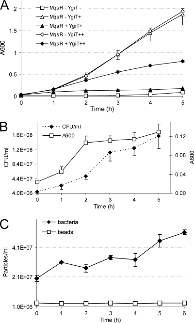
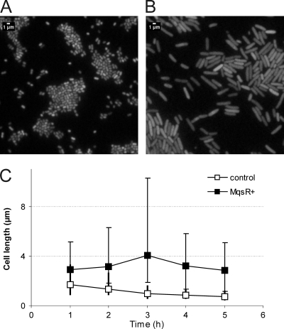
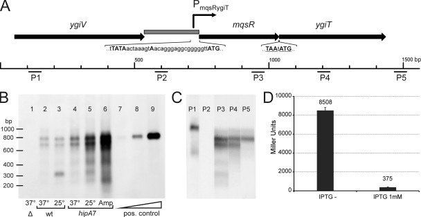
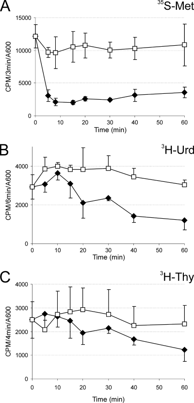
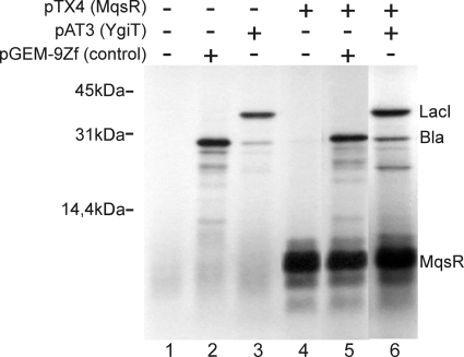
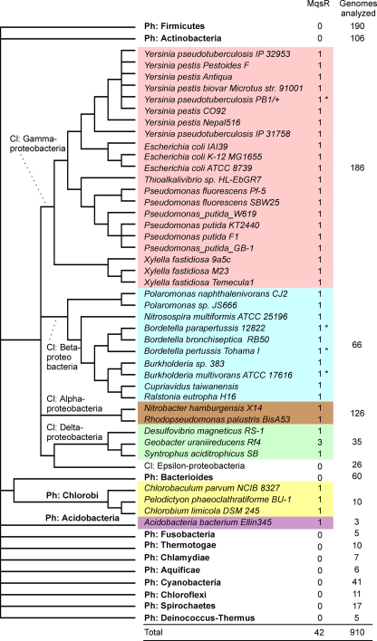
Toxin-antitoxin (TA) systems are plasmid- or chromosome-encoded protein complexes composed of a stable toxin and a short-lived inhibitor of the toxin. In cultures of Escherichia coli, transcription of toxin-antitoxin genes was induced in a nondividing subpopulation of bacteria that was tolerant to bactericidal antibiotics. Along with transcription of known toxin-antitoxin operons, transcription of mqsR and ygiT, two adjacent genes with multiple TA-like features, was induced in this cell population. Here we show that mqsR and ygiT encode a toxin-antitoxin system belonging to a completely new family which is represented in several groups of bacteria. The mqsR gene encodes a toxin, and ectopic expression of this gene inhibits growth and induces rapid shutdown of protein synthesis in vivo. ygiT encodes an antitoxin, which protects cells from the effects of MqsR. These two genes constitute a single operon which is transcriptionally repressed by the product of ygiT. We confirmed that transcription of this operon is induced in the ampicillin-tolerant fraction of a growing population of E. coli and in response to activation of the HipA toxin. Expression of the MqsR toxin does not kill bacteria but causes reversible growth inhibition and elongation of cells.


Figures

FIG. 1.
FIG. 1.
MqsR induces growth arrest which is reversible by expression of YgiT. HM21Δ contained plasmid pAT3 for IPTG-inducible ygiT expression and plasmid pTX3 for l-arabinose-inducible mqsR expression. (A) Effect on bacterial growth as measured by A600. Bacteria were grown without IPTG (squares) (YgiT−), in the presence of 50 μM IPTG (triangles) (YgiT+), or in the presence of 5 mM IPTG (diamonds) (YgiT++). At time zero, some cultures (filled symbols) (MqsR+) were supplemented with 1 mM l-arabinose. (B) Colony formation after induction of mqsR. Bacteria were grown in the presence of 50 μM IPTG, and mqsR was induced at time zero by addition of 1 mM l-arabinose. At the time points indicated, samples were taken, the A600 was measured, and appropriate dilutions were plated onto solid medium supplemented with 0.2% glucose and 50 μM IPTG. Colonies were counted after 24 h, and the number of CFU per ml of culture was calculated. (C) Counting of individual bacteria after induction of mqsR. Bacteria were grown, mqsR was induced, and samples were taken as described above for the plating experiments. Bacteria were stained with the SYTO BC dye and mixed with a constant number of standard microspheres. Particles were counted by flow cytometry. All results are averages of three independent experiments. The error bars indicate standard deviations.
FIG. 2.
FIG. 2.
MqsR causes elongation of bacterial cells. Expression of mqsR was induced in the logarithmic growth phase at time zero by addition of 1 mM l-arabinose to a culture of HM21Δ cells bearing plasmids pAT3 (ygiT expression) and pTX3 (mqsR expression). An uninduced culture of the same strain was used as a control. Samples were taken at the times indicated, and bacteria were stained with the SYTO BC dye and examined by epifluorescence microscopy. Control bacteria (A) and the MqsR-arrested bacteria (B) were observed 4 h after induction of mqsR. (C) Average lengths of 100 bacterial cells. Filled squares, MqsR-expressing bacteria; open squares, uninduced control. The vertical bars indicate the limits of cell length (the longest and shortest bacteria in a set of cells).
FIG. 3.
FIG. 3.
Organization and transcription of the mqsR-ygiT operon. (A) Map of the mqsR-ygiT region in the E. coli K-12 chromosome. The arrows represent the open reading frames. The stop codon of mqsR and the start codon of ygiT are underlined. The gray box represents the PmqsR-ygiT promoter region cloned in front of the lacZ reporter in the pPTX3 promoter probe plasmid. The Pribnow (−10) box, the 5′ end of the mqsR-ygiT transcript, and the start codon of mqsR are indicated by uppercase letters in the nucleotide sequence of PmqsR-ygiT. P1 to P5 indicate the locations of oligoprobes used in Northern hybridization. (B) Northern analysis of mqsR-ygiT transcription in different strains and under different conditions. Lane 1, HM21Δ [Δ(mqsR ygiT)]; lanes 2 and 3, HM21 (wt); lanes 4, 5, and 6, HM22 (hipA7); lanes 7, 8, and 9, 5-fold serial dilutions of the in vitro-synthesized mqsR-ygiT transcript. Bacteria were grown at 37°C for 2.5 h (to the exponential growth phase), and the cultures used for lanes 3 and 5 were transferred to 25°C and incubated for an additional 1.5 h. Ampicillin (100 μg ml−1) was added to the culture used for lane 6, and the culture was incubated at 37°C for additional 3 h to induce lysis of the sensitive bacteria and isolate the ampicillin-tolerant subpopulation. Total RNA was extracted from each sample, and 10-μg aliquots were subjected to electrophoresis, transferred to a membrane, and hybridized with fluorescein-labeled oligoprobe P3. Hybrids were detected using antifluorescein-AP conjugate and chemiluminescent detection. (C) Mapping of mqsR-ygiT transcripts by Northern oligoprobe hybridization. RNA was extracted from HM21 cells grown at 37°C for 2.5 h, and 10-μg portions were subjected to electrophoresis and transferred to a membrane. The membrane was cut into strips and hybridized with different fluorescein-labeled probes. P1 to P5 indicate the probes used for hybridization. (D) Repression of transcription from the PmqsR-ygiT promoter by YgiT. E. coli HM21Δ2 PTX lacZ carries a single copy of a Pmqs-RygiT::lacZ transcriptional fusion in the chromosome and was transformed with plasmid pAT3 for IPTG-inducible expression of YgiT. Overnight cultures were diluted 100-fold in fresh medium lacking IPTG (IPTG−) or containing 1 mM IPTG, and β-galactosidase levels were determined after 4 h. The values are the averages of results from two assays. The error bars indicate the standard errors.
FIG. 4.
FIG. 4.
Effect of mqsR expression on macromolecular synthesis in vivo. Pulse-labeling experiments showed that there was incorporation of radiolabeled precursors into protein (A), RNA (B), and DNA (C). Ectopic expression of mqsR (filled symbols) was induced at time zero by addition of l-arabinose (1 mM) to HM21Δ cultures bearing plasmids pAT3 (for ygiT expression) and pTX3 (for mqsR expression). Open symbols indicate the results for the negative control (no l-arabinose added). Samples were taken at the times indicated, radioactive precursors were added, and samples were incubated at 37°C for 3 min (A), 6 min (B), and 4 min (C) for optimal labeling. Incorporation of the label was stopped by adding trichloroacetic acid (TCA). The rates of synthesis are expressed as the number of cpm incorporated during a pulse-labeling reaction divided by the A600 of the bacterial culture. The values are the averages of three independent experiments; the error bars indicate the standard deviations.
FIG. 5.
FIG. 5.
MqsR does not inhibit protein synthesis in vitro. Coupled transcription-translation was carried out using the E. coli T7 S30 extract system for circular DNA. Templates for expression of β-lactamase as a positive control (pGEM9Zf) (lanes 2 and 5), MqsR (pTX4) (lanes 4 to 6), and YgiT plus LacI (pAT3) (lanes 3 and 6) were added. In vitro-synthesized proteins were labeled with [35S]methionine and analyzed by SDS-PAGE using a 14% polyacrylamide gel. The positions of synthesized proteins are indicated on the right.
FIG. 6.
FIG. 6.
Phyletic distribution of MqsR homologues on a cladistic tree of bacteria. Phyla for which only one or two genomes have been sequenced are not shown, and the total number of genomes analyzed was 914. Asterisks indicate MqsR variants with a ∼40-amino-acid N-terminal truncation. The cladistic tree was constructed by using a deeper branching order (phylum [Ph] and class [Cl] levels) than an NCBI microbial taxonomic tree (Genome Browser taxonomic tree). The more subtle branching order of genomes which contained toxin-antitoxin pairs is based on a 16S rRNA alignment retrieved from RDP II (13) and was computed by using neighbor-joining tools of MEGA4 (32).
FIG. 7.
FIG. 7.
Biofilm formation by wild-type and mutant E. coli K-12 strains. Bacteria with the genotypes indicated were diluted from an overnight culture and grown in LB medium in 96-well polystyrene microtiter dishes at 30°C without shaking. Eight wells were inoculated using a single overnight culture. After 48 h, the microtiter plates were rinsed and stained with crystal violet. The surface-bound stain was dissolved and quantified by measuring the adsorption at 590 nm. The averages of three to five independent experiments are shown for each strain. The error bars indicate the standard errors. kanr, kanamycin resistant (contains resistance cassette acquired during disruption of mqsR); kans, kanamycin sensitive (resistance cassette eliminated). wt, wild type.

Similar articles

Cited by

References

    1. Allen, G. C., Jr., and A. Kornberg. 1991. Fine balance in the regulation of DnaB helicase by DnaC protein in replication in Escherichia coli. J. Biol. Chem. 266:22096-22101. - PubMed
    1. Altschul, S. F., T. L. Madden, A. A. Schaffer, J. Zhang, Z. Zhang, W. Miller, and D. J. Lipman. 1997. Gapped BLAST and PSI-BLAST: a new generation of protein database search programs. Nucleic Acids Res. 25:3389-3402. - PMC - PubMed
    1. Amitai, S., Y. Yassin, and H. Engelberg-Kulka. 2004. MazF-mediated cell death in Escherichia coli: a point of no return. J. Bacteriol. 186:8295-8300. - PMC - PubMed
    1. Baba, T., T. Ara, M. Hasegawa, Y. Takai, Y. Okumura, M. Baba, K. A. Datsenko, M. Tomita, B. L. Wanner, and H. Mori. 2006. Construction of Escherichia coli K-12 in-frame, single-gene knockout mutants: the Keio collection. Mol. Syst. Biol. 2:2006.0008. - PMC - PubMed
    1. Balaban, N. Q., J. Merrin, R. Chait, L. Kowalik, and S. Leibler. 2004. Bacterial persistence as a phenotypic switch. Science 305:1622-1625. - PubMed

Publication types

MeSH terms

LinkOut - more resources