. 2010 Jun;192(12):2950-64.
doi: 10.1128/JB.01642-09. Epub 2010 Mar 16.

Cyclic-di-GMP-mediated repression of swarming motility by Pseudomonas aeruginosa: the pilY1 gene and its impact on surface-associated behaviors

Affiliations

Cyclic-di-GMP-mediated repression of swarming motility by Pseudomonas aeruginosa: the pilY1 gene and its impact on surface-associated behaviors

S L Kuchma et al. J Bacteriol. 2010 Jun.

Abstract

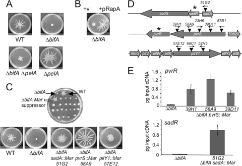
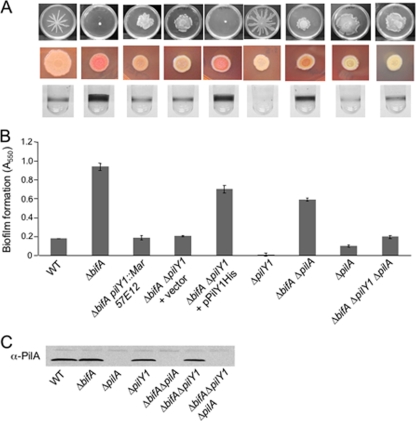
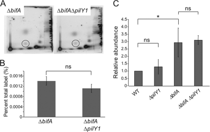
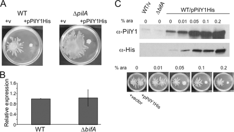
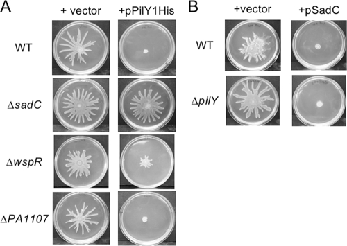
The intracellular signaling molecule cyclic-di-GMP (c-di-GMP) has been shown to influence surface-associated behaviors of Pseudomonas aeruginosa, including biofilm formation and swarming motility. Previously, we reported a role for the bifA gene in the inverse regulation of biofilm formation and swarming motility. The bifA gene encodes a c-di-GMP-degrading phosphodiesterase (PDE), and the Delta bifA mutant exhibits increased cellular pools of c-di-GMP, forms hyperbiofilms, and is unable to swarm. In this study, we isolated suppressors of the Delta bifA swarming defect. Strains with mutations in the pilY1 gene, but not in the pilin subunit pilA gene, show robust suppression of the swarming defect of the Delta bifA mutant, as well as its hyperbiofilm phenotype. Despite the ability of the pilY1 mutation to suppress all the c-di-GMP-related phenotypes, the global pools of c-di-GMP are not detectably altered in the Delta bifA Delta pilY1 mutant relative to the Delta bifA single mutant. We also show that enhanced expression of the pilY1 gene inhibits swarming motility, and we identify residues in the putative VWA domain of PilY1 that are important for this phenotype. Furthermore, swarming repression by PilY1 specifically requires the diguanylate cyclase (DGC) SadC, and epistasis analysis indicates that PilY1 functions upstream of SadC. Our data indicate that PilY1 participates in multiple surface behaviors of P. aeruginosa, and we propose that PilY1 may act via regulation of SadC DGC activity but independently of altering global c-di-GMP levels.


Figures

FIG. 1.
FIG. 1.
Genetic analyses of the ΔbifA swarming defect. (A) Impact of Pel polysaccharide on the ΔbifA swarming impairment. Representative images of swarms formed by the WT, the ΔbifA mutant, the ΔbifA ΔpelA double mutant, and the ΔpelA mutant swarms after 16 h at 37°C on 0.5% swarming agar are shown. (B) Impact of expression of the RapA PDE on swarming motility of the ΔbifA mutant. Representative images of swarms of the ΔbifA mutant carrying the pMQ72 vector control (left) and pMQ72 expressing the RapA c-di-GMP phosphodiesterase (right) are shown. The plates contained 0.2% (wt/vol) arabinose and were incubated for 16 h at 37°C. (C) Genetic screen for suppressors of the ΔbifA swarming impairment. The top image shows a swarm plate from the screen of ΔbifA mariner transposon mutants, with filled arrows indicating the WT control (top right) and the ΔbifA mutant (top left). The open arrow indicates a candidate ΔbifA mariner suppressor mutant. The lower panel shows typical images of swarms of the WT, the ΔbifA mutant, and the ΔbifA sadA::Mar, ΔbifA pvrS::Mar, and ΔbifA pilY1::Mar suppressor mutants. The plates were incubated for 16 h at 37°C. (D) Genetic loci of ΔbifA swarm suppressors. The schematic diagram shows the approximate locations of mariner transposon mutations (▾) in the sadA, pvrS, and pilY1 genes. Arrows above transposons indicate the direction of transcription from the mariner Ptac promoter. Asterisks denote genes encoding predicted c-di-GMP phosphodiesterases. (E) qRT-PCR analysis of pvrR expression in ΔbifA pvrS::Mar suppressor mutants (top panel) and sadR expression in the ΔbifA sadA::Mar suppressor mutant (bottom panel) relative to the ΔbifA single mutant. Expression is plotted as picograms input cDNA for each strain. Error bars indicate standard deviations.
FIG. 2.
FIG. 2.
Phenotypes of the bifA suppressor mutants. (A) pilY1 mutations suppress multiple defects of the ΔbifA mutant. Top panel, typical swarm images for (left to right) the WT, the ΔbifA mutant, a representative ΔbifA pilY1::Mar mutant (57E12), the ΔbifA ΔpilY1 double mutant carrying either empty vector or the pPilY1His complementing plasmid, and the ΔpilY1 mutant. Also shown are swarm images of the ΔbifA ΔpilA double mutant, the ΔpilA single mutant, and the ΔbifA ΔpilA ΔpilY1 triple mutant. The plates were incubated for 16 h at 37°C. The middle panel shows CR binding for each strain. CR assay plates were incubated for 24 h at 37°C followed by 48 h at room temperature. The bottom panel shows representative wells of a 96-well dish from a biofilm assay for each strain. Strains were grown in M63 with glucose, MgSO4, and CAA for 24 h at 37°C prior to crystal violet staining. (B) Quantification of crystal violet-stained biofilms. Crystal violet was solubilized in 30% glacial acetic acid, and the absorbance was measured at 550 nm. Error bars indicate standard deviations. (C) Western blot analysis showing PilA protein in crude lysates prepared from the indicated strain.
FIG. 3.
FIG. 3.
Quantification of global c-di-GMP pools. (A) Shown are autoradiographs of representative two-dimensional TLC plates used to separate [32P]orthophosphate-labeled, acid-extracted whole-cell extracts prepared from the ΔbifA mutant and the ΔbifA ΔpilY1 double mutant. The circles indicate the position of c-di-GMP. (B) Quantification of c-di-GMP levels determined by analyzing autoradiographs using the Storm 860 and ImageQuant software (v5.1). The graph shows a representative experiment with results for three replicates of each strain plotted as a percentage of the total 32P label incorporated into c-di-GMP. ns, no statistically significant difference between the ΔbifA mutant and the ΔbifA ΔpilY1 double mutant using the criterion of a P value of <0.05. (C) Quantification of c-di-GMP levels as measured using liquid chromatography-tandem mass spectrometry. The graph depicts the relative abundance of c-di-GMP for three replicates. *, statistically significant difference; ns, no significant difference (using the criterion of a P value of <0.05 by the t test). Error bars indicate standard deviations.
FIG. 4.
FIG. 4.
Increased expression of PilY1 represses swarming motility. (A) Enhanced expression of pilY1 represses swarming in a pilus-independent manner. Shown are representative images of the WT (left panel) and the ΔpilA mutant (right panel) carrying either empty vector or the pPilY1His arabinose-inducible expression plasmid. Swarm plates contained 0.2% arabinose and were incubated for 16 h at 37°C. (B) qRT-PCR analysis of pilY1 expression in the ΔbifA mutant relative to the WT. Expression data were normalized to the WT, and the results shown are the average from two independent experiments. Error bars indicate standard deviations. (C) Assessment of PilY1 levels and impact on swarming phenotype. Shown in the upper panels are Western blots showing PilY1 protein levels in the WT carrying empty vector, the ΔbifA mutant, and the WT carrying the pPilY1His plasmid. Concentrations of arabinose (wt/vol) added during culturing of each sample are indicated. PilY1 protein was detected using anti-PilY1 antibodies (top panel) or anti-His antibody to detect His-tagged PilY1 (middle panel) expressed from the pPilY1His plasmid. Shown in the lower panel are swarm plates containing arabinose at the indicated concentrations. On the left side of each plate is the WT carrying the empty vector control, and on the right is the WT carrying pPilY1His.
FIG. 5.
FIG. 5.
PilY1 functions genetically upstream of the SadC diguanylate cyclase. (A) Shown are representative swarms for the WT and the ΔsadC, ΔwspR, and ΔPA1107 DGC mutants carrying either the empty vector (left column) or the pPilY1His plasmid (right column). Plates contained arabinose at a concentration of 0.2%. (B) Swarms of the WT and ΔpilY1 mutant when carrying either the vector (left column) or pSadC expression plasmid (right column). Plates contained arabinose at a concentration of 0.2%.
FIG. 6.
FIG. 6.
PilY1 cellular localization. Cellular fractions of the WT, ΔpilY1 mutant, and ΔpilA mutant were separated by SDS-PAGE. Fractions are indicated as supernatant (Sup), cell-associated (CA), whole-cell (WC), soluble cytoplasmic (Cyt), total membrane (TM), inner membrane (IM), and outer membrane (OM) fractions. The cell-associated fraction refers to proteins weakly associated with the cell surface that can be released by brief vortexing. Western analysis was performed using the following antibodies as indicated: anti-PilY1, anti-SecY, anti-OprF or anti-PilA antibody. SecY (∼50 kDa) served as a control for inner membrane localization, and OprF (∼37 kDa) served as an outer membrane marker. The protein size marker lanes are indicated (M), with sizes in kDa. The arrow indicates the position of the PilY1 protein at ∼110 kDa. The molecular mass of WT PilY1 remains constant across all cellular fractions, as confirmed by SDS-PAGE and Western blotting (not shown).
FIG. 7.
FIG. 7.
Structure/function analysis of PilY1. (A) A schematic diagram of the PilY1 protein. Numbers indicate amino acids, and predicted regions of PilY1 are denoted as follows: signal sequence (SS), N terminus (NT), von Willebrand factor A (VWA), and the domain with similarity to PilC of Neisseria species (PilC). The table lists the pilY1 deletion constructs made in the pMQ80 vector (left column), the amino acids deleted from PilY1 for each construct (middle column), and the protein stability (right column) for each of the expressed proteins relative to the WT protein as assessed by Western blot analysis using the anti-His antibody. WT PilY1 and all PilY1 variants are C-terminally His tagged as indicated in the diagram. (B) Representative images of swarms for the WT carrying either the vector (v), pPilY1His (left), pPilY1HisΔPilC (middle), or the pPilY1His-MTAA mutant construct (right). Plates contained 0.2% arabinose and were incubated for 16 h at 37°C. (C) Partial complementation of the ΔbifA ΔpilY1 mutant by pPilY1His-MTAA. Shown are images of swarms for the ΔbifA ΔpilY1 mutant carrying the vector (left), pPilY1His (middle), or pPilY1His-MTAA (right). Plates contained 0.2% arabinose and were incubated 16 h at 37°C. (D) CLUSTALW-generated amino acid sequence alignment of the PilY1 protein from representative strains listed on the left: P. aeruginosa strains PA14, PAO1, and PA7; Pseudomonas mendocina, Pseudomonas stutzeri, Nitrosomonas eutropha, and Nitrococcus mobilis. Asterisks indicate identical amino acids conserved across the PilY1 proteins shown in the alignment. Bold indicates conserved regions (“TPL” and “MTDG”), with underlined amino acids mutated to alanine residues. (E) Western blot analysis showing cellular localization of PilY1 in the WT carrying either the vector, pPilY1, or pPilY1-MTAA. Cellular fractions are as follows: supernatant (Sup), cell-associated (CA), whole-cell (WC), soluble cytoplasmic (Cyt), total membrane (TM), inner membrane (IM), and outer membrane (OM) fractions. Fractions were separated by SDS-PAGE, and Western blots were probed with the anti-PilY1 polyclonal antibody. The protein size marker (M) is indicated on the top left panel with sizes in kDa. Note that the PilY1 protein expressed from its native promoter can be detected in the TM and IM fractions (Fig. 6) but is not observed in those fractions in this figure due to the shorter exposure times used to detect the PilY1His protein expressed from the arabinose-inducible plasmid. Furthermore, endogenously expressed PilY1 was not detected in the OM fraction (Fig. 6) but can be detected when PilY1 is overexpressed from this plasmid.

Similar articles

Cited by

References

    1. Akiyama, Y., and K. Ito. 1985. The SecY membrane component of the bacterial protein export machinery: analysis by new electrophoretic methods for integral membrane proteins. EMBO J. 4:3351-3356. - PMC - PubMed
    1. Aldridge, P., R. Paul, P. Goymer, P. Rainey, and U. Jenal. 2003. Role of the GGDEF regulator PleD in polar development of Caulobacter crescentus. Mol. Microbiol. 47:1695-1708. - PubMed
    1. Alm, R. A., J. P. Hallinan, A. A. Watson, and J. S. Mattick. 1996. Fimbrial biogenesis genes of Pseudomonas aeruginosa: pilW and pilX increase the similarity of type 4 fimbriae to the GSP protein-secretion systems and pilY1 encodes a gonococcal PilC homologue. Mol. Microbiol. 22:161-173. - PubMed
    1. Alm, R. A., and J. S. Mattick. 1995. Identification of a gene, pilV, required for type 4 fimbrial biogenesis in Pseudomonas aeruginosa, whose product possesses a pre-pilin-like leader sequence. Mol. Microbiol. 16:485-496. - PubMed
    1. Alm, R. A., and J. S. Mattick. 1996. Identification of two genes with prepilin-like leader sequences involved in type 4 fimbrial biogenesis in Pseudomonas aeruginosa. J. Bacteriol. 178:3809-3817. - PMC - PubMed

Publication types

Substances

LinkOut - more resources