. 2010 May 27;115(21):4293-301.
doi: 10.1182/blood-2009-05-222190. Epub 2010 Mar 16.

NK cells mediate reduction of GVHD by inhibiting activated, alloreactive T cells while retaining GVT effects

Affiliations

NK cells mediate reduction of GVHD by inhibiting activated, alloreactive T cells while retaining GVT effects

Janelle A Olson et al. Blood. .

Abstract

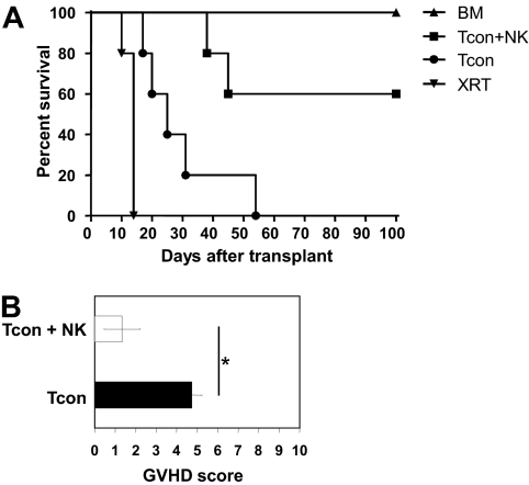
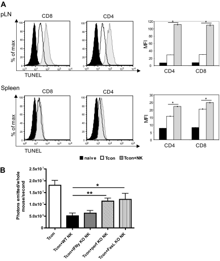
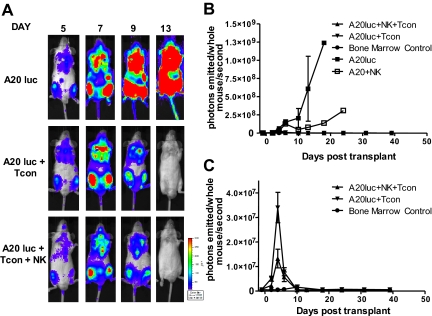
Natural killer (NK) cells suppress graft-versus-host disease (GVHD) without causing GVHD themselves. Our previous studies demonstrated that allogeneic T cells and NK cells traffic similarly after allogeneic bone marrow transplantation (BMT). We therefore investigated the impact of donor NK cells on donor alloreactive T cells in GVHD induction. Animals receiving donor NK and T cells showed improved survival and decreased GVHD score compared with controls receiving donor T cells alone. Donor T cells exhibited less proliferation, lower CD25 expression, and decreased interferon-gamma (IFN-gamma) production in the presence of NK cells. In vivo, we observed perforin- and Fas ligand (FasL)-mediated reduction of donor T cell proliferation and increased T cell apoptosis in the presence of NK cells. Further, activated NK cells mediated direct lysis of reisolated GVHD-inducing T cells in vitro. The graft-versus-tumor (GVT) effect was retained in the presence of donor NK cells. We demonstrate a novel mechanism of NK cell-mediated GVHD reduction whereby donor NK cells inhibit and lyse autologous donor T cells activated during the initiation of GVHD.


Figures

Figure 1
Figure 1
Donor NK cells improve survival and reduce GVHD severity after allogeneic BMT. (A) Donor natural killer (NK) cells prolong survival of mice receiving donor conventional T cells (Tcon + NK) compared with Tcon only (Tcon). Mice were sacrificed because of morbidity according to euthanasia criteria at the indicated time points. Mice receiving irradiation plus T cell–depleted bone marrow (TCD-BM) only and mice receiving irradiation only (XRT) are shown as controls. Tcon + NK animals had significantly prolonged survival compared with Tcon mice (P < .05). Data are from 5 mice per group, and are representative of 3 experiments. (B) Mice were scored for GVHD symptoms after transplantation. Tcon + NK mice had significantly lower average graft-versus-host disease (GVHD) scores than Tcon mice (*P < .05). Data are shown for day 20 after T-cell transplantation, and are representative of 2 experiments.
Figure 2
Figure 2
Donor Tcon proliferation is reduced in the presence of donor NK cells. (A) Average photons emitted from luc+ donor T cells in animals receiving TCD-BM, TCD-BM plus luc+ Tcons, or TCD-BM plus luc+ Tcons and donor NK cells; n = 4-5 animals per group, representative of 2 experiments. Signal intensity is higher in luc+ Tcon compared with luc+ Tcon + NK animal (P < .05, day 6). Error bars represent SE. (B) Donor Tcons reisolated from spleen and lymph nodes of mice that underwent transplantation are gated on the donor marker CD45.1, and CD4 and CD8. (C) CFSE histograms of donor Tcons reisolated from pLN on day 4 in the presence (Tcon + NK) or absence (Tcon) of donor NK cells. More CD4+ and CD8+ cells have divided in the Tcon group. Percentage of undivided cells is indicated. One representative FACS of 4 pooled mice is shown, representative of 2 independent experiments. (D) Mean fluorescence intensity of CFSE staining for CD4+ (top) and CD8+ (bottom) cells reisolated from pLN on day 4 in the presence (Tcon + NK) or absence (Tcon) of donor NK cells. A significantly lower mean fluorescence intensity (MFI) (*P < .001 for CD4+ and CD8+) was observed in Tcon groups.
Figure 3
Figure 3
NK cells reduce activated and IFN-γ–secreting donor T cells. (A) CD25 expression on CD45.1+ Foxp3 and CD4+ or CD8+ donor T cells reisolated from spleen, pLN, or mLN of Tcon (white histograms) or Tcon + NK mice (gray histograms) on day 4, or a naive control mouse (dotted line). Percentage of T cells from Tcon or Tcon + NK animals expressing CD25 is quantified in the bar graphs. CD25 expression is reduced in Tcon + NK animals in the spleen, mLN, and pLN (*P < .05, **P < .01, ***P < .001). (B) Intracellular cytokine staining demonstrates decreased IFN-γ secretion from Tcon + NK (gray histograms) compared with Tcon (white histograms) animals in the spleen on day 3. Graphic representation of summary statistics with SE appear to the right of histograms (*P < .05, IL-2 was NS). Unstimulated staining controls are depicted in the dotted lines. (C) Quantification of absolute cell numbers reisolated from Tcon and Tcon + NK animals on day 4. Donor CD4+ and CD8+ numbers are reduced in Tcon + NK mice from pLN and spleen (*P < .05, **P < .01). For all, data are averaged from 3 to 4 mice and are representative of 2 to 3 independent experiments. Error bars represent SE.
Figure 4
Figure 4
Increased ratio of splenic donor Tregs to total donor Tcons in the presence of NK cells. (A) The percentage of donor Tregs (CD45.1+CD4+CD25+Foxp3+) reisolated from Tcon + NK groups was significantly higher in spleen and mLN (***P < .001 and *P < .05, respectively; pLN was NS) compared with Tcon. (B) Absolute donor Treg cell numbers were higher in the Tcon group compared with the Tcon + NK group (spleen, **P < .01; mLN, NS; pLN, *P < .05). Inset shows the same mLN and pLN data as large graph at higher magnification to visualize relative differences. (C) Ratios of donor Tregs/total donor Tcons (CD4+ and CD8+) were significantly higher in Tcon + NK animals than Tcon animals in the spleen (***P < .001). Results were mixed in the lymph node (mLN, NS; pLN, *P < .05). All data are from day 4 after T-cell transplantation, are averaged from 4 mice, and are representative of 3 independent experiments.
Figure 5
Figure 5
Increased donor T-cell apoptosis in lymphoid organs and perforin- and FasL-mediated reduction of donor Tcons in the presence of donor NK cells. (A) FACS-based TUNEL staining on reisolated donor Tcons on day 4 after transplantation. Histograms indicate CD8+ and CD4+ reisolated from pLNs and spleens from Tcon + NK animals (gray filled histograms) stained significantly higher for TUNEL than the Tcon group (white filled histograms) group. TUNEL staining on freshly isolated T cells is shown as a control (black filled histograms). Quantification of the mean fluorescence intensity (right panels) shows significantly increased TUNEL stain in the Tcon + NK group (*P < .001). Error bars indicate SE. Data are pooled from 3 mice and are representative of 3 independent experiments. (B) Average photons emitted from animals receiving luc+ Tcon together with NK cells from wild-type donors or donors deficient for either IFNγ (IFN-γ KO), perforin (perf KO), or FasL (FasL KO) on day 4 after T-cell transplantation. Bioluminescent signal was significantly greater in animals receiving perforin−/− or FasL−/− NK cells compared with wild-type NK cells, implicating these molecules in the mechanism of NK cell–mediated reduction of donor Tcons (*P < .05, **P < .01). One-way analysis of variance was also significant (P = .001) and Dunnett multiple comparison test showed that only the Tcon + WT NK and Tcon + perf KO NK groups were significantly different from control. Data are an average of 5 mice per group, and are representative of 2 experiments.
Figure 6
Figure 6
NK cells directly lyse activated, donor Tcons in an NKG2D-dependent manner. (A) Activated NK cells alone (solid line) or incubated with isotype control antibody (dashed line) lysed donor Tcon reisolated from spleen and lymph node of transplant recipients. Lysis was significantly reduced with NKG2D-blocking antibody at a 20:1 effector to target ratio and for LN at a 5:1 E/T ratio (*P < .01, **P < .001, 5:1 spleen was NS). Data are an average of triplicates. (B) Expression of the NKG2D ligand Rae1γ was higher on donor CD4+ and CD8+ cells reisolated from lymph node and spleen on day 5 than naive, freshly isolated T cells as detected by FACS analysis. Data are representative of 3 experiments. (C) The MFI of Rae1γ NKG2D ligand expression was significantly higher on activated CD69+ than CD69 CD4+ and CD8+ cells from spleen and lymph node (***P < .001). Data are pooled from 3 mice per group and are representative of 2 experiments.
Figure 7
Figure 7
NK cells maintain GVT response to A20. (A) Ventral BLI images of animals that received a transplant of TCD-BM plus A20-luc+/yfp, A20-luc+/yfp plus Tcons, or A20-luc+/yfp plus Tcons and NK cells. Both Tcon and Tcon + NK groups reject the tumor within 2 weeks after transplantation. (B) Quantification of BLI images from panel A. Compared with A20 controls, Tcon and Tcon + NK groups have significantly lower BLI signal. NK cells alone provide some antitumor capability but fail to clear the tumor. (C) BLI quantification shown without the A20 control. TCD-BM–only recipients are shown as controls. Tcons + NK have significantly lower BLI at day 4 after T-cell transplantation (P < .05). Data points are average BLI from 4 to 5 mice, and are representative of 2 experiments. Error bars represent SE.

Similar articles

Cited by

References

    1. Copelan EA. Hematopoietic stem-cell transplantation. N Engl J Med. 2006;354(17):1813–1826. - PubMed
    1. Ferrara JL, Deeg HJ. Graft-versus-host disease. N Engl J Med. 1991;324(10):667–674. - PubMed
    1. Yokoyama WM, Kim S, French AR. The dynamic life of natural killer cells. Annu Rev Immunol. 2004;22:405–429. - PubMed
    1. Asai O, Longo DL, Tian ZG, et al. Suppression of graft-versus-host disease and amplification of graft-versus-tumor effects by activated natural killer cells after allogeneic bone marrow transplantation. J Clin Invest. 1998;101(9):1835–1842. - PMC - PubMed
    1. Ruggeri L, Capanni M, Urbani E, et al. Effectiveness of donor natural killer cell alloreactivity in mismatched hematopoietic transplants. Science. 2002;295(5562):2097–2100. - PubMed

Publication types

MeSH terms