. 2010 Jun 3;115(22):4478-87.
doi: 10.1182/blood-2009-12-257261. Epub 2010 Mar 16.

The pan-HDAC inhibitor vorinostat potentiates the activity of the proteasome inhibitor carfilzomib in human DLBCL cells in vitro and in vivo

Affiliations

The pan-HDAC inhibitor vorinostat potentiates the activity of the proteasome inhibitor carfilzomib in human DLBCL cells in vitro and in vivo

Girija Dasmahapatra et al. Blood. .

Retraction in

Abstract

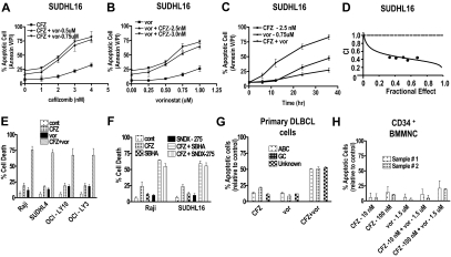
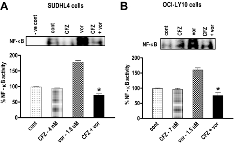
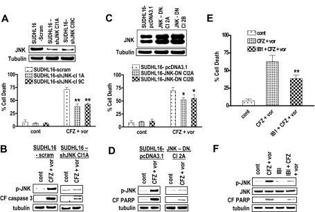
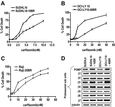
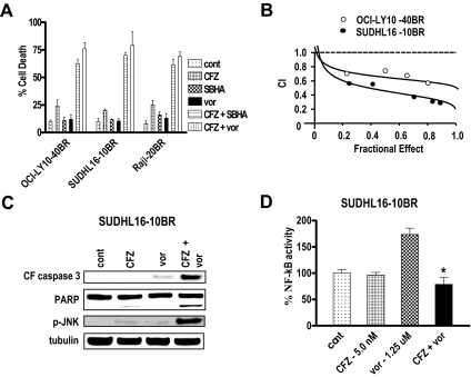
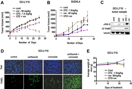
Interactions between histone deacetylase inhibitors (HDACIs) and the novel proteasome inhibitor carfilzomib (CFZ) were investigated in GC- and activated B-cell-like diffuse large B-cell lymphoma (ABC-DLBCL) cells. Coadministration of subtoxic or minimally toxic concentrations of CFZ) with marginally lethal concentrations of HDACIs (vorinostat, SNDX-275, or SBHA) synergistically increased mitochondrial injury, caspase activation, and apoptosis in both GC- and ABC-DLBCL cells. These events were associated with Jun NH2-terminal kinase (JNK) and p38MAPK activation, abrogation of HDACI-mediated nuclear factor-kappaB activation, AKT inactivation, Ku70 acetylation, and induction of gammaH2A.X. Genetic or pharmacologic JNK inhibition significantly diminished CFZ/vorinostat lethality. CFZ/vorinostat induced pronounced lethality in 3 primary DLBCL specimens but minimally affected normal CD34(+) hematopoietic cells. Bortezomib-resistant GC (SUDHL16) and ABC (OCI-LY10) cells exhibited partial cross-resistance to CFZ. However, CFZ/vorinostat dramatically induced resistant cell apoptosis, accompanied by increased JNK activation and gammaH2A.X expression. Finally, subeffective vorinostat doses markedly increased CFZ-mediated tumor growth suppression and apoptosis in a murine xenograft OCI-LY10 model. These findings indicate that HDACIs increase CFZ activity in GC- and ABC-DLBCL cells sensitive or resistant to bortezomib through a JNK-dependent mechanism in association with DNA damage and inhibition of nuclear factor-kappaB activation. Together, they support further investigation of strategies combining CFZ and HDACIs in DLBCL.


Figures

Figure 1
Figure 1
Cotreatment with CFZ and HDACIs leads to synergistic induction of cell death in DLBCL (both GC and ABC) cells and primary DLBCL cells, but not in normal hematopoietic cells. (A) SUDHL16 cells were treated with various CFZ concentrations (1.0-4.0nM) in conjunction with fixed vorinostat (0.5 or 0.75mM) concentrations for 36 hours, after which cell death was monitored by flow cytometry and annexin V/propidium iodide staining. (B) SUDHL16 cells were treated with various vorinostat (0.25-1.0μM) concentrations in the presence or absence of fixed concentrations of CFZ (2.5 or 3.0nM) for 36 hours, after which cell death was monitored by flow cytometry and annexin V/propidium iodide staining. (C) SUDHL16 cells were treated with CFZ 2.5nM with or without vorinostat 0.75μM for the indicated intervals, after which cell death was monitored by flow cytometry and annexin V/propidium iodide staining. (D) Fractional effect values were determined by comparing results obtained for untreated controls and treated cells after exposure to agents administered at a fixed ratio, after which median dose effect analysis was used to characterize the nature of the interaction. Combination index (CI) values less than 1.0 denote a synergistic interaction. (E) Cells were treated with minimally toxic concentrations of CFZ (Raji 5nM, SUDHL4 4nM, OCI-LY10 7nM, OCI-LY3 5nM) in the presence or absence of vorinostat (Raji 2.0μM, SUDHL4, OCI-LY10, OCI-LY3 1.5μM) for 48 hours, after which cell death was monitored by 7-AAD and 3,3-dihexyloxacarbocyanine iodide (DiOC6) staining. (F) Raji and SUDHL16 cells were treated with minimally toxic concentrations of CFZ (SUDHL16 3nM, Raji 5nM), SBHA (SUDHL16 30μM, Raji 50μM), and SNDX-275 (1.0μM) for 36 to 48 hours, after which cell death was monitored by 7-AAD and DiOC6 staining. (G) Primary human DLBCL cells were isolated as described in “Methods” and resuspended in medium containing 10% fetal calf serum at a cell density of 0.75 × 10⁶/mL cells. They were then treated with CFZ (ABC sample 2nM, GC sample 100nM, unknown type 4nM) with or without vorinostat (ABC sample 0.5μM, GC sample 1.0μM, unknown type 0.75μM) for 16 hours. The percentage of apoptotic cells was monitored by annexin V/propidium iodide staining, and the percentage of dead cells was normalized to controls. Viability of the 3 primary specimens without treatment was 60% to 70%, 80% to 85%, and 70% to 75% for the ABC, the GC, and the unknown types, respectively. (H) CD34+ cells were collected from the bone marrow, isolated by an immunomagnetic bead separation technique as described in “Methods,” and exposed to CFZ with or without vorinostat as indicated for 48 hours. Cell death was monitored by annexin V/propidium iodide staining, and the percentage of apoptotic cells was normalized to controls. For all studies, values represent the means for 3 experiments performed in triplicate plus or minus SD.
Figure 2
Figure 2
Combined exposure of SUDHL16 and OCI-LY10 cells to CFZ and vorinostat leads to a pronounced increase in caspase activation, mitochondrial damage, JNK activation, and DNA damage. SUDHL16 cells were treated with CFZ (3.0nM) with or without vorinostat (0.75μM) for 14 hours. (A) Cytosolic (S-100) fractions were obtained as described in “S-100 fractions,” and the expression of cytochrome C and apoptosis inducing factor (AIF) was monitored by Western blot. (B-E) Proteins from whole-cell lysates were prepared, and expression of the indicated proteins was determined by Western blotting after drug exposure identical to that described in panel A. Expression of acetylated Ku70 and acetylated Ku86 was determined by immunoprecipitation with Ku70 and Ku86 antibodies followed by Western blotting with acetyl-Lys primary antibody. (F) OCI-LY10 cells were treated with CFZ (7.0nM) with or without vorinostat (1.5μM) for 24 hours. Cells were lysed, sonicated, proteins denatured, and subjected to Western blot analysis using the indicated primary antibodies. Each lane was loaded with 20 μg of protein; blots were stripped and reprobed with antibodies directed against tubulin or actin to ensure equivalent loading and transfer. Results are representative of 3 separate experiments.
Figure 3
Figure 3
CFZ blocks HDACI-mediated NF-κB activation in both SUDHL4 and OCI-LY10 DLBCL cells. (A) SUDHL4 DLBCL cells were treated with CFZ (4.0nM) with or without vorinostat (1.5μM), and (B) OCI-LY10 DLBCL cells were treated with CFZ (7.0nM) with or without vorinostat (1.5μM) for 18 hours. Nuclear protein was extracted using Nuclear Extract Kit (Active Motif), and NF-κB activity was determined using an ELISA TransAM NF-κB p65 Transcription Factor Assay Kit (Active Motif), as described in “NF-κB activity.” Inset: After identical treatment, the same nuclear proteins were subjected to EMSA gel shift assays to assess NF-κB DNA binding as described in “NF-κB activity.” Values represent the means for triplicate determinations for 3 separate experiments. *Significantly less than values for vorinostat alone (P < .002).
Figure 4
Figure 4
Pharmacologic and genetic interruption of the JNK pathways significantly diminishes CFZ/vorinostat lethality in SUHDL16 cells. (A) SUDHL16 cells stably transfected with JNK shRNA or vectors encoding a scrambled sequence were exposed to CFZ (3.0nM) plus vorinostat (0.75μM). After 36 hours of drug exposure, cell death was monitored by 7-AAD staining and flow cytometry. Inset: Relative expression of JNK protein in SUDHL16-scrambled sequence and shJNK clones. (B) After 14 hours of drug exposure as in panel A, Western blot analysis was used to monitor protein expression of phospho-JNK and the cleavage fragment of caspase-3 (CF caspase-3). (C) SUDHL16 cells stably transfected with JNK-DN cDNA or empty vector (pcDNA3.1) were exposed to CFZ (3.0nM) plus vorinostat (0.75μM). After 36 hours of drug exposure, cell death was monitored by 7-AAD staining and flow cytometry. Inset: Expression of JNK protein in SUDHL16 empty vector and JNK-DN clones. (D) After 14 hours of drug exposure as in panel C, Western blot analysis was used to monitor protein expression of phospho-JNK and cleaved PARP. (E) SUDHL16 cells pretreated with the selective JNK inhibitor IB1 (ALX 159-600; 10μM) for 2 hours were exposed to CFZ (3.0nM) plus vorinostat (0.75μM) for 36 hours. At the end of drug exposure, cell death was monitored by 7-AAD staining and flow cytometry. (F) After 14 hours of drug exposure as described in panel E, Western blot analysis was used to monitor protein expression of phospho-JNK, JNK, and cleaved PARP. For all studies, blots were stripped and reprobed with antitubulin antibodies to ensure equal loading and transfer of proteins. Each lane was loaded with 20 μg of protein. All values represent the means of triplicate experiments performed on 3 separate occasions plus or minus SD. (A,E) **Significantly less than values for scrambled sequence clone or control (P < .01). (C) *Significantly less than values for empty-vector controls (P < .05).
Figure 5
Figure 5
Bortezomib-resistant SUDHL16-10BR, OCI-LY10-40BR, and Raji –20BR cells exhibit partial cross-resistance to CFZ and up-regulation of proteasomal subunits. SUDHL16 and SUDHL16-10BR (A), OCI-LY10 and OCI-LY10-40BR (B), and Raji and Raji-20BR (C) cells were treated with the indicated concentration of CFZ for 36, 48, and 48 hours, respectively, after which cell death was monitored by 7-AAD staining by flow cytometry. Values represent the means for experiments performed in triplicate on 3 separate occasions plus or minus SD. (D) Logarithmically growing SUDHL16, SUDHL16-10BR, OCI-LY10, and OCI-LY10-40BR cells were harvested and proteins extracted. Western blot analysis was then performed using the indicated antibodies. Blots were stripped and reprobed with antitubulin antibodies to ensure equal loading and transfer of protein. Each lane was loaded with 20 μg of protein. Two additional experiments yielded equivalent results.
Figure 6
Figure 6
The CFZ/vorinostat regimen potently induces apoptosis in bortezomib-resistant SUDHL16-10BR, Raji 20-BR, and OCY-LY10-40BR cells. (A) OCI-LY10-40BR, Raji-20BR, and SUDHL16-10BR cells were treated with minimally toxic concentrations of CFZ and either vorinostat or SBHA for 48, 48, or 36 hours, respectively. Concentrations were as follows: (OCI-LY10-40BR) CFZ (20nM) with or without SBHA (40μM), or vorinostat (1.5μM); (Raji-20BR) CFZ (15nM) with or without SBHA (40μM) or vorinostat (2.0μM); (SUDHL16-10BR) CFZ (5nM) with or without SBHA (30μM) or vorinostat (1.25μM). Cell death was monitored by 7-AAD/DiOC6 staining and flow cytometry. (B) Fractional effect values were determined for the combination treatments, after which median dose effect analysis was used to characterize the nature of the interactions. Combination index (CI) values less than 1.0 denote a synergistic interaction. (C) SUDHL16-10 BR cells were exposed to the indicated concentrations of CFZ and vorinostat as described in panel A for 24 hours, after which Western blot analysis was performed using the indicated antibodies. Blots were stripped and reprobed with antitubulin antibodies to ensure equal loading and transfer of protein. Each lane was loaded with 20 μg of protein. Two additional experiments yielded equivalent results. (D) SUDHL16-10BR cells were treated with the indicated concentrations of CFZ and vorinostat for 24 hours. Nuclear protein was extracted using the Nuclear Extract Kit (Active Motif), and NF-κB activity was determined using an ELISA TransAM NF-κB p65 Transcription Factor Assay Kit (Active Motif), as described in “NF-κB activity.” *Significantly less than values for cells treated with vorinostat alone (P < .002). In all cases, values represent the means for experiments performed in triplicate on 3 separate occasions plus or minus SD.
Figure 7
Figure 7
Vorinostat potentiates CFZ-mediated DNA damage, apoptosis, and tumor growth suppression in an in vivo OCI-LY10 xenograft model. NIH-III nude mice were injected in the flank with (A) 10 × 106 OCI-LY10 cells or (B) 10 × 106 SUDHL4 cells and treated with the indicated doses CFZ with or without vorinostat twice weekly on days 1 and 2 as described in “Animal studies.” Tumor volumes were measured twice every week, and mean tumor volume was plotted against days of treatment. (C) Tumor samples were extracted from mice and lysed with lysis buffer followed by sonication. Western blotting was performed using the extracted proteins, which were then probed with the indicated primary antibodies. Each lane was loaded with 20 μg of protein; blots were subsequently stripped and reprobed with antibodies to tubulin to ensure equivalent loading and transfer. (D) Tumor samples were extracted after the 25th day of treatment and fixed to slides as described in “TUNEL assays of tissue sections.” TUNEL assays were performed on the fixed cells, which were also counterstained with 4,6-diamidino-2-phenylindole. Photomicrographs were obtained with an Olympus fluorescence microscope (original magnification ×20). (E) Weights of each mouse after various treatment regimens were monitored weekly, and the mean weight of each group was plotted against days of treatment.

Comment in

  • Findings of Research Misconduct.
    [No authors listed] [No authors listed] NIH Guide Grants Contracts (Bethesda). 2015 Dec 18:NOT-OD-16-040. NIH Guide Grants Contracts (Bethesda). 2015. PMID: 26693581 Free PMC article. No abstract available.
  • Findings of Research Misconduct.
    [No authors listed] [No authors listed] Fed Regist. 2015 Dec 10;80(237):76703-76704. Fed Regist. 2015. PMID: 27737268 Free PMC article. No abstract available.

Similar articles

Cited by

References

    1. Bea S, Zettl A, Wright G, Salaverria I, et al. Diffuse large B-cell lymphoma subgroups have distinct genetic profiles that influence tumor biology and improve gene-expression-based survival prediction. Blood. 2005;106(9):3183–3190. - PMC - PubMed
    1. Davis RE, Brown KD, Siebenlist U, Staudt LM. Constitutive nuclear factor kappaB activity is required for survival of activated B cell-like diffuse large B cell lymphoma cells. J Exp Med. 2001;194(12):1861–1874. - PMC - PubMed
    1. Coiffier B, Lepage E, Briere J, et al. CHOP chemotherapy plus rituximab compared with CHOP alone in elderly patients with diffuse large-B-cell lymphoma. N Engl J Med. 2002;346(4):235–242. - PubMed
    1. Compagno M, Lim WK, Grunn A, et al. Mutations of multiple genes cause deregulation of NF-kappaB in diffuse large B-cell lymphoma. Nature. 2009;459(7247):717–721. - PMC - PubMed
    1. Dunleavy K, Pittaluga S, Czuczman MS, et al. Differential efficacy of bortezomib plus chemotherapy within molecular subtypes of diffuse large B-cell lymphoma. Blood. 2009;113(24):6069–6076. - PMC - PubMed

Publication types

MeSH terms