. 2010 May 28;285(22):17112-22.
doi: 10.1074/jbc.M110.116830. Epub 2010 Mar 16.

Structure of a DNA polymerase alpha-primase domain that docks on the SV40 helicase and activates the viral primosome

Affiliations

Structure of a DNA polymerase alpha-primase domain that docks on the SV40 helicase and activates the viral primosome

Hao Huang et al. J Biol Chem. .

Abstract

DNA polymerase alpha-primase (pol-prim) plays a central role in DNA replication in higher eukaryotes, initiating synthesis on both leading and lagging strand single-stranded DNA templates. Pol-prim consists of a primase heterodimer that synthesizes RNA primers, a DNA polymerase that extends them, and a fourth subunit, p68 (also termed B-subunit), that is thought to regulate the complex. Although significant knowledge about single-subunit primases of prokaryotes has accumulated, the functions and regulation of pol-prim remain poorly understood. In the SV40 replication model, the p68 subunit is required for primosome activity and binds directly to the hexameric viral helicase T antigen, suggesting a functional link between T antigen-p68 interaction and primosome activity. To explore this link, we first mapped the interacting regions of the two proteins and discovered a previously unrecognized N-terminal globular domain of p68 (p68N) that physically interacts with the T antigen helicase domain. NMR spectroscopy was used to determine the solution structure of p68N and map its interface with the T antigen helicase domain. Structure-guided mutagenesis of p68 residues in the interface diminished T antigen-p68 interaction, confirming the interaction site. SV40 primosome activity of corresponding pol-prim mutants decreased in proportion to the reduction in p68N-T antigen affinity, confirming that p68-T antigen interaction is vital for primosome function. A model is presented for how this interaction regulates SV40 primosome activity, and the implications of our findings are discussed in regard to the molecular mechanisms of eukaryotic DNA replication initiation.


Figures
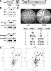
FIGURE 1.
FIGURE 1.
The p68 N terminus interacts directly with the Tag AAA+ domain. A, diagram of the p68 subunit of human pol-prim. CDK, cluster of sites phosphorylated by cyclin A-dependent kinase (15); CTD, C-terminal domain composed of an oligonucleotide-oligosaccharide binding (OB) subdomain and a phosphoesterase (PE) subdomain (13). B, purified His-tagged full-length p68 (lanes 1–5), p68(1–107) (lanes 6–10), and p68(108–598) (lanes 11–15) were incubated with anti-Tag beads in the presence (+) or absence (−) of purified Tag. Bound proteins were analyzed by Western blotting with an anti-His or anti-Tag antibody as indicated. C, yeast two-hybrid analysis of p68 interaction with Tag. Left panel, control plate −Leu, −Trp; right panel, selective plate −Leu, −Trp, −His, −Adenine. The numbered sectors are identified in the table below. D, p68N contains the structured region of p68 1–107. 15N-1H HSQC spectra were collected for p68 1–107 (left panel) and p68 1–78 (p68N) (right panel). The disperse peaks are present in both spectra, and the peaks absent in the p68N spectrum are all found in the central region of p68 1–107. This indicates that only flexible residues were removed in truncating from 107 to 78 residues, whereas the structured region remains intact. IPs, immunoprecipitates.
FIGURE 2.
FIGURE 2.
p68N adopts a compact, helical fold. A, stereo view of the backbone heavy atom trace of the 20 p68N structures with the lowest violation energies. B, ribbon diagram of the structure closest to the mean. C, structural alignment of p68N (cyan) with residues 1–75 of the pol ϵ B-subunit, Dpoe2NT (salmon) (Protein Data Bank code 2v6z (37)). D, electrostatic surface potential of p68N (top panel) and Dpoe2NT (bottom panel) with red representing negative charge and blue representing positive charge. The orientation on the left is the same as shown in A–C.
FIGURE 3.
FIGURE 3.
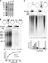
Loss of the p68 N terminus abolishes initiation of SV40 DNA replication and primosome activity. A, purified WT, Δ1–107, and Δ1–78 pol-prim were separated by SDS-PAGE and Coomassie-stained. M, protein size markers. B, initiation of SV40 replication was assayed in monopolymerase reactions containing increasing amounts of WT (lanes 1–4) or Δ1–107 (lanes 5–8) pol-prim. Control reactions lacked pol-prim (lane 9) or Tag (lane 10). The reaction products were resolved by alkaline electrophoresis, visualized by autoradiography, and quantified (lower panel). DNA size markers are indicated (M). C, primosome activity of 160, 320, or 640 ng of WT (lanes 1–3) or Δ1–107 pol-prim (lanes 6–8) on ssDNA precoated with RPA was stimulated by Tag. Control reactions lacked Tag (lanes 4 and 9) or both Tag and RPA (lanes 5 and 10). The reaction products were separated by alkaline electrophoresis, visualized by autoradiography, and quantified (lower panel).
FIGURE 4.
FIGURE 4.
p68N interacts with Tag 303–627. A, isothermal titration calorimetry titration of p68N into Tag 303–627. The data were fit to a single-site binding model, which provided a Kd of 6 ± 1 μm. B, NMR differential line broadening assay. Unlabeled Tag 303–627 was added to 15N-labeled p68N. The relative peak intensity after addition was plotted versus residue number to identify selectively broadened residues. The dashed line indicates the mean plus one standard deviation. C, the selectively broadened residues map to a hydrophobic patch (yellow) surrounded by negative charge (red). This surface represents a potential binding site for Tag.
FIGURE 5.
FIGURE 5.
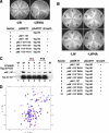
Structure-guided mutational analysis of the Tag-binding surface of p68N. A and B, single residue substitutions in p68(1–107) were screened in yeast two-hybrid assays for interaction with GST-TagHD. Left panel, control plate; right panel, growth on the selective plate indicates interaction. The numbered sectors are identified in the tables below. C, glutathione (G) beads were incubated with 10 μg of WT His-tagged p68(1–107) or mutants I14A, I14V, F15A, and F15Y as indicated in the presence (+) or absence (−) of GST-TagHD (lanes 1–6). Proteins bound to the beads were detected by Western blotting with anti-His or anti-GST antibody. Lane 7, input (IN) p68(1–107) (200 ng). D, the p68N I14A domain maintains the WT fold. 15N-1H HSQC spectra were collected for WT (red) and I14A (blue) p68N. The spectra overlay very well, indicating that no major structural perturbation was induced by the I14A mutation.
FIGURE 6.
FIGURE 6.
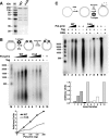
A single residue substitution in the Tag-interacting surface of p68N diminishes SV40 primosome activity. A, purified pol-prim containing p68 WT or substitution I14A were separated by SDS-PAGE and stained with Coomassie. B, SV40 initiation was assayed in monopolymerase reactions containing increasing amounts of WT (lanes 1–4) or I14A pol-prim (lanes 5–8) and analyzed by alkaline electrophoresis and autoradiography. DNA size markers are shown (M). Reaction products were quantified; background (lanes 9 and 10) was subtracted from incorporation in lanes 1–8 (lower panel). C, primosome activity of 200, 400, or 600 ng of WT (lanes 1–3) or I14A pol-prim (lanes 6–8) was assayed in the presence of 600 ng of Tag on ssDNA precoated with 1 μg RPA. Control reactions lacked Tag (lanes 4 and 9), both Tag and RPA (lanes 5 and 10), or pol-prim (lane 11). The reaction products were analyzed by alkaline electrophoresis and autoradiography. DNA size markers are shown (M). The reaction products were quantified; incorporation in the negative control reaction (lane 11) was subtracted from that in lanes 1–10. Incorporation in lanes 1–4 and 6–9 is graphed as a fraction of that in lanes 5 and 10, respectively.
FIGURE 7.
FIGURE 7.
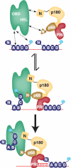
A working model for regulation of SV40 primosome activity via p68N docking with Tag. Top panel, the three major proteins in the SV40 primosome: Tag hexamer (green) containing the origin DNA- and RPA-binding domains (OBD) and helicase domains (HEL); RPA (blue) with four oligonucleotide-oligosaccharide binding fold domains A–D bound with 5′ to 3′ polarity to ssDNA (red) (occluding 28–30 nucleotides) and the RPA32C domain (cyan) tethered to oligonucleotide-oligosaccharide binding domain D; pol-prim with the p180 DNA polymerase (orange), p68 subunit (brown), and p58-p48 primase (PRI, red). Wavy solid lines represent flexible linkers between folded domains. Dotted lines with arrowheads denote transient physical interactions among protein modules. Middle panel, ssDNA is transiently exposed by Tag contacts with RPA that remodel it into a more compact, lower affinity ssDNA binding mode and stabilize it as a ternary complex (23–25). The p68N domain of pol-prim is proposed to dock on the AAA+ domain of the Tag hexamer. The N-terminal region of the p180 subunit also docks on TagHD at an unidentified site; the reported interaction of p48/p58 with Tag has not been mapped. A single pol-prim complex binds to a Tag hexamer in solution (28). The ensemble of these interactions is proposed to position primase on the naked template to synthesize an RNA primer (red) and transfer it to p180 for extension (bottom panel). Thus, p68N docking on Tag hexamer effectively directs primase activity in the SV40 primosome.

Similar articles

Cited by

References

    1. Hamdan S. M., Richardson C. C. (2009) Annu. Rev. Biochem. 78, 205–243 - PubMed
    1. Langston L. D., Indiani C., O'Donnell M. (2009) Cell Cycle 8, 2686–2691 - PMC - PubMed
    1. Kunkel T. A., Burgers P. M. (2008) Trends Cell Biol. 18, 521–527 - PMC - PubMed
    1. Copeland W. C., Wang T. S. (1993) J. Biol. Chem. 268, 26179–26189 - PubMed
    1. Kuchta R. D., Stengel D. (2010) Biochim. Biophys. Acta 1804, 1180–1189 - PMC - PubMed

Publication types

LinkOut - more resources