. 2010 Apr;120(4):1337-44.
doi: 10.1172/JCI41305. Epub 2010 Mar 15.

Analysis of mutations from SCID and Omenn syndrome patients reveals the central role of the Rag2 PHD domain in regulating V(D)J recombination

Affiliations

Analysis of mutations from SCID and Omenn syndrome patients reveals the central role of the Rag2 PHD domain in regulating V(D)J recombination

Chrystelle Couëdel et al. J Clin Invest. 2010 Apr.

Abstract

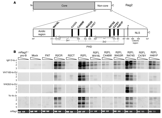
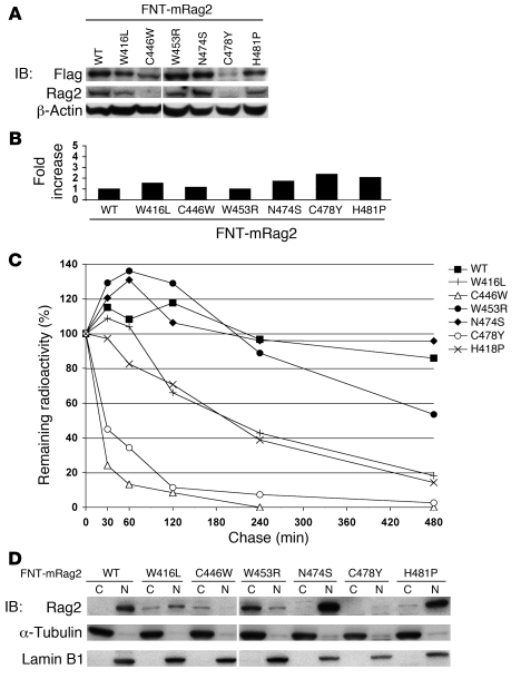
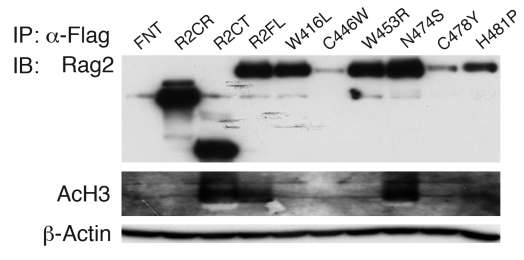
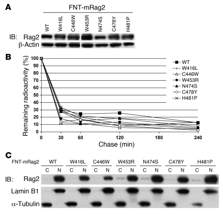
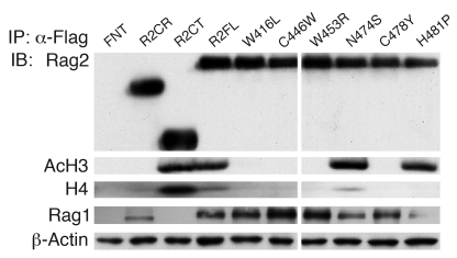
Rag2 plays an essential role in the generation of antigen receptors. Mutations that impair Rag2 function can lead to severe combined immunodeficiency (SCID), a condition characterized by complete absence of T and B cells, or Omenn syndrome (OS), a form of SCID characterized by the virtual absence of B cells and the presence of oligoclonal autoreactive T cells. Here, we present a comparative study of a panel of mutations that were identified in the noncanonical plant homeodomain (PHD) of Rag2 in patients with SCID or OS. We show that PHD mutant mouse Rag2 proteins that correspond to those found in these patients greatly impaired endogenous recombination of Ig gene segments in a Rag2-deficient pro-B cell line and that this correlated with decreased protein stability, impaired nuclear localization, and/or loss of the interaction between Rag2 and core histones. Our results demonstrate that point mutations in the PHD of Rag2 compromise the functionality of the entire protein, thus explaining why the phenotype of cells expressing PHD point mutants differs from those expressing core Rag2 protein that lacks the entire C-terminal region and is therefore devoid of the regulation imposed by the PHD. Together, our findings reveal the various deleterious effects of PHD Rag2 mutations and demonstrate the crucial role of this domain in regulating antigen receptor gene assembly. We believe these results reveal new mechanisms of immunodeficiency in SCID and OS.


Figures

Figure 1
Figure 1. T-B-SCID/OS Rag2PHD mutants fail to support recombination at the endogenous Ig loci.
(A) Schematic of the Rag2 protein showing the core and non-core regions and enhancement of the C-terminal non-core region that includes the acidic hinge region, the PHD, and the noncanonical NLS overlapping the phosphorylation site at residue T490 (thin black vertical bar) and the cationic region between residues 499–508. Thick black vertical bars indicate residues mutated in OS/SCID patients; mutations analyzed in this study are in bold. Anchor residues forming the noncanonical PHD finger are indicated, and related interactions with Zinc ions (Zn1 and Zn2) are represented by gray bars; L1 and L2 indicate segments forming loops between pairs of zinc-coordinating residues, as previously described (18). C and H represent Zn+2 coordinating cysteine and histidine residues present in the Rag2 PHD domain. (B) A mouse Rag2–/– pro-B cell line was retrovirally transduced and selected to express constructs of interest: empty vector (Mock), FNT tag, FNT-tagged R2CR, C terminus of Rag2 (R2CT), R2FL, or full-length T-B-SCID/OS Rag2 mutants. Genomic DNA was harvested from the above cell lines, and Southern Blot analysis was performed on PCR-amplified products of IgH D-to-J, VH7183-to-DJ, VHQ52-to-DJ, and Vκ-to-Jκ rearrangements, as indicated to the left of each panel. Numbers of the left side of the figure (1 to 5) represent different recombination products. Triangles at the top of panels indicate PCR amplification of 400 ng, 200 ng, and 25 ng of DNA template. A fragment of Rag1 gene was amplified as a loading control. For each type of rearrangement, samples were loaded on the same gel, and detection intensity was identical. PCR from nontransduced cells was also used as control. The results shown are representative of 2 independent experiments. mRag2–/–, mouse Rag2–/–.
Figure 2
Figure 2. T-B-SCID/OS Rag2PHD mutations affect Rag2 stability and cellular localization in pro-B cells.
A mouse Rag2–/– pro-B cell line was retrovirally transduced to express FNT-tagged full-length wild-type Rag2 or T-B-SCID/OS mutants. (A) Western blot analysis was performed on whole cell extracts from retrovirally transduced pro-B cell lines for Flag, Rag2, and β-actin. Samples were loaded on the same gel, and the length of exposure was identical. The vertical line between FNT–mouse Rag2–C446W and –W453R mutants indicates a sample that was excluded from our study. (B) Total mRNA was isolated from retrovirally transduced pro-B cell lines, and Rag2 transcription levels were determined by Q-PCR. The average of 2 independent experiments are graphed as fold increase compared with mRNA expression of FNT–mouse Rag2. (C) Retrovirally transduced pro-B cell lines were pulse labeled with [35S] methionine/cysteine to determine degradation of wild-type Rag2 and T-B-SCID/OS mutants. Radiolabeled proteins were IP at various times after pulse with anti-Rag2 antibody, fractionated by SDS PAGE, and quantified on a PhosphorImager. Data were normalized to the radioactivity levels at the end of pulse. The graph presents the average from 2 independent experiments. (D) Cellular localization of Rag2 in retrovirally transduced pro-B cell lines was determined by Western blotting analysis of fractionated cytoplasm (C) and nuclear (N) extracts (top). Purity and loading of the fractions were analyzed by Western blotting for α-tubulin (middle) and Lamin B1 (bottom). The vertical line between FNT–mouse Rag2–C446W and –W453R is as in A.
Figure 3
Figure 3. T-B-SCID/OS Rag2PHD mutations disrupt Rag2-histone interaction in pro-B cells.
Anti-Flag IPs were performed on whole cell extracts from retrovirally transduced pro-B cells expressing constructs of interest: FNT tag, FNT-tagged core Rag2, C terminus of Rag2, R2FL, or full-length T-B-SCID/OS Rag2 mutants. Western immunoblotting analysis is shown for Rag2 and acetylated Histone H3 (AcH3). Flag IP loading was analyzed by Western blotting for β-actin on 1:1,000 input volume.
Figure 4
Figure 4. T-B-SCID/OS Rag2PHD mutants showed wild-type stability and cellular localization in 293T cells.
(A) Whole cell extracts from 24-hour posttransfection 293T cells, coexpressing full-length mouse Rag1 and FNT-tagged full-length wild-type mouse Rag2 or full-length T-B-SCID/OS Rag2 mutants were analyzed by Western blotting for Rag2 and β-actin. The vertical line between FNT–mouse Rag2–C446W and –W453R is as in Figure 2A. (B) Transfected 293T cells were pulsed labeled with [35S] methionine/cysteine to determine degradation of wild-type Rag2 and T-B-SCID/OS mutants. Radiolabeled proteins were IP at various times after pulse with anti-Rag2 antibody, fractionated by SDS PAGE, and quantified on a PhosphorImager. Data are normalized to the radioactivity levels at the end of pulse. (C) Cellular localization of Rag2 and T-B-SCID/OS mutants in 293T cells was determined by Western blotting analysis of fractionated cytoplasm and nuclear extracts (top). Purity and loading of the fractions were controlled by Western blotting for Lamin B1 (middle) and α-tubulin (bottom).
Figure 5
Figure 5. T-B-SCID/OS Rag2PHD mutations disrupt Rag2-histone interaction in 293T cells.
Anti-Flag IPs were performed on whole cell extracts from 48-hour posttransfection cells coexpressing full-length Rag1 and constructs of interest: FNT tag, FNT-tagged core Rag2, C terminus of Rag2, R2FL, or full-length T-B-SCID/OS Rag2 mutants. Flag IPs were analyzed by Western immunoblotting for Rag2, acetylated Histone H3, Histone H4 (H4), or Rag1 and 1:1,000 input volume with anti–β-actin. Samples were loaded on the same gel, and the length of exposure was identical. The vertical line between mutants FNT–mouse Rag2–C446W and –aW453R is as in Figure 2A.

References

    1. Schatz DG, Oettinger MA, Baltimore D. The V(D)J recombination activating gene, RAG-1. Cell. 1989;59(6):1035–1048. doi: 10.1016/0092-8674(89)90760-5. - DOI - PubMed
    1. Oettinger MA, Schatz DG, Gorka C, Baltimore D. RAG-1 and RAG-2, adjacent genes that synergistically activate V(D)J recombination. Science. 1990;248(4962):1517–1523. doi: 10.1126/science.2360047. - DOI - PubMed
    1. Soulas-Sprauel P, et al. V(D)J and immunoglobulin class switch recombinations: a paradigm to study the regulation of DNA end-joining. Oncogene. 2007;26(56):7780–7791. doi: 10.1038/sj.onc.1210875. - DOI - PubMed
    1. Puebla-Osorio N, Zhu C. DNA damage and repair during lymphoid development: antigen receptor diversity, genomic integrity and lymphomagenesis. Immunol Res. 2008;41(2):103–122. doi: 10.1007/s12026-008-8015-3. - DOI - PubMed
    1. Alt FW, et al. Ordered rearrangement of immunoglobulin heavy chain variable region segments. EMBO J. 1984;3(6):1209–1219. - PMC - PubMed

Publication types