. 2010 Apr;120(4):1111-24.
doi: 10.1172/JCI40269. Epub 2010 Mar 15.

Chemotherapy enhances tumor cell susceptibility to CTL-mediated killing during cancer immunotherapy in mice

Affiliations

Chemotherapy enhances tumor cell susceptibility to CTL-mediated killing during cancer immunotherapy in mice

Rupal Ramakrishnan et al. J Clin Invest. 2010 Apr.

Abstract

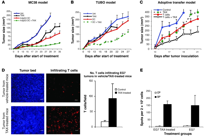
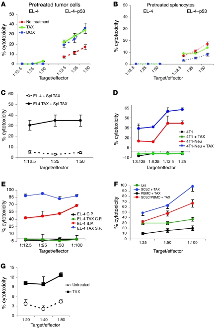
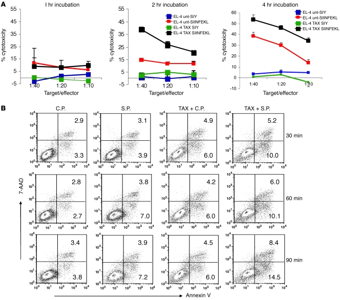
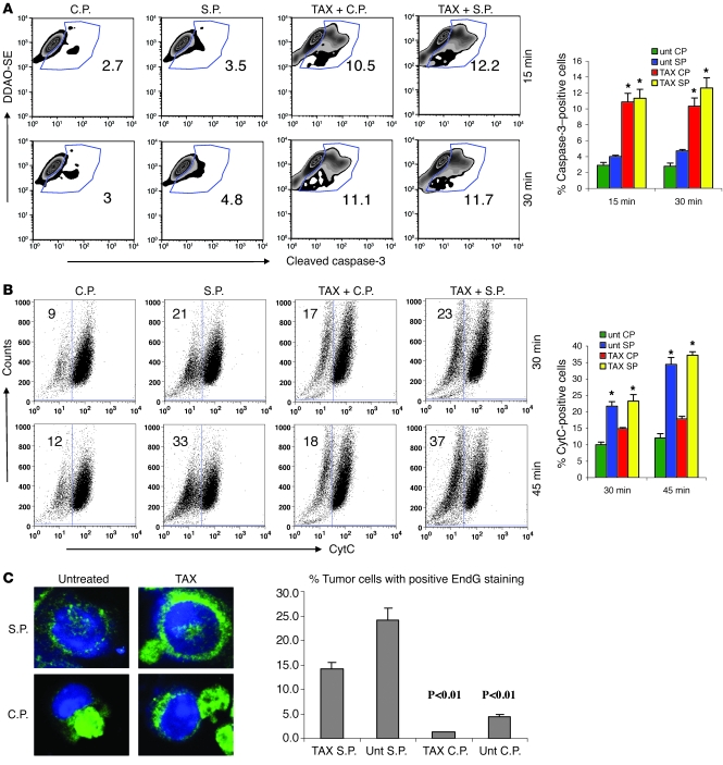
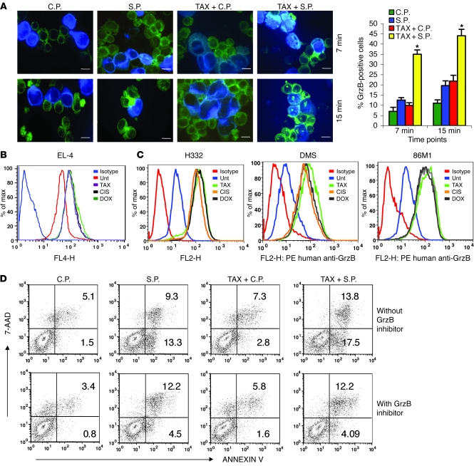
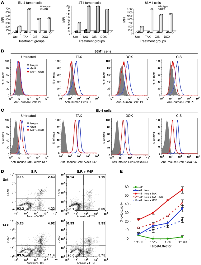
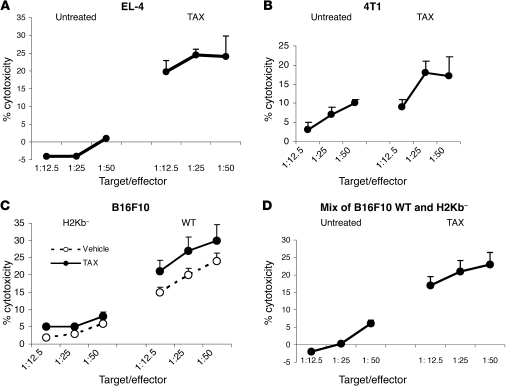
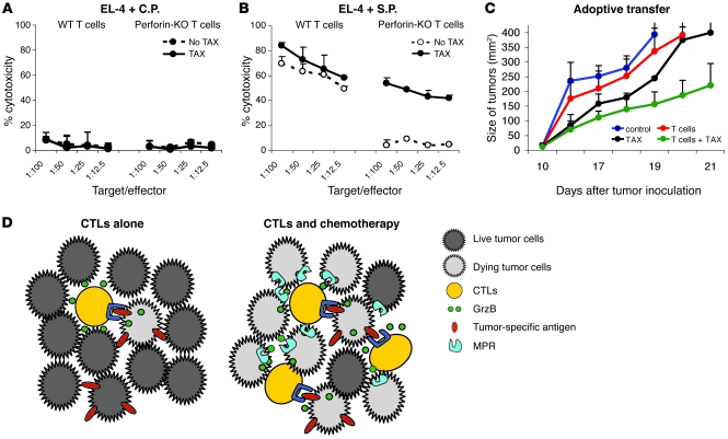
Cancer immunotherapy faces a serious challenge because of low clinical efficacy. Recently, a number of clinical studies have reported the serendipitous finding of high rates of objective clinical response when cancer vaccines are combined with chemotherapy in patients with different types of cancers. However, the mechanism of this phenomenon remains unclear. Here, we tested in mice several cancer vaccines and an adoptive T cell transfer approach to cancer immunotherapy in combination with several widely used chemotherapeutic drugs. We found that chemotherapy made tumor cells more susceptible to the cytotoxic effect of CTLs through a dramatic perforin-independent increase in permeability to GrzB released by the CTLs. This effect was mediated via upregulation of mannose-6-phosphate receptors on the surface of tumor cells and was observed in mouse and human cells. When combined with chemotherapy, CTLs raised against specific antigens were able to induce apoptosis in neighboring tumor cells that did not express those antigens. These data suggest that small numbers of CTLs could mediate a potent antitumor effect when combined with chemotherapy. In addition, these results provide a strong rationale for combining these modalities for the treatment of patients with advanced cancers.


Figures

Figure 1
Figure 1. Combined effect of chemotherapy and immunotherapy.
(A) Murine colon carcinoma tumors were established in C57BL/6 mice by s.c. injection of MC38 tumor cells. Treatment was started 5 days after tumor inoculation. Mice in treatment groups (DC, DC + TAX) were injected s.c. with 5 × 10⁵ DCs transduced with recombinant adenovirus containing the mouse wild-type p53 gene (Adp53). Immunizations were repeated twice at 7-day intervals. Treatment with TAX (12.5 mg/kg) was started 3 days after the second immunization. Tumor size was calculated by multiplying the 2 longest dimensions. n = 5 mice group. The experiment was repeated twice with the same results. Unt, untreated. (B) Mammary carcinoma TUBO was established s.c. in BALB/c mice. The treatment times and intervals were the same as in A. DCs used for immunizations were loaded with 10 μg/ml Neu-derived peptide. n = 5 mice group. The experiment was repeated twice with the same results. (C) T cells from mice immunized with OVA-derived peptide SIINFEKL were transferred to EG7 tumor–bearing C57BL/6 mice by i.v. injection of 5 × 10⁶ cells. The treatment protocol for the treatment with TAX and adoptive transfer is described in Methods. n = 4 mice group. The experiment was repeated once with the same results. (D) EG7 tumors were established s.c. in C57BL/6 mice. On day 16, the mice were treated with TAX (12.5 mg/kg) i.p. Three days later, they were administered 5 × 10⁶ CMAC-labeled T cells from mice immunized with SIINFEKL. The tumors were removed 24 hours later, and cryosections were prepared. The slides were observed under a Leica fluorescence microscope, and 20 high-power (×400) fields were counted per slide. Right: Number of T cells per tumor field in 3 mice per group. P < 0.05, 2-sided t test. Scale bars: 5 μm. In AD, data are shown as mean ± SEM. (E) EG7 tumors were established by s.c. injection of 3 × 10⁵ cells. When tumor reached 1.5 cm in diameter, 5 × 10⁶ OT-T cells were injected i.v. in each mouse. After 3 days, half of the mice received 12.5 mg/kg BW TAX i.p. Splenocytes were collected 6 days later and restimulated with control (CP) or specific (SP) peptides. IFN-γ production was evaluated by ELISPOT assay. The number of spots per 2 × 10⁵ lymph node cells are shown. Each point represents mean ± SD of 6 replicates.
Figure 2
Figure 2. Chemotherapy sensitizes tumor cells to the cytotoxic effect of CTLs.
(A) Tumor-free C57BL/6 mice were immunized with Kb-bound p53-derived peptide. Splenocytes were isolated, restimulated with the specific peptide for 6 days, and used as effectors in a CTL assay against EL-4 target cells loaded with specific (p53) or control peptides. EL-4 cells were pretreated overnight with 1.5 μg/ml DOX or 12.5 nM TAX. Standard 4-hour 51Cr-release assay was performed. For AE, data are shown as mean ± SEM. Each experiment was performed 3 times with the same results. (B) Splenocytes from immunized mice were pretreated overnight with DOX or TAX at doses described above and used as effectors against EL-4 target cells loaded with specific or control peptides in a Cr51 release assay. (C) Results of a CTL assay wherein both effectors and targets were pretreated with TAX as described above. Spl, splenocytes. (D) T cells from mice immunized with Neu-derived p66 peptide were used as effectors against either nonmodified 4T1 cells or 4T1-Neu cells transfected with Neu-expressing plasmid. The targets were pretreated with TAX for 18 hours. (E) T cells from 2C-transgenic mice were used as effectors against EL-4 cells loaded with the specific (S.P.) or control (C.P.) peptides. The target cells were pretreated with TAX overnight. (F) Cytotoxicity against H332 SCLC cells was measured in duplicate in a standard 6-hour chromium release assay as detailed in Methods. PBMCs stimulated with SCLC tumor cells lysates were used as effectors. The viability of target cells and PBMCs in all tests was similar and greater than 85% at onset of assay. UNT, SCLC and PBMCs were not treated with TAX; SCLC+TAX, tumor cells were pretreated for 18 hours with 100 nM TAX; PBMC+TAX, PBMCs were pretreated for 18 hours with 100 nM TAX; SCLC/PBMC+TAX, tumor cells and PBMCs were treated with TAX. (G) Survivin-specific T cells were generated by 2 rounds of stimulation of mononuclear cells from HLA-A2+ healthy volunteers as described elsewhere (18). The T cells were column purified and added to the tumor cells at indicated ratios. Primary tumor from HLA-A2+ patient with non-SCLC cancer was used as target. The tumor cells were treated with 50 nM TAX 18 hours prior to 51Cr release assay.
Figure 3
Figure 3. Kinetics of apoptosis in combination therapy.
(A) OT-1 T cells were mixed with EL-4 cells that had been loaded either with specific SIINFEKL or a control SIYRYYG (SIY) peptide. Target cells were either pretreated with TAX overnight or were left untreated. Chromium release assay was performed in duplicate 1, 2, and 4 hours after start of the incubation. The 2h and 4h data were obtained from different reproducible experiments. (B) For the detection of early apoptosis, the effector OT-1 T cells were labeled with DDAO-SE and incubated with target EL-4 cells loaded either with specific or control peptides at a 20:1 ratio. After the indicated incubation time, cells were stained with Annexin V–FITC and 7-AAD. The proportion of Annexin V–positive cells was measured within the population of tumor cells by flow cytometry. Typical result of 1 of 3 performed experiments is shown.
Figure 4
Figure 4. Mechanism of apoptosis induced by CTLs and TAX.
(A) Cleaved caspase-3 in tumor cells. Untreated and TAX-treated EL-4 targets were labeled with DDAO-SE and loaded with control or specific peptide. The tumor cells were incubated with purified OT-1 T cells at a 1:10 ratio. After the indicated incubation time, cells were permeabilized and labeled with PE-conjugated antibody against cleaved caspase-3. Target cells were gated, and the levels of cleaved caspase-3 in the target cells were analyzed by flow cytometry. *P < 0.05 versus untreated EL-4 cells. (B) CytC in tumor cells. Experiments were performed as described in Figure 2B. The cells were permeabilized, fixed, and labeled with anti-CytC antibody. *P < 0.05 versus untreated EL-4 cells. In A and B, typical results of 1 experiment (left) and mean ± SEM of 5 experiments (right) are shown. (C) EndG in tumor cells. EL-4 cells were treated with TAX for 18 hours and loaded with control or specific peptides as described above. OT-1 T cells were labeled with Po-Pro-3 iodide to distinguish them from tumor cells and incubated with target EL-4 cells at a 10:1 ratio for 1 hour. The cells were fixed and stained with anti-EndG antibody and DAPI. Visualization of the staining in the nuclei was performed using a Leica confocal microscope. The images were analyzed with Image Pro Plus 6.2 software. One hundred tumor cells were counted, and the proportion of those with positive nuclear staining was determined. The P values were calculated using Fisher 2-tailed exact test. Data are presented as mean ± SEM.
Figure 5
Figure 5. The effect of chemotherapy on permeability of tumor cells to GrzB.
(A) EL-4 cells were treated with TAX and loaded with control or specific peptides as described above. Cells were labeled with CMAC and mixed with OT-1 cells at a 1:10 ratio and incubated for 7 and 15 minutes, then fixed and stained with anti-GrzB monoclonal antibody, followed by FITC-conjugated secondary antibody. The proportion of GrzB+ cells among blue target cells was calculated in triplicate by counting 200 target cells. Scale bars: 10 μm. Data represent mean ± SEM of 4 experiments. *P < 0.05 versus untreated tumor cells loaded with control peptide. (B) CMAC-labeled TAX-, DOX-, or CIS-treated EL-4 cells were incubated with 1 μg/ml recombinant mouse GrzB for 30 minutes. Cells were fixed and stained with anti–mouse GrzB antibody. The presence of GrzB was detected by flow cytometry. Histogram overlays represent isotype control, untreated EL-4 cells, and TAX-, CIS-, or DOX-treated cells. Each experiment was repeated 3 times with the same results. (C) The above experimental procedure was used for detecting the presence of GrzB+ cells in human cell lines. Human recombinant GrzB and PE-conjugated anti–human GrzB antibody were used. Tumor cells were treated overnight with 12.5 nM TAX, 25 ng/ml DOX, or 25 ng/ml CIS. These doses did not decrease cell viability after overnight treatment by more than 5% but significantly reduced the cell growth of tumor cells. After 48 hours, these doses caused apoptosis in more than 90% of cells. Isotype control IgG was used in all samples and showed similar values. Isotype control of TAX-treated cells is shown. Histogram overlays represent isotype, untreated tumor cells, and TAX-, CIS-, or DOX-treated cells. Each experiment was repeated twice with the same results. (D) OT-1 T cells were pretreated with GrzB inhibitor I (Z-AAD-CMK) prior to incubation with EL-4 cells. The target cells were labeled with CMAC; the effectors were labeled with DDAO-SE. The target population was assessed for apoptosis using Annexin V–FITC/7AAD staining and analyzed using flow cytometry. Apoptosis was measured among tumor cells. Data represent 2 experiments with the same results.
Figure 6
Figure 6. The role of MPR in the synergistic effect of chemotherapy and CTLs.
(A) EL-4, 4T1, or 86M1 tumor cells were treated with 12.5 nM TAX, 25 ng/ml CIS, or 25 ng/ml DOX for 18 hours. Cells were washed, blocked with 10% mouse or human serum for 20 minutes at 4°C, and incubated with 1:100 vol/vol cation-independent MPR antibody (Abcam) followed by staining with goat anti-rabbit IgG Alexa Fluor 647. The cells were washed and acquired on a FACSCalibur flow cytometer. The MFI is shown. Data from 1 of 2 experiments with the same results are shown. (B and C) To block MPR, M6P (Sigma-Aldrich) was used at a concentration of 20 mM. 86M1 (B) or EL-4 (C) cells were treated with TAX, CIS, or DOX as described above. The cells were incubated with M6P for 15 minutes at room temperature. After 2 washes, the cells were incubated with recombinant human (B) or mouse (C) GrzB for 2 hours. The cells were permeabilized and labeled with anti–mouse GrzB–Alexa 647 or anti–human GrzB–PE. Two experiments with the same results were performed. (D) EL-4 cells were treated with TAX and loaded with control or specific peptides as described in Methods. In the last 15 minutes of peptide loading, one-half of the cells from each treatment group were incubated with 20 mM M6P. The cells were washed and incubated with DDAO-SE–labeled activated OT-1 T cells at a 1:15 ratio. After 1.5 hours, incubated cells were labeled with Annexin V–FITC and 7AAD. DDAO-SE–negative tumor cells were gated and apoptosis measured by flow cytometry. Three experiments with the same results were performed. (E) Neu-specific T cells were generated by immunization of mice as described in Methods and used as effector cells in CTL assay. 4T1 and 4T1-Neu tumor cells were treated with TAX and M6P as described above and used as targets in 51Cr release assay. Experiments were performed in duplicate. Data are presented as mean ± SEM.
Figure 7
Figure 7. Mechanism of synergistic antitumor activity of CTLs and chemotherapy.
(A) EL-4 cells loaded with control peptide and labeled with 51Cr were mixed at a 1:1 ratio with unlabeled EL-4 cells loaded with specific peptide. The mixture of target cells was incubated with OT-1 T cells at the indicated ratios. Pretreatment of target cells with TAX was performed overnight. Standard 4-hour chromium release assay was performed in duplicate. Experiments were repeated 3 times with the same results. Appropriate positive controls were set up with each experiment (data not shown). (B) The experiment was performed essentially as described in Figure 4A. As target cells, chromium-labeled 4T1 cells mixed with unlabeled 4T1-Neu cells were used. Effector T cells were obtained from splenocytes of BALB/c mice immunized with Neu-derived peptide as described in Methods. (C) Wild-type and B16F10Kb tumor cells were used as targets in chromium release assay. Labeled target cells treated overnight with TAX were incubated in duplicate with T cells isolated from mice immunized with TRP-2–derived peptide as described in Methods. Two experiments with similar results were performed. (D) Unlabeled wild-type B16F10 tumor cells were mixed at 1:1 ratio with 51Cr labeled B16F10 cells with deleted H2Kb. These target cells were incubated with T cells from TRP-2–immunized mice, and cytotoxicity was evaluated in standard 4-hour 51Cr release assay. Two experiments with similar results were performed. Data are presented as mean ± SEM.
Figure 8
Figure 8. Perforin is not required for CTL activity if targets are treated with chemotherapy.
(A and B) Perforin-KO and C57BL/6 mice were immunized with SIINFEKL. Purified T cells from the spleens of these mice were used as effectors in 51Cr release assay. EL-4 cells loaded with control or specific peptide were used as targets. Half of the target cells were pretreated with TAX overnight. Two experiments with the same results were performed. (C) An adoptive transfer experiment using T cells from SIINFEKL-immunized wild-type or perforin-KO mice was performed according to the method described in Figure 1C. The treatment protocol is described in Methods. Tumor size was measured and presented as described in Figure 1C. Data are presented as mean ± SEM. (D) Schematic representation of the advantage of combination therapy in targeting tumor cells over the use of immunotherapy alone. The figure is a depiction of the possible mechanism of bystander lysis of nonspecific targets within tumors. Red bars indicate the presence of specific antigen on tumor cells.

Comment in

Similar articles

Cited by

References

    1. Rosenberg SA, Yang JC, Restifo NP. Cancer immunotherapy: moving beyond current vaccines. Nat Med. 2004;10(9):909–915. doi: 10.1038/nm1100. - DOI - PMC - PubMed
    1. Gribben JG, et al. Unexpected association between induction of immunity to the universal tumor antigen CYP1B1 and response to next therapy. Clin Cancer Res. 2005;11(12):4430–4436. doi: 10.1158/1078-0432.CCR-04-2111. - DOI - PubMed
    1. Antonia SJ, et al. Combination of p53 cancer vaccine with chemotherapy in patients with extensive stage small cell lung cancer. Clin Cancer Res. 2006;12(3 Pt 1):878–887. doi: 10.1158/1078-0432.CCR-05-2013. - DOI - PubMed
    1. Arlen PM, et al. A randomized phase II study of concurrent docetaxel plus vaccine versus vaccine alone in metastatic androgen-independent prostate cancer. Clin Cancer Res. 2006;12(4):1260–1269. doi: 10.1158/1078-0432.CCR-05-2059. - DOI - PMC - PubMed
    1. Schlom J, Arlen PM, Gulley JL. Cancer vaccines: moving beyond current paradigms. Clin Cancer Res. 2007;13(13):3776–3782. doi: 10.1158/1078-0432.CCR-07-0588. - DOI - PMC - PubMed

Publication types