. 2010 Apr;120(4):1253-64.
doi: 10.1172/JCI41615. Epub 2010 Mar 15.

CNS-targeted gene therapy improves survival and motor function in a mouse model of spinal muscular atrophy

Affiliations

CNS-targeted gene therapy improves survival and motor function in a mouse model of spinal muscular atrophy

Marco A Passini et al. J Clin Invest. 2010 Apr.

Abstract

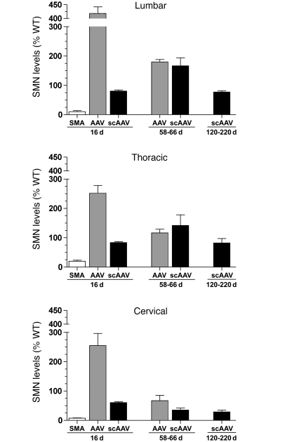
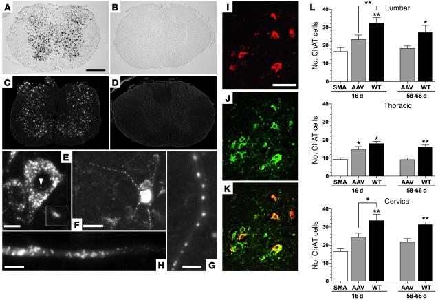
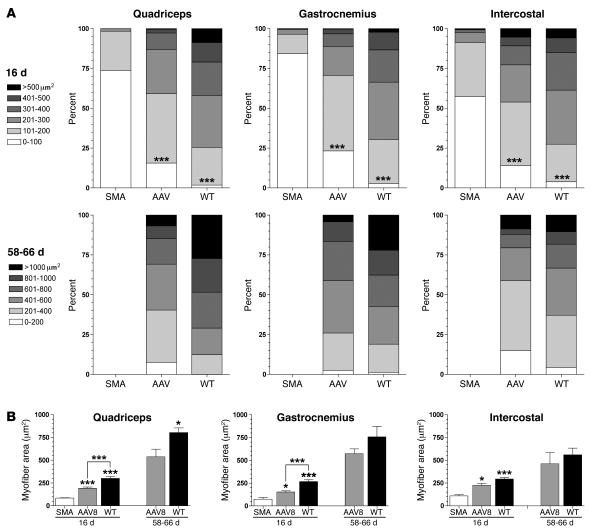
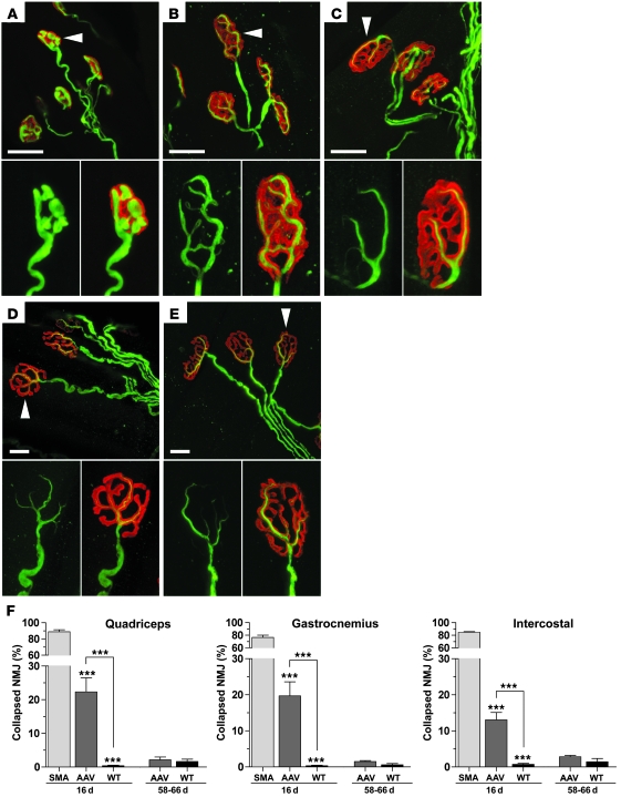
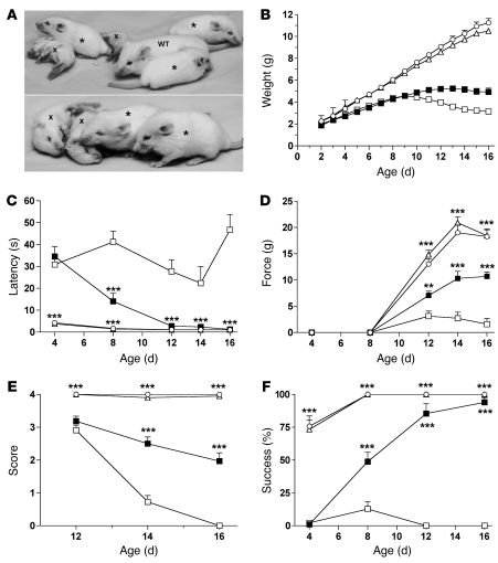
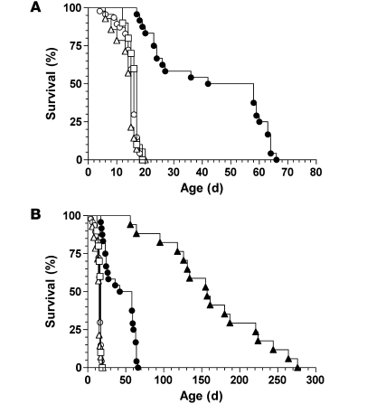
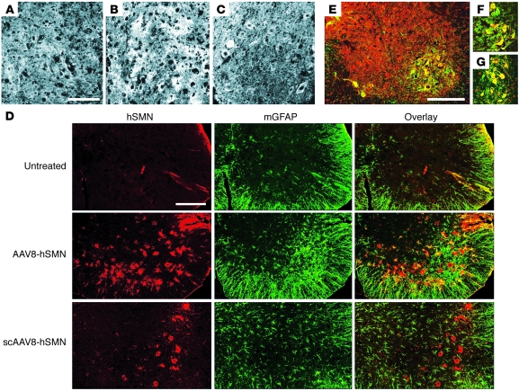
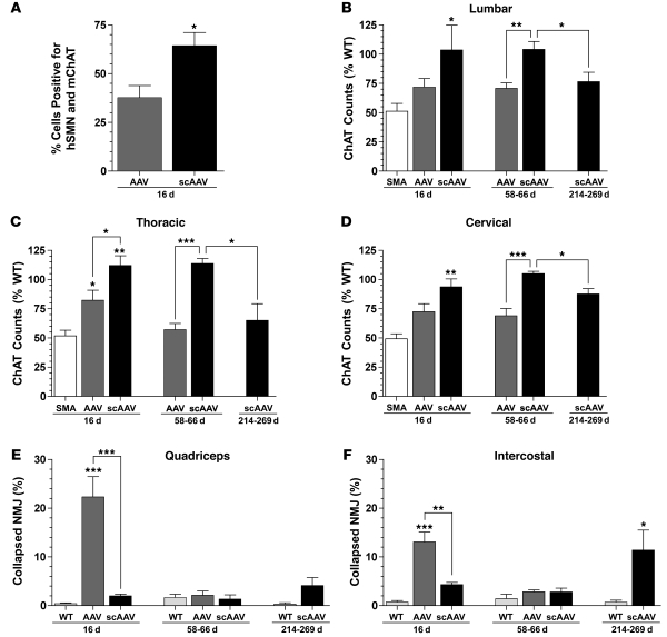
Spinal muscular atrophy (SMA) is a neuromuscular disease caused by a deficiency of survival motor neuron (SMN) due to mutations in the SMN1 gene. In this study, an adeno-associated virus (AAV) vector expressing human SMN (AAV8-hSMN) was injected at birth into the CNS of mice modeling SMA. Western blot analysis showed that these injections resulted in widespread expression of SMN throughout the spinal cord, and this translated into robust improvement in skeletal muscle physiology, including increased myofiber size and improved neuromuscular junction architecture. Treated mice also displayed substantial improvements on behavioral tests of muscle strength, coordination, and locomotion, indicating that the neuromuscular junction was functional. Treatment with AAV8-hSMN increased the median life span of mice with SMA-like disease to 50 days compared with 15 days for untreated controls. Moreover, injecting mice with SMA-like disease with a human SMN-expressing self-complementary AAV vector - a vector that leads to earlier onset of gene expression compared with standard AAV vectors - led to improved efficacy of gene therapy, including a substantial extension in median survival to 157 days. These data indicate that CNS-directed, AAV-mediated SMN augmentation is highly efficacious in addressing both neuronal and muscular pathologies in a severe mouse model of SMA.


Figures

Figure 1
Figure 1. Treatment with gene therapy increased SMN levels in the spinal cord.
Western blots were performed on the lumbar, thoracic, and cervical segments at 16, 58–66, and 120–220 days after injection. The Western blots from the 3 segments were quantified and, to control for protein levels, SMN was normalized to β-tubulin and plotted as a percentage of age-matched WT. SMA, untreated knockout (n = 5 at 16 days); AAV, AAV8-hSMN–treated SMA mice (n = 7 at 16 days; n = 5 at 58–66 days); scAAV, scAAV8-hSMN–treated SMA mice (n = 5 at each time point). Data represent the mean ± SEM.
Figure 2
Figure 2. Human SMN is expressed in the proper intracellular compartments and in motor neurons with AAV8-hSMN treatment.
In situ hybridization (A and B) and immunohistochemistry (CK) of the lumbar segment in AAV8-hSMN–treated (A, C, EK) and untreated (B and D) SMA mice at 16 days (AD) and 58–66 days (EK) after injection. The low level of endogenous hSMN in untreated SMA mice was below the threshold of detection using these assays (B and D). Vector-derived hSMN was abundantly detected in the cytoplasm and in the nucleus of transduced cells, as illustrated by the pair of gem-like structures in the nucleus (E, the arrowhead points to the hSMN immunosignal magnified in the inset). hSMN was also detected in the dendrites (F and G) and in the axons (H) of neurons. The colocalization of hSMN (I) with mChAT (J) showed that a subset of transduced cells consisted of motor neurons (K). Shown are the average numbers of ChAT immunopositive cell counts from the lumbar, thoracic, and cervical segments at both time points (L). SMA (n = 8 at 16 days); AAV (n = 8 at 16 days, n = 5 at 58–66 days); WT, untreated WT (n = 8 at 16 days, n = 5 at 58–66 days). Values represent the mean ± SEM. Scale bars: 500 microns (AD); 10 microns (E and H); 50 microns (F); 12 microns (G), 100 microns (IK). Statistical comparisons were performed with 1-way ANOVA and Bonferroni’s multiple post hoc tests at 16 days and with unpaired 2-tailed Student’s t tests at 58–66 days (L). *P < 0.05; **P < 0.01.
Figure 3
Figure 3. The myofiber cross-section area was increased with AAV8-hSMN treatment.
Stacked graphs of the quadriceps, gastrocnemius, and intercostal muscles from 16 days (first row) and 58–66 days (second row) showed that the relative distribution of myofiber sizes was similar between the AAV8-hSMN–treated SMA mice and the untreated WT controls (A). Furthermore, the number of myofibers with a cross-section area of less than 100 μm2 was significantly reduced with AAV8-hSMN treatment (A). The overall mean of the myofiber cross-section area was higher with treatment compared with untreated SMA at 16 days (B). In addition, at 58–66 days, the average myofiber size in treated SMA mice approached WT levels, particularly in the gastrocnemius and intercostal muscles (B). SMA (n = 8 at 16 days); AAV (n = 8 at 16 days; n = 5 at 58–66 days); WT (n = 8 at 16 days; n = 5 at 58–66 days). Values represent the mean ± SEM. Statistical comparisons were performed with 1-way ANOVA and Bonferroni’s multiple post hoc tests at 16 days (A and B) and with unpaired 2-tailed Student’s t tests at 58–66 days (B). *P < 0.05; ***P < 0.001.
Figure 4
Figure 4. The NMJ in the quadriceps, gastrocnemius, and intercostal muscles was improved with gene therapy.
Shown are the NMJ from the quadriceps of untreated SMA (A), AAV8-hSMN–treated SMA (B and D), and untreated WT (C and E) mice at 16 days (AC) and at 58–66 days (D and E) after injection. The pre- and postsynaptic NMJ was labeled with a neurofilament antibody (green) and with α-bungarotoxin staining (red). The arrowhead in each main panel points to the NMJ that is highlighted in the inset below. The percentage of NMJs that contained a collapsed structure similar to that shown in panel A was determined (F). WT (n = 8 at 16 days: n = 5 at 58–66 days); SMA (n = 8 at 16 days) (n = 8 at 16 days; n = 5 at 58–66 days), WT (n = 8 at 16 days; n = 5 at 58–66 days). Data represent the mean ± SEM. Statistical comparisons were performed using 1-way ANOVA and Bonferroni’s multiple post hoc tests for each muscle group at 16 days and with unpaired 2-tailed Student’s t tests at 58–66 days (F). ***P < 0.001. Scale bars: 20 microns (AE).
Figure 5
Figure 5. Treated SMA mice showed significant improvements on behavioral tests.
Treated SMA (asterisk) and untreated WT mice were substantially fitter than untreated SMA mice (labeled x) at 16 days (A). Treated SMA mice were also significantly heavier than untreated SMA controls from day 11 and onwards (B). Treated SMA mice performed significantly better than untreated SMA mice on the righting reflex (C), grip-strength (D), hind-limb splay (E), and negative-geotaxis (F) tests. Treated SMA mice were statistically similar to WT and heterozygote mice on the righting reflex and negative-geotaxis tests at 12–16 days (C and F). Untreated SMA mice (n = 11, open squares); AAV8-hSMN–treated SMA mice (n = 16, closed squares); untreated heterozygote mice (n = 16, open triangles); untreated WT mice (n = 16, open circles). Data represent the mean ± SEM. Statistical comparisons were performed for each time point using 1-way ANOVA and Bonferroni’s multiple post hoc tests. **P < 0.01; ***P < 0.001.
Figure 6
Figure 6. Gene therapy increased longevity of SMA mice.
Untreated SMA mice (n = 34, open circles) had a median survival of 15 days (A). SMA mice treated at P0 with either saline (n = 14, open triangles) or AAV8-null (n = 10, open square) had median survivals of 15 and 17 days, respectively (P > 0.05). SMA mice treated at P0 with AAV8-hSMN (n = 24, closed circles) had a median survival of 50 days (P < 0.0001), which was a 233% increase in longevity compared with untreated SMA mice (A). The survival curve revealed 2 groups of AAV8-hSMN–treated SMA mice, a first group that was found dead by 27 days and a second group that was sacrificed at 58–66 days (A). Treatment with scAAV8-hSMN showed an even greater increase in survival (B). SMA mice treated at P0 with scAAV8-hSMN (n = 17, closed triangles) had a median life span of 157 days (P < 0.0001) compared with 16 days in untreated SMA mice (n = 47, open circles) (B). As a reference, the survival plots of the SMA mice treated with AAV8-hSMN, AAV8-null, and saline were also included (B).
Figure 7
Figure 7. Comparison of AAV vector tropism in the lumbar spinal cord.
Human SMN immunostaining was performed on frozen tissue sections from untreated SMA (A and D), AAV8-hSMN–treated SMA (B, D, and F), and scAAV8-hSMN–treated SMA (CE, and G) mice at 16 days (AD, F, and G) and 157 days (E) after injection. A diffuse hSMN immunostaining pattern consistent with glial cell morphology was observed at 16 days with AAV8-hSMN (B and D). Doubling immunolabeling (yellow) of hSMN (red) and mGFAP (green) confirmed that a subset of the AAV8-hSMN–transduced cells were astrocytes (D). In contrast, scAAV8 treatment resulted in hSMN expression only in distinct cell bodies with neuronal morphology (C), which did not colocalize with GFAP (D). Double immunolabeling (yellow) of hSMN (red) and the motor neuron marker mChAT (green) confirmed that a subset of cells transduced by scAAV8-hSMN (E and G) and AAV8-hSMN (F) were motor neurons. hSMN expression (red) was also observed in the interneuronal cell layers of the spinal cord with both viral vectors, as exemplified by scAAV8-hSMN at 157 days (E). Scale bars: 100 microns (AC); 200 microns (D and E).
Figure 8
Figure 8. scAAV8-hSMN expression increased motor neuron counts and improved the NMJ in SMA mice.
Shown is the percentage of mChAT immunopositive cells that colocalized with hSMN expression in the thoracic-lumbar region at 16 days after injection (A). Shown are the average numbers of mChAT immunopositive cells in the lumbar (B), thoracic (C), and cervical (D) segments and the average percentages of collapsed NMJs in the quadriceps (E) and intercostal (F) muscles at 16, 58–66, and 214–269 days. As a reference for panels E and F, 75%–90% of NMJ in the quadriceps and intercostal muscles of untreated SMA mice contained an aberrant collapsed structure at 16 days (see Figure 4F). SMA (n = 8 at 16 days), AAV, AAV8-hSMN (n = 8 at 16 days; n = 5 at 58–66 days); scAAV (n = 5 at each time point); WT (n = 8 at 16 days; n = 5 each at 58–66 and 216–269 days). Data represent the mean ± SEM. Statistical comparisons were performed with 1-way ANOVA and Bonferroni’s multiple post hoc tests at 16 days (BF). The unpaired 2-tailed Student’s t tests compared (a) the 2 vectors to each other at 16 days (A) and 58–66 days (BD); (b) the relative number of ChAT cells in the 58- to 66-day and 214- to 269-day groups with scAAV8-hSMN treatment (BD); (c) the relative number of abnormal NMJs between the age-matched untreated WT and scAAV8-hSMN–treated SMA mice at 214–269 days (E and F). *P < 0.05; **P < 0.01; ***P < 0.001.

Similar articles

Cited by

References

    1. Lefebvre S, et al. Identification and characterization of a spinal muscular atrophy-determining gene. Cell. 1995;80(1):155–165. doi: 10.1016/0092-8674(95)90460-3. - DOI - PubMed
    1. Sumner CJ. Therapeutics development for spinal muscular atrophy. NeuroRx. 2006;3(2):235–245. doi: 10.1016/j.nurx.2006.01.010. - DOI - PMC - PubMed
    1. Samaha FJ, et al. Pulmonary function in spinal muscular atrophy. J Child Neurol. 1994;9(3):326–329. doi: 10.1177/088307389400900321. - DOI - PubMed
    1. Iannaccone ST. Modern management of spinal muscular atrophy. J Child Neurol. 2007;22(8):974–978. doi: 10.1177/0883073807305670. - DOI - PubMed
    1. Monani UR, et al. A single nucleotide difference that alters splicing patterns distinguishes the SMA gene SMN1 from the copy gene SMN2. Hum Mol Genet. 1999;8(7):1177–1183. doi: 10.1093/hmg/8.7.1177. - DOI - PubMed

Substances