

Isoflurane-induced neuroapoptosis in the neonatal rhesus macaque brain
- PMID: 20234312
- PMCID: PMC3962067
- DOI: 10.1097/ALN.0b013e3181d049cd
Isoflurane-induced neuroapoptosis in the neonatal rhesus macaque brain
Abstract
Background: Brief isoflurane anesthesia induces neuroapoptosis in the developing rodent brain, but susceptibility of non-human primates to the apoptogenic action of isoflurane has not been studied. Therefore, we exposed postnatal day 6 (P6) rhesus macaques to a surgical plane of isoflurane anesthesia for 5 h, and studied the brains 3 h later for histopathologic changes.
Method: With the same intensity of physiologic monitoring typical for human neonatal anesthesia, five P6 rhesus macaques were exposed for 5 h to isoflurane maintained between 0.7 and 1.5 end-tidal Vol% (endotracheally intubated and mechanically ventilated) and five controls were exposed for 5 h to room air without further intervention. Three hours later, the brains were harvested and serially sectioned across the entire forebrain and midbrain, and stained immunohistochemically with antibodies to activated caspase-3 for detection and quantification of apoptotic neurons.
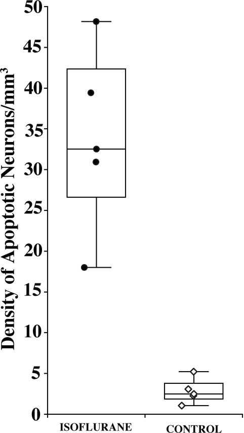
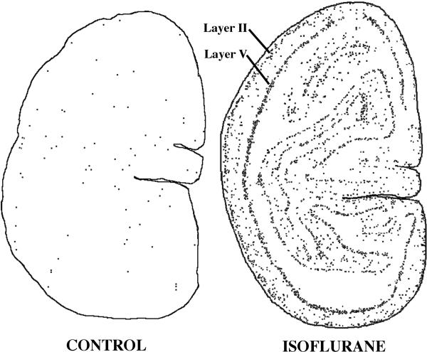
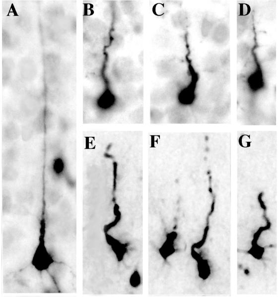
Results: Quantitative evaluation of brain sections revealed a median of 32.5 (range, 18.0-48.2) apoptotic cells/mm of brain tissue in the isoflurane group and only 2.5 (range, 1.1-5.2) in the control group (difference significant at P = 0.008). Apoptotic neuronal profiles were largely confined to the cerebral cortex. In the control brains, they were sparse and randomly distributed, whereas in the isoflurane brains they were abundant and preferentially concentrated in specific cortical layers and regions.
Conclusion: The developing non-human primate brain is sensitive to the apoptogenic action of isoflurane and displays a 13-fold increase in neuroapoptosis after 5 h exposure to a surgical plane of isoflurane anesthesia.
Figures

Comment in
-
Neurotoxicity of anesthetic agents and the developing brain in rodents and primates: the time has come to focus on human beings.Anesthesiology. 2010 Nov;113(5):1244-5; author reply 1245-6. doi: 10.1097/ALN.0b013e3181f71092. Anesthesiology. 2010. PMID: 20966669 No abstract available.
-
Isoflurane-induced neuroapoptosis in the neonatal rhesus macaque brain: isoflurane or ischemia-reperfusion?Anesthesiology. 2010 Nov;113(5):1245; author reply 1245-6. doi: 10.1097/ALN.0b013e3181f710a4. Anesthesiology. 2010. PMID: 20966672 No abstract available.
References
-
- Ikonomidou C, Bosch F, Miksa M, Bittigau P, Vöckler J, Dikranian K, Tenkova T, Stevoska V, Turski L, Olney JW. Blockade of NMDA receptors and apoptotic neurodegeneration in the developing brain. Science. 1999;283:70–4. - PubMed
-
- Ikonomidou C, Bittigau P, Ishimaru MJ, Wozniak DF, Koch C, Genz K, Price MT, Stefovska V, Hörster F, Tenkova T, Dikranian K, Olney JW. Ethanol-induced apoptotic neurodegeneration and fetal alcohol syndrome. Science. 2000;287:1056–60. - PubMed
-
- Olney JW, Tenkova T, Dikranian K, Qin YQ, Labruyere J, Ikonomidou C. Ethanol-induced apoptotic neurodegeneration in the developing C57BL/6 mouse brain. Dev Brain Res. 2002;133:115–26. - PubMed
Publication types
MeSH terms
Substances
Grants and funding
LinkOut - more resources
Full Text Sources
Other Literature Sources
Medical
Research Materials
