. 2009 Aug 17:2:13-27.
doi: 10.4137/cgm.s2814.

Molecular Imaging of Proteases in Cancer

Molecular Imaging of Proteases in Cancer

Yunan Yang et al. Cancer Growth Metastasis. .

Abstract

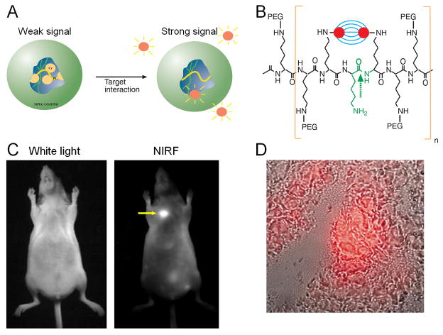
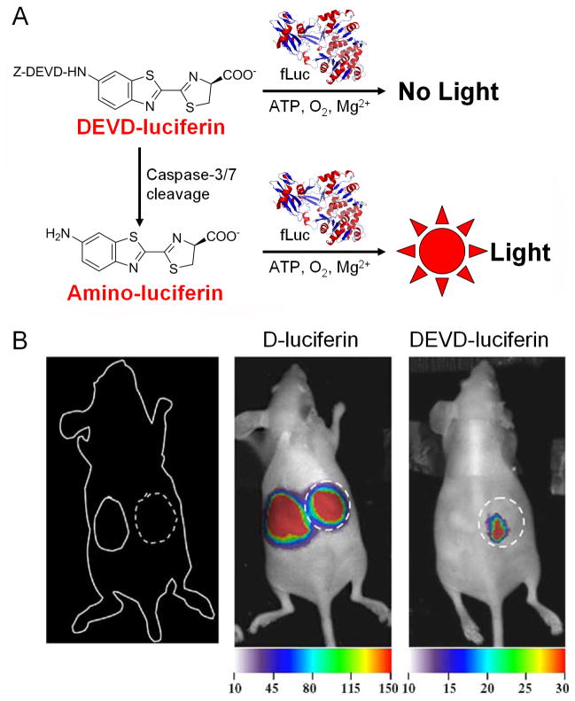
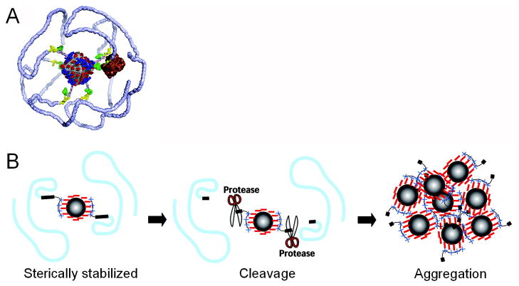
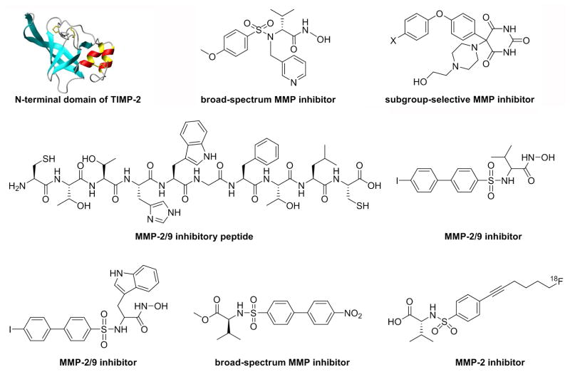
Proteases play important roles during tumor angiogenesis, invasion, and metastasis. Various molecular imaging techniques have been employed for protease imaging: optical (both fluorescence and bioluminescence), magnetic resonance imaging (MRI), single-photon emission computed tomography (SPECT), and positron emission tomography (PET). In this review, we will summarize the current status of imaging proteases in cancer with these techniques. Optical imaging of proteases, in particular with fluorescence, is the most intensively validated and many of the imaging probes are already commercially available. It is generally agreed that the use of activatable probes is the most accurate and appropriate means for measuring protease activity. Molecular imaging of proteases with other techniques (i.e. MRI, SPECT, and PET) has not been well-documented in the literature which certainly deserves much future effort. Optical imaging and molecular MRI of protease activity has very limited potential for clinical investigation. PET/SPECT imaging is suitable for clinical investigation; however the optimal probes for PET/SPECT imaging of proteases in cancer have yet to be developed. Successful development of protease imaging probes with optimal in vivo stability, tumor targeting efficacy, and desirable pharmacokinetics for clinical translation will eventually improve cancer patient management. Not limited to cancer, these protease-targeted imaging probes will also have broad applications in other diseases such as arthritis, atherosclerosis, and myocardial infarction.


Figures

Figure 1
Figure 1
Fluorescence imaging of tumors with a protease-activated NIRF probe. A) A schematic diagram of probe activation. The initial proximity of the fluorochrome to each other results in signal quenching. B) Chemical structure of the probe. Green arrow indicates the enzymatic degradation site. C) Images of a LX-1 tumor implanted into the mammary fat pad of a nude mouse after probe injection. The arrow indicates the tumor. D) NIRF image (in red) superimposed onto the correlative phase contrast microscopy image of the excised tumor. Adapted from .
Figure 2
Figure 2
Fluorescence imaging of cathepsin activity using quenched activity-based probes (qABPs). A) Mechanism of covalent inhibition of a protease by an acyloxymethyl ketone. B) Activity-dependent labeling of a protease target by a qABP. Covalent modification of the target results in loss of the quenching group, resulting in production of a fluorescently labeled enzyme. C) Optical imaging of tumors in live mice. The red circles indicate the tumor (top) and an area (bottom) used for the measurement of background signal. Adapted from , .
Figure 3
Figure 3
Bioluminescence imaging of protease activity in vivo. A) DEVD-luciferin, which is not a substrate for firefly luciferase (fLuc), becomes a fLuc substrate upon caspase-3/7 cleavage of the peptide. B) Left: A cartoon of the tumor implantation sites in mice. Dotted circle: a caspase-positive tumor. Center: The mouse was imaged for fLuc activity and tumor progression with D-luciferin injection. Right: Twenty-four hours later, the same mouse was injected with DEVD-luciferin and imaged. Note the different scale for the two images. Adapted from .
Figure 4
Figure 4
Protease-sensitive agents for MRI. A) The agent consists of a 5 nm magnetite core (gray) covered by a negatively charged citrate shell (red). The peptide-mPEG copolymers are electrostatically bound by the positively charged coupling domains (blue). The mPEG polymers (light blue) are linked to the coupling domain via the cleavage domain (yellow) and a linker peptide. A model of MMP-9 (brown) is shown for size comparison at the cleavage domain. Green: fluorescein dyes. B) When the sterically stabilized agent is exposed to a protease, the peptide-mPEG linkage is cut at the cleavage domain, resulting in a loss of sterical stabilization. This leads to particle aggregation and enhanced MR signal. Adapted from .
Figure 5
Figure 5
Representative MMPIs that have been radiolabeled for SPECT/PET applications.

Similar articles

Cited by

References

    1. Steeg PS. Tumor metastasis: mechanistic insights and clinical challenges. Nat Med. 2006;12:895–904. - PubMed
    1. Chambers AF, Groom AC, MacDonald IC. Dissemination and growth of cancer cells in metastatic sites. Nat Rev Cancer. 2002;2:563–72. - PubMed
    1. Egeblad M, Werb Z. New functions for the matrix metalloproteinases in cancer progression. Nat Rev Cancer. 2002;2:161–74. - PubMed
    1. Turk B. Targeting proteases: successes, failures and future prospects. Nat Rev Drug Discov. 2006;5:785–99. - PubMed
    1. Garcia Boy R, Knapp EM, Eisenhut M, Haberkorn U, Mier W. Enzymes/transporters. Handb Exp Pharmacol. 2008:131–43. - PubMed

LinkOut - more resources