

Ventricular dyssynchrony patterns in left bundle branch block, with and without heart failure
- PMID: 20234808
- PMCID: PMC2836215
Ventricular dyssynchrony patterns in left bundle branch block, with and without heart failure
Abstract
Background: Assessment of ventricular dyssynchrony in patients with heart failure is used for selecting candidates for cardiac resynchronization therapy (CRT). The patterns of regional distribution of dyssynchrony in a population with LBBB with and without heart failure have not been well delineated. This aspect forms the object of the study.
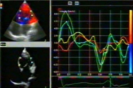
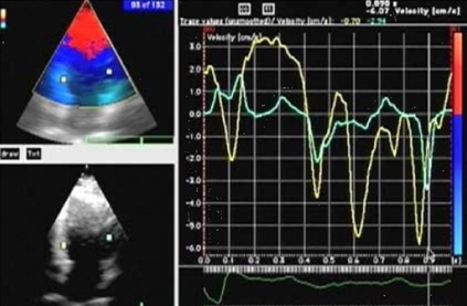
Methods: Tissue Doppler Imaging (TDI) data of consecutive patients with heart failure and LBBB (Group A) was compared with those with LBBB and normal LV function (Group B). All patients had standard 2D-echocardigraphic examination and TDI. Tissue velocity curves obtained by placing sample volumes in opposing basal and mid segments of septal, lateral, inferior, anterior and posterior walls were analyzed. Inter ventricular dyssynchrony (IVD) was assessed by the difference between aortic and pulmonary pre ejection intervals. LV dyssynchrony (LVD) was assessed by the difference in times to peak velocity. A delay of >/= 40 msec was considered significant for presence of IVD and LVD.
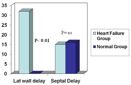
Results: There were 103 patients in Group A and 25 in Group B. The mean QRS duration and PR intervals respectively were 146 +/- 25 vs. 152+/-20 msec and 182+/- 47 vs. 165+/-36 msec. (p=NS) LVEF in the 2 groups were (32 +/- 6 % vs. 61+/- 11%; p< 0.01). Prevalence of dyssynchrony in the HF group compared to Group B was 72% vs. 16%, (P< 0.01). Lateral wall dyssynchrony in the 2 groups was 37% vs. 0% (p< 0.01) while septal dyssynchrony was 16% vs. 16% (p- NS).
Conclusions: 72% of heart failure patients with LBBB have documented dyssynchrony on TDI, which has a heterogeneous regional distribution. Dyssynchrony may be seen in LBBB and normal hearts but it is does not involve the lateral wall. Septal dyssynchrony in heart failure patients may not have the same significance as lateral wall delay.
Keywords: Dyssynchrony; Heart Failure; LBBB; Normal Heart; Tissue Doppler Imaging.
Figures
Similar articles
-
Patterns of ventricular dyssynchrony in congestive heart failure- comparison with controls and implications for patient selection for cardiac resynchronization therapy.Indian Heart J. 2010 Jul-Aug;62(4):308-12. Indian Heart J. 2010. PMID: 21280469
-
Comparison of Echocardiographic Markers of Cardiac Dyssynchrony and Latest Left Ventricular Activation Site in Heart Failure Patients with and without Left Bundle Branch Block.J Tehran Heart Cent. 2013 Apr;8(2):95-100. Epub 2013 Apr 28. J Tehran Heart Cent. 2013. PMID: 23967031 Free PMC article.
-
Diminished left ventricular dyssynchrony and impact of resynchronization in failing hearts with right versus left bundle branch block.J Am Coll Cardiol. 2007 Oct 9;50(15):1484-90. doi: 10.1016/j.jacc.2007.07.011. Epub 2007 Sep 24. J Am Coll Cardiol. 2007. PMID: 17919569
-
Assessment of mechanical dyssynchrony in cardiac resynchronization therapy.Dan Med J. 2014 Dec;61(12):B4981. Dan Med J. 2014. PMID: 25441737 Review.
-
Understanding the Application of Mechanical Dyssynchrony in Patients with Heart Failure Considered for CRT.J Cardiovasc Dev Dis. 2024 Feb 17;11(2):64. doi: 10.3390/jcdd11020064. J Cardiovasc Dev Dis. 2024. PMID: 38392278 Free PMC article. Review.
Cited by
-
Intraventricular Dyssynchrony among patients with left bundle branch block.Pak J Med Sci. 2018 Mar-Apr;34(2):390-392. doi: 10.12669/pjms.342.14103. Pak J Med Sci. 2018. PMID: 29805414 Free PMC article.
-
Left Ventricular "Longitudinal Rotation" and Conduction Abnormalities-A New Outlook on Dyssynchrony.J Clin Med. 2023 Jan 17;12(3):745. doi: 10.3390/jcm12030745. J Clin Med. 2023. PMID: 36769391 Free PMC article.
-
Association between left ventricular mechanical dyssynchrony with myocardial perfusion and functional parameters in patients with left bundle branch block.J Nucl Cardiol. 2013 Apr;20(2):253-61. doi: 10.1007/s12350-013-9673-7. Epub 2013 Jan 25. J Nucl Cardiol. 2013. PMID: 23354659
References
-
- Cazeau S, et al. Effects of multisite biventricular pacing in patients with heart failure and intraventricular conduction delay. N Engl J Med. 2001;344:873. - PubMed
-
- Abraham WT, et al. Cardiac resynchronization in chronic heart failure. N Engl J Med. 2002;346:1845. - PubMed
-
- Bristow MR, et al. Cardiac resynchronization therapy with or without an implantable defibrillator in advanced chronic heart failure. N Engl J Med. 2004;350:2140. - PubMed
-
- Cleland JG, et al. Cardiac Resynchronization Heart Failure (CARE-HF) Study. The effect of cardiac resynchronization on morbidity and mortality in heart failure. N Engl J Med. 2005;352:1539. - PubMed
-
- Pitzalis MV, et al. Cardiac resynchronization therapy tailored by echocardiographic evaluation of ventricular asynchrony. J Am Coll Cardiol. 2002;40:1615. - PubMed
LinkOut - more resources
Full Text Sources
Research Materials
Miscellaneous