Review
. 2010 Apr 13;49(14):2987-98.
doi: 10.1021/bi902153g.

Practically useful: what the Rosetta protein modeling suite can do for you

Affiliations
Free PMC article
Review

Practically useful: what the Rosetta protein modeling suite can do for you

Kristian W Kaufmann et al. Biochemistry. .
Free PMC article

Abstract

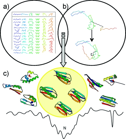
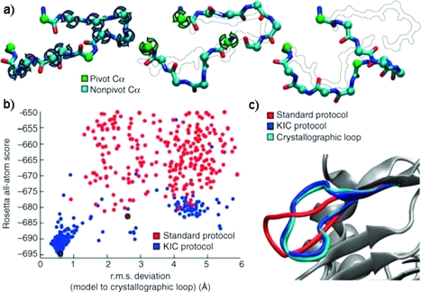
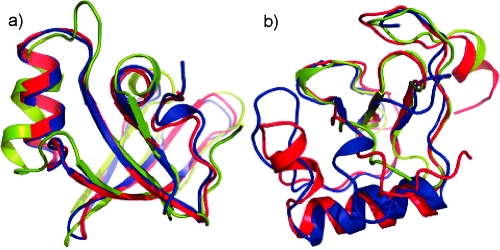
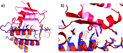
The objective of this review is to enable researchers to use the software package Rosetta for biochemical and biomedicinal studies. We provide a brief review of the six most frequent research problems tackled with Rosetta. For each of these six tasks, we provide a tutorial that illustrates a basic Rosetta protocol. The Rosetta method was originally developed for de novo protein structure prediction and is regularly one of the best performers in the community-wide biennial Critical Assessment of Structure Prediction. Predictions for protein domains with fewer than 125 amino acids regularly have a backbone root-mean-square deviation of better than 5.0 A. More impressively, there are several cases in which Rosetta has been used to predict structures with atomic level accuracy better than 2.5 A. In addition to de novo structure prediction, Rosetta also has methods for molecular docking, homology modeling, determining protein structures from sparse experimental NMR or EPR data, and protein design. Rosetta has been used to accurately design a novel protein structure, predict the structure of protein-protein complexes, design altered specificity protein-protein and protein-DNA interactions, and stabilize proteins and protein complexes. Most recently, Rosetta has been used to solve the X-ray crystallographic phase problem.


Figures

Figure 1
Figure 1
De novo folding algorithm. Rosetta starts from (a) fragment libraries with sequence-dependent (φ and ψ) angles that capture the local conformational space accessible to a sequence. (b) Combining different fragments from the libraries folds the protein through optimization of non-local contacts. The low-resolution energy function depicted in panel c smoothes the rough energy surface, resulting in a deep, broad minimum for the native conformation. Metropolis Monte Carlo minimization drives the structure toward the global minimum.
Figure 2
Figure 2
Kinematic loop closure. (a) The kinematic loop closure algorithm samples φ and ψ angles at the cyan Cα spheres from a residue specific Ramachandran map. The φ and ψ angles at green Cα spheres are determined analytically to close the loop. (b) The energy vs rmsd plot shows accuracies for the prediction of loop conformation better than 1 Å achieved through the improved sampling offered by the kinematic closure protocol. (c) The kinematic closure prediction (blue) closely resembles the crystallographic conformation (cyan). From (24) reprinted with permission from Nature Methods.
Figure 3
Figure 3
Comparative modeling CASP performance. Raman and colleagues demonstrated that comparative models refined with Rosetta improved upon the best template structure available for several CASP targets, for example, (a) T0492 and (b) T0464. The native structure is colored blue, the best submitted Rosetta model red, and the best template structure green. The Rosetta models for T0492 resulted in atomic-level accuracy for side chains in the core of the protein. For T0464, a 25-residue insertion was predicted which resulted in models that were significantly improved over the best templates available. One of the models was further improved in the model refinement category. From (19) reprinted with permission from Proteins.
Figure 4
Figure 4
De novo protein structure prediction from sparse EPR data. Alexander et al. demonstrated that approximately one low-resolution distance restraint for every four residues is sufficient to determine a model of T4 lysozyme that is accurate at an atomic level of detail. The distance restraints had been determined using SDSL-EPR experiments. The native T4 lysozyme structure is colored gray, while the model with an rmsd of 1.0 Å is shown with a rainbow coloring scheme. Side chain conformations in the core of the protein are accurately represented in the model. From (39) reprinted with permission from Structure.
Figure 5
Figure 5
Crysallographic phase problem. Qian et al. demonstrated that Rosetta-predicted protein models can be used in conjunction with automated molecular replacement algorithms to determine phases for electron density maps. The coordinates of BH3980 from Bacillus halodurans [PDB entry 2hh6 (unpublished), colored red] fit well into the isosurface of the electron density determined by molecular replacement using a Rosetta prediction for T0283 at CASP 7. From (26) reprinted with permission from Nature.
Figure 6
Figure 6
Protein interface prediction. High-resolution CAPRI prediction of the colicin D−immunity protein D interface. Both rigid-body orientation and side chain conformation were modeled. The crystal structure is colored red and orange, and the Rosetta model is colored blue. (a) Whole protein complex. (b) The interface shows the side chains of catalytic residue H611 and additional positively charged residues that are thought to bind to the RNA, as well as their matching negatively charged residues in the immunity protein. From (54) reprinted with permission from Proteins: Structure, Function, and Bioinformatics.
Figure 7
Figure 7
Complex of the human serotonin transporter with its substrate. The color scheme of serotonin displays the differential sensitivity of human and Drosophila serotonin transporter (SERT) for serotonin derivatives as dervied from a QSAR study. Blue indicates a higher sensitivity in dSERT, while red indicates a higher sensitivity in hSERT. The QSAR data indicate that the docking pose predicted by RosettaLigand is plausible. From (65) reprinted with permission from Proteins.
Figure 8
Figure 8
Design of a novel protein fold. (a) The experimentally determined structure of the Top7 (red) fold displays an rmsd of 1.17 Å with respect to the model that had been previously designed for this protein (blue). (b) In the core of the protein, side chain conformations have been designed to atomic-detail accuracy. From (12) reprinted with permission from AAAS.
Figure 9
Figure 9
Design of a novel protein interface. Comparison of the designed specificity switch in the colicin E7 DNase−Im7 immunity complex with the experimentally determined structure. (a) Experimentally determined coordinates, including a density map for computationally designed residues. (b) The computational design (yellow) is superimposed on an experimental structure (orange). (c and d) Side-by-side comparison of the designed and experimentally determined hydrogen bond networks. (e) Hydrogen bonding connectivity in the context of the interface region. From (78) reprinted with permission from Journal of Molecular Biology.

References

    1. Berman H. M.; Battistuz T.; Bhat T. N.; Bluhm W. F.; Bourne P. E.; Burkhardt K.; Feng Z.; Gilliland G. L.; Iype L.; Jain S.; Fagan P.; Marvin J.; Padilla D.; Ravichandran V.; Schneider B.; Thanki N.; Weissig H.; Westbrook J. D.; Zardecki C. (2002) The Protein Data Bank. Acta Crystallogr. D58, 899–907. - PubMed
    1. Bernstein F. C.; Koetzle T. F.; Williams G. J.; Meyer E. F. Jr.; Brice M. D.; Rodgers J. R.; Kennard O.; Shimanouchi T.; Tasumi M. (1977) The Protein Data Bank: A computer-based archival file for macromolecular structures. J. Mol. Biol. 112, 535–542. - PubMed
    1. Wang C.; Bradley P.; Baker D. (2007) Protein-protein docking with backbone flexibility. J. Mol. Biol. 373, 503–519. - PubMed
    1. Simons K. T.; Kooperberg C.; Huang E.; Baker D. (1997) Assembly of protein tertiary structures from fragments with similar local sequences using simulated annealing and Bayesian scoring functions. J. Mol. Biol. 268, 209–225. - PubMed
    1. Bystroff C.; Simons K. T.; Han K. F.; Baker D. (1996) Local sequence-structure correlations in proteins. Curr. Opin. Biotechnol. 7, 417–421. - PubMed