. 2010 Jun 7;7(3):619-29.
doi: 10.1021/mp9001377.

Adenoviral transduction of enterocytes and M-cells using in vitro models based on Caco-2 cells: the coxsackievirus and adenovirus receptor (CAR) mediates both apical and basolateral transduction

Affiliations

Adenoviral transduction of enterocytes and M-cells using in vitro models based on Caco-2 cells: the coxsackievirus and adenovirus receptor (CAR) mediates both apical and basolateral transduction

Filippos Kesisoglou et al. Mol Pharm. .

Abstract

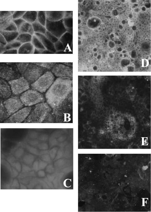
Understanding virus-cell interaction is a key to the design of successful gene delivery vectors. In the present study we investigated Ad5 transduction of enterocytes and M-cells utilizing differentiated Caco-2 cells and cocultures of Caco-2 cells with lymphocytes. Transduction inhibition studies showed that CAR is the major receptor mediating apical and basolateral virus entry in differentiated Caco-2 cells. Integrins and heparan sulfate glycosaminoglycans do not appear to play a significant role. Immunofluorescence localized CAR to sites of cell-cell contact, with staining mostly observed on the cell perimeter. Staining was observed even in nonpermeabilized monolayers, suggesting apical accessibility of the receptor. Cocultures with mouse Peyer's patch lymphocytes or Raji B human lymphocytes were more susceptible to transduction than Caco-2 cells, and the effects were dose-dependent. Similar to Caco-2 cells, CAR and not integrins mediated apical transduction. In conclusion, contrary to other epithelial cell lines, both apical and basolateral transduction of absorptive enterocytes and M-cells is mediated by binding to CAR. The coculture system can be used to study the interactions between M-cells and gene delivery vectors.


Figures

Figure 1
Figure 1. Transduction inhibition in fully differentiated Caco-2 monolayers: A) after apical application of the vectors; B) after basolateral application of the vectors
Caco-2 cell monolayers grown for 2 weeks on Transwell inserts were pre-incubated for 2h at room temperature with the following: RmcB anti-CAR monoclonal antibody (1:50 ascites), an RGD peptide (GRGDSP, 1mM) that blocks integrin binding, or culture medium (control). Subsequently cells were exposed to Ad5CMV-luc(loxP) (MOI 200) for 2h at 37°C. Basolateral application of solutions/vectors was carried out on inverted inserts. Luminescence activity was assayed after 48h. *** = p<0.001, ** = p<0.05 compared to control. Values are mean ± SE of 4 duplicates/triplicates.
Figure 2
Figure 2. Effect of blocking heparan sulfate glycosaminoglycan binding on transduction of fully differentiated Caco-2 monolayers: A) after apical application of the vectors; B) after basolateral application of the vectors
Vectors were pre-incubated with a 10 μg/ml solution of sodium heparin in DMEM with 0.1% FBS for 1h at 37°C. The vector was subsequently applied to the cells which were incubated at 4°C for 1hr. At the end of the incubation, medium was replenished and cells were returned to the 37°C incubator for 48h before assessing transduction efficiency (n=3-6 wells).
Figure 3
Figure 3. Immunofluorescence for CAR on fully differentiated Caco-2 monolayers
A) anti-CAR RmcB mAb on permeabilized monolayer; B) MOPC control immunoglobulin on permeabilized monolayer; C) PBS (no primary mAb) on permeabilized monolayer; D) anti-CAR RmcB mAb on permeabilized monolayer (low magnification); E) anti-CAR RmcB mAb on non-permeabilized monolayer (low magnification); F) MOPC control immunoglobulin on non-permeabilized monolayer (low magnification). Immunofluorescence was performed with standard techniques using an antibody to CAR (RmcB, 1:100 stock) or a control immunoglobulin (MOPC) followed by detection with an Alexa Fluor® 488 labeled secondary anti-mouse immunoglobulin mAb. Cells were imaged using an Olympus BX-51 epi-fluorescence microscope equipped with a digital camera.
Figure 4
Figure 4. Confocal microscope image of a permeabilized Caco-2 monolayer stained for CAR with the RmcB mAb
Immunofluorescence was performed with standard techniques, using an antibody to CAR (RmcB, 1:100 stock) followed by detection with an Alexa Fluor® 488 labeled secondary anti-mouse immunoglobulin mAb. Cells were imaged using a Bio-Rad MRC600 confocal microscope. Slice reconstruction was done in ImageJ imaging software.
Figure 5
Figure 5. Characterization of Caco-2 cell/mouse Peyer's patch lymphocyte co-cultures
A) Transmission electron micrograph of a Caco-2 cell monolayer co-cultured for three days with mouse Peyer's patch lymphocytes. A lymphocyte (L) can be seen within the Caco-2 monolayer. The epithelial cells above the lymphocyte exhibit a more sparse and less organized brush-border as well as less dense cytoplasm compared to neighboring epithelial cells not in contact with lymphocytes. B) TEER is unchanged in the co-cultures, confirming that tight junctions remain intact. Results of two experiments are shown, each with triplicate wells. C) Alkaline phosphatase activity assayed using p-nitrophenylphosphate (2.86 mM) as substrate. Absorbances at 405nm after a 1.5h incubation at 37°C were 0.454 ± 0.018 and 0.603 ± 0.024 (mean ± SD) for co-cultures and Caco-2 monocultures, respectively (n=6; p<0.0001). D) Transcytosis of fluorescent 0.2 μm microbeads from apical to basolateral medium, assessed by fluorescence microscopy. Images representative of two experiments, n=3-6.
Figure 6
Figure 6. Differences in transduction efficiency of Caco-2 cells and Caco-2/PP lymphocyte co-cultures
Cell monolayers were exposed to Ad5CMV-luc(loxP) for 2h at 37°C. Additional maintenance medium was added to the cells and infection was allowed to occur for 24 or 48h. Data are mean photon counts±SE, n=8-10 cultures, ** = p<0.01 for the comparison with Caco-2 monocultures at the same MOI.
Figure 7
Figure 7. Transduction inhibition experiments with an anti-CAR mAb (RmcB) on Caco-2 cells and Caco-2/PP lymphocyte co-cultures
Cell monolayers were pre-incubated for 2h with 1:50 dilution of RmcB mAb or MOPC control immunoglobulin. Subsequently cells were transduced with Ad5CMV-luc(loxP) for 2h at 37°C. Luminescence activity was assayed after 48h. ***=p<0.001, *=p<0.05 compared to control. Values are mean ± SD of 8-10 cultures.
Figure 8
Figure 8. Transduction inhibition experiments with an integrin-binding peptide on Caco-2 cells and Caco-2/PP lymphocyte co-cultures
Cell monolayers were pre-incubated with the RGD peptide GRGDSP (1mM in culture medium). Subsequently cells were exposed to Ad5CMV-luc(loxP) for 2h. Luminescence activity was assayed after 48h. Values are mean ± SD of 6-7 cultures.
Figure 9
Figure 9. Caco-2/Raji B cell co-cultures
A) assessment of tight junction integrity by TEER. Results of one representative experiment are shown, with ten replicate wells. B) Differences in transduction efficiency of Caco-2 cells and Caco-2/Raji B cell co-cultures. Cell monolayers were exposed to Ad5CMV-luc(loxP) for 2h at 37°C. Additional maintenance medium was added to the cells and infection was allowed to occur for 48h. Data are mean photon counts±SE; n=4 experiments, each with 2-3 replicate cultures; ** = p<0.005, * = p<0.05 for the comparison with Caco-2 monocultures at the same MOI.

Similar articles

Cited by

References

    1. Hidalgo IJ, Raub TJ, Borchardt RT. Characterization of the human colon carcinoma cell line (Caco-2) as a model system for intestinal epithelial permeability. Gastroenterology. 1989;96:736–749. - PubMed
    1. Walter E, Croyle MA, Davidson BL, Roessler BJ, Hilfinger JM, Amidon GL. Adenovirus mediated gene transfer to intestinal epithelial cells as a potential approach for oral delivery of peptides and proteins. J. Control. Release. 1997;46:75–87.
    1. Walter E, Croyle MA, Roessler BJ, Amidon GL. The absence of accessible vitronectin receptors in differentiated tissue hinders adenoviral-mediated gene transfer to the intestinal epithelium in vitro. Pharm. Res. 1997;14:1216–1222. - PubMed
    1. Croyle MA, Walter E, Janich S, Roessler BJ, Amidon GL. Role of integrin expression in adenovirus-mediated gene delivery to the intestinal epithelium. Hum. Gene Ther. 1998;9:561–573. - PubMed
    1. Bergelson JM, Cunningham JA, Droguett G, Kurt-Jones EA, Krithivas A, Hong JS, Horwitz MS, Crowell RL, Finberg RW. Isolation of a common receptor for Coxsackie B viruses and adenoviruses 2 and 5. Science. 1997;275:1320–1323. - PubMed

Publication types

MeSH terms

Substances

LinkOut - more resources